©The Author(s) 2025.
World J Nephrol. Mar 25, 2025; 14(1): 99555
Published online Mar 25, 2025. doi: 10.5527/wjn.v14.i1.99555
Published online Mar 25, 2025. doi: 10.5527/wjn.v14.i1.99555
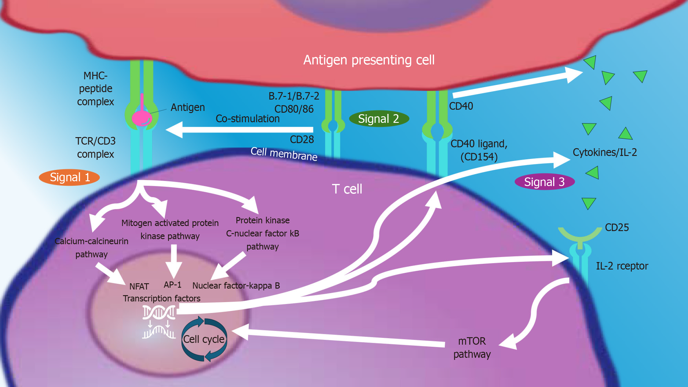
Figure 1 Three signal model of T-cell activation.
This figure presents a general overview of the key signaling events linking the production of a T-cell response. Signal 1 occurs when antigen-presenting cells (APCs) such as monocytes, macrophages, dendritic cells present antigen via major histocompatibility complex peptide complex on the APC cell surface to their respective T cell receptors on the surface of T cells and that is transduced through the CD3 complex. For Signal 1 to induce clonal expansion of naive T cells, the costimulatory signal 2 with B.7-1 (CD80) and B.7-2 (CD86) of dendritic cells binding with T-cell CD28 must also occur. Intracellular CD28 signaling also upregulates binding of CD40 by CD40 ligand (CD154) that also activates the APC to secrete proinflammatory molecules/and express B.7 molecules stimulating further T cell proliferation. These signals trigger three transduction pathways: Calcium-calcineurin, rat sarcoma virus-mitogen-activated protein kinase, and protein kinase C-nuclear factor kappa B. These pathways activate transcription factors that upregulate molecules such as interleukin-2 receptor genes and cytokines such as interleukin-2, supporting cell proliferation (signal 3), and activating the mammalian target of rapamycin pathway. Cell images obtained from reactome, an open-source, open access database. TCR: T cell receptor; MHC: Major histocompatibility complex; IL-2: Interleukin-2; mTOR: Mammalian target of rapamycin; NFAT: Nuclear factor of activated T cells; AP-1: Activating protein-1.
- Citation: Belal AA, Santos Jr AH, Kazory A, Koratala A. Providing care for kidney transplant recipients: An overview for generalists. World J Nephrol 2025; 14(1): 99555
- URL: https://www.wjgnet.com/2220-6124/full/v14/i1/99555.htm
- DOI: https://dx.doi.org/10.5527/wjn.v14.i1.99555


