©The Author(s) 2021.
World J Nephrol. Sep 25, 2021; 10(5): 101-108
Published online Sep 25, 2021. doi: 10.5527/wjn.v10.i5.101
Published online Sep 25, 2021. doi: 10.5527/wjn.v10.i5.101
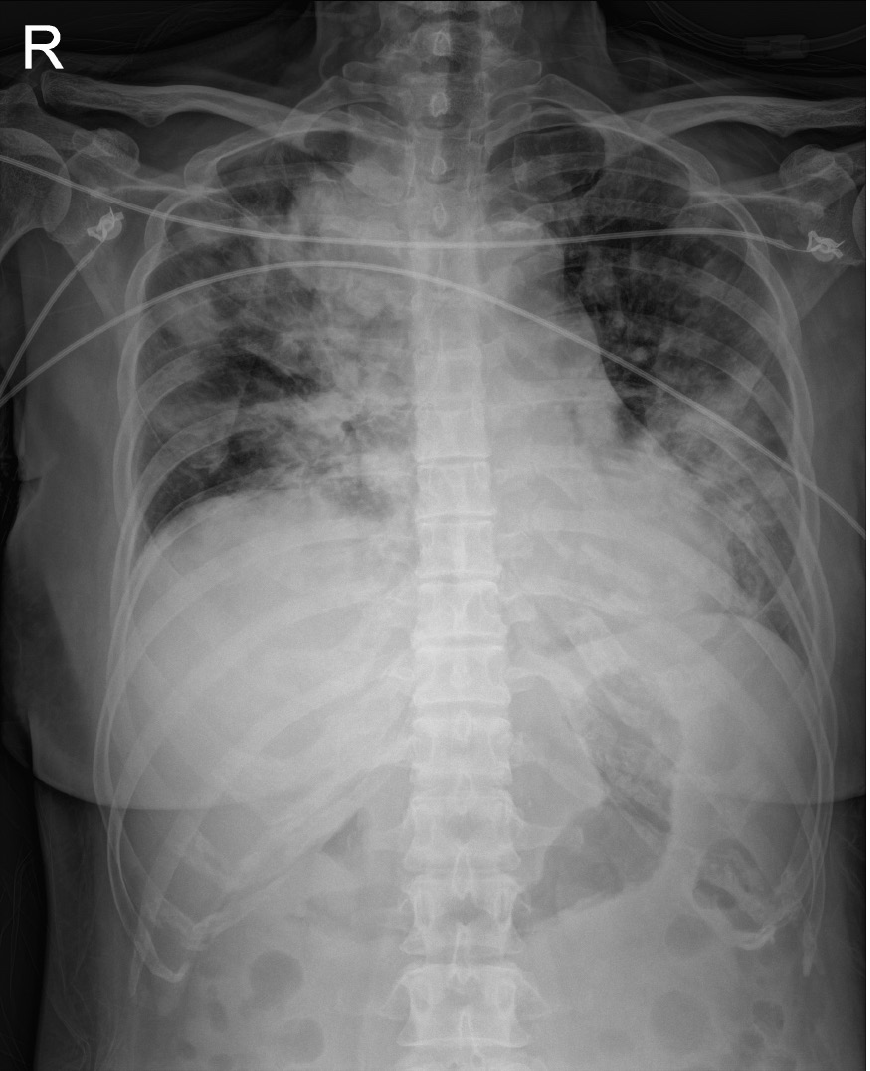
Figure 1
Chest radiography showed multiple patchy infiltrations at both lungs.
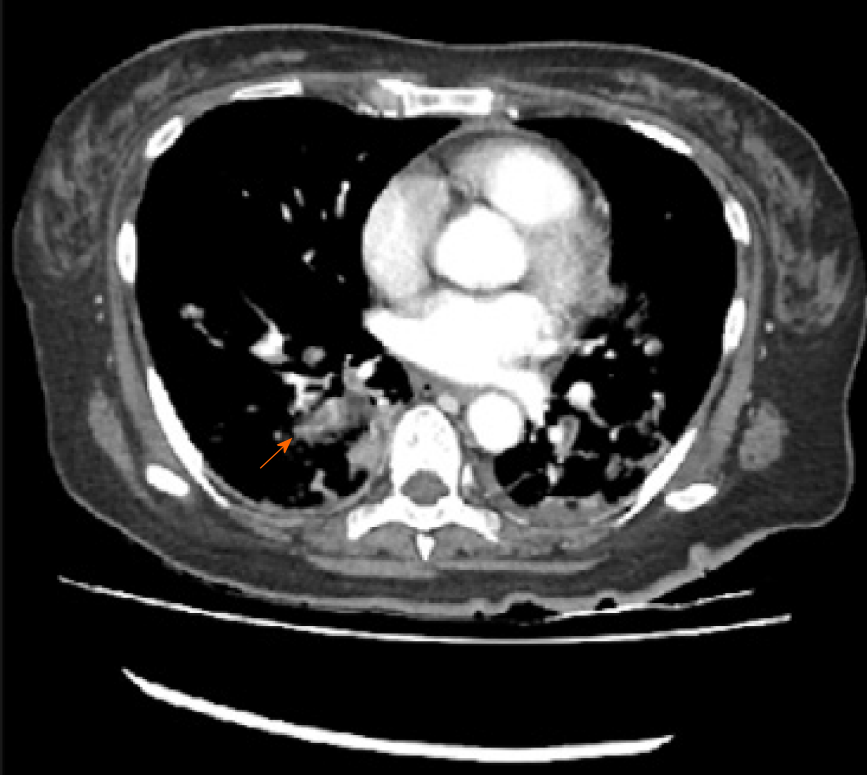
Figure 2
Computed tomography scan of the chest showed suspicious pulmonary thromembolism in segmental and subsegmental pulmonary arteries of right lower lobe (orange arrow).
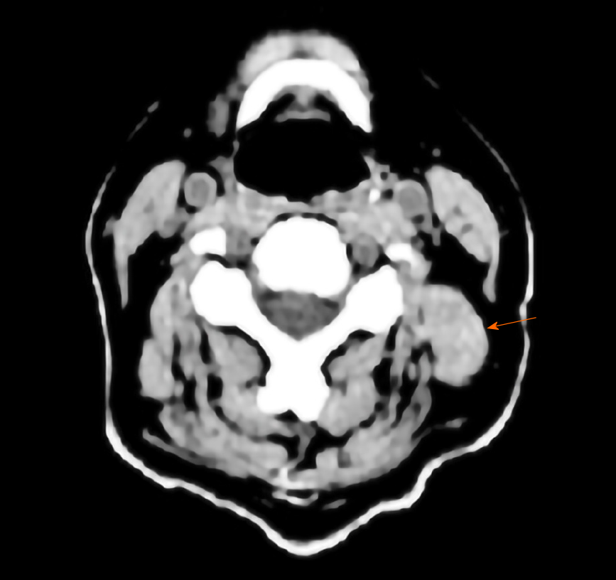
Figure 3
Computed tomography scan of the neck showed the 13 mm × 10 mm size nodular lesion (orange arrow) in left parotid gland.
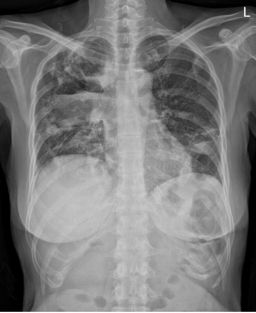
Figure 4
Chest radiography showed large cavitary consolidation with internal air-fluid level in right upper and middle lobes.
- Citation: Hwang SY, Shin SJ, Yoon HE. Lemierre's syndrome caused by Klebsiella pneumoniae: A case report. World J Nephrol 2021; 10(5): 101-108
- URL: https://www.wjgnet.com/2220-6124/full/v10/i5/101.htm
- DOI: https://dx.doi.org/10.5527/wjn.v10.i5.101


