Minireviews Open Access
Copyright ©The Author(s) 2021. Published by Baishideng Publishing Group Inc. All rights reserved.
World J Transplant. Nov 18, 2021; 11(11): 466-479
Published online Nov 18, 2021. doi: 10.5500/wjt.v11.i11.466
Exercise training in heart transplantation
Christos Kourek, Eleftherios Karatzanos, Serafim Nanas, Stavros Dimopoulos, Clinical Ergospirometry, Exercise & Rehabilitation Laboratory, Evaggelismos Hospital, Athens 10676, Attica, Greece
Andreas Karabinis, Stavros Dimopoulos, Cardiac Surgery Intensive Care Unit, Onassis Cardiac Surgery Center, Athens 17674, Greece
ORCID number: Christos Kourek (0000-0003-4348-2153); Eleftherios Karatzanos (0000-0002-6735-4183); Serafim Nanas (0000-0003-4666-4550); Andreas Karabinis (0000-0002-5666-7221); Stavros Dimopoulos (0000-0003-2199-3788).
Author contributions: Kourek C and Karatzanos E reviewed the literature; and Kourek C drafted the manuscript; Nanas S and Karabinis A revised the manuscript; Dimopoulos S designed the research study and revised the drafted manuscript; and all authors have read and approve the final manuscript.
Conflict-of-interest statement: Authors declare no conflict of interests for this article.
Open-Access: This article is an open-access article that was selected by an in-house editor and fully peer-reviewed by external reviewers. It is distributed in accordance with the Creative Commons Attribution NonCommercial (CC BY-NC 4.0) license, which permits others to distribute, remix, adapt, build upon this work non-commercially, and license their derivative works on different terms, provided the original work is properly cited and the use is non-commercial. See: http://creativecommons.org/Licenses/by-nc/4.0/
Corresponding author: Stavros Dimopoulos, MD, PhD, Consultant Physician-Scientist, Doctor, Postdoc, Research Scientist, Senior Researcher, Staff Physician, Clinical Ergospirometry, Exercise & Rehabilitation Laboratory, Evaggelismos Hospital, 45-47 Ipsilantou str., Athens 10676, Attica, Greece. stdimop@gmail.com
Received: March 1, 2021
Peer-review started: March 1, 2021
First decision: July 29, 2021
Revised: August 12, 2021
Accepted: October 27, 2021
Article in press: October 27, 2021
Published online: November 18, 2021
Processing time: 256 Days and 3 Hours

Abstract

Heart transplantation remains the gold standard in the treatment of end-stage heart failure (HF). Heart transplantation patients present lower exercise capacity due to cardiovascular and musculoskeletal alterations leading thus to poor quality of life and reduction in the ability of daily self-service. Impaired vascular function and diastolic dysfunction cause lower cardiac output while decreased skeletal muscle oxidative fibers, enzymes and capillarity cause arteriovenous oxygen difference, leading thus to decreased peak oxygen uptake in heart transplant recipients. Exercise training improves exercise capacity, cardiac and vascular endothelial function in heart transplant recipients. Pre-rehabilitation regular aerobic or combined exercise is beneficial for patients with end-stage HF awaiting heart transplantation in order to maintain a higher fitness level and reduce complications afterwards like intensive care unit acquired weakness or cardiac cachexia. All hospitalized patients after heart transplantation should be referred to early mobilization of skeletal muscles through kinesiotherapy of the upper and lower limbs and respiratory physiotherapy in order to prevent infections of the respiratory system prior to hospital discharge. Moreover, all heart transplant recipients after hospital discharge who have not already participated in an early cardiac rehabilitation program should be referred to a rehabilitation center by their health care provider. Although high intensity interval training seems to have more benefits than moderate intensity continuous training, especially in stable transplant patients, individualized training based on the abilities and needs of each patient still remains the most appropriate approach. Cardiac rehabilitation appears to be safe in heart transplant patients. However, long-term follow-up data is incomplete and, therefore, further high quality and adequately-powered studies are needed to demonstrate the long-term benefits of exercise training in this population.

Key Words: Heart transplantation; Endothelial dysfunction; Exercise training; High intensity interval training; Moderate intensity continuous training; Cardiac rehabilitation

Core Tip: Heart transplantation is the gold standard treatment of end-stage heart failure (HF). Heart transplantation patients present lower exercise capacity due to cardiac, vascular and skeletal muscle abnormalities. Exercise training improves exercise capacity, cardiac and vascular endothelial function in heart transplant recipients. Pre-rehabilitation regular aerobic or combined exercise is beneficial for patients with end-stage HF awaiting heart transplantation. All heart transplant recipients either hospitalized or after hospital discharge should be referred to a cardiac rehabilitation program. Individualized training still remains the most applicable approach despite the fact that high intensity interval training seems to have more benefits than moderate intensity continuous training.


INTRODUCTION

Heart transplantation is the gold standard treatment of end-stage heart failure (HF), although important advances in the field of mechanical circulatory support and technology have been noticed during the last 30 years. Since the first human-to-human heart transplant operation by a cardiac surgeon called Christiaan Neethling Barnard back in 1967, many adult heart transplants have been performed worldwide, especially in patients with end-stage HF. A continuous improvement in morbidity and mortality of transplanted recipients has been noticed despite the fact that they may be older with higher risk[1]. Heart transplantation remains, however, a difficult operation with significant short-term and long-term post-surgery outcomes including graft-related complications such as early graft dysfunction, acute allograft rejection and cardiac allograft vasculopathy, and non-graft-related complications such as infections, acute and chronic renal injury and malignancies[2]. All these complications usually lead to higher morbidity and mortality[3].

Despite the fact that donor and recipient age and comorbidity are being increased over the last years, heart transplantation survival rates seem to have progressively improved. It is estimated that, worldwide, almost 5000 transplants are being performed each year. The median survival for adult heart transplant recipients varies and ranges from 10.7 to 12.2 years approximately, with 82% one-year survival and 69% at five-years[1]. Survival for women is slightly better compared to men[1]. The highest incidence of mortality most often occurs within the first 6 mo after the transplant[1]. After 12 mo, the mortality rate decreases to 3.4% per year[1].

Heart transplantation patients present lower exercise capacity due to cardiac, vascular and skeletal muscle abnormalities leading thus to poor quality of life and reduction in the ability of daily self-service[4]. Impaired vascular function and diastolic dysfunction cause lower cardiac output (CO) while decreased skeletal muscle oxidative fibers, enzymes and capillarity cause arteriovenous oxygen difference, leading thus to decreased peak oxygen uptake (VO2) in heart transplant recipients which is lower at about 40% to 50% than age, sex, and activity matched healthy controls[4-6]. Exercise has been proven to improve exercise capacity and vascular endothelial function in patients with chronic HF and thus in patients with vascular endothelial impairment[7-10]. Exercise also improves aerobic capacity via the suppression of the oxidative stress, the increase of the bioavailability of nitric oxide (NO) and the induction of vasodilation[11,12].

The aim of this narrative review was to demonstrate the existing knowledge on the training protocols and highlight the benefits of exercise training in patients after heart transplantation.

CARDIOVASCULAR AND MUSCULOSKELETAL ALTERATIONS IN HEART TRANSPLANT RECIPIENTS
Cardiovascular alterations

One possible complication after heart transplantation is that the donor heart is surgically denervated and loses efferent and afferent autonomic connections. As a result, the regulation and function of the cardiovascular system is being affected and its reflex reactions are reduced[13,14] (Figure 1). Hypertension and peripheral vasoconstriction are usually the first signs in heart transplant recipients[14]. A possible explanation could be the permanent denervation of low-pressure cardiopulmonary baroreceptors in the heart and the permanent enhancement of sympathetic vasomotion due to lack of afferent impulses[15,16]. Left ventricular (LV) mass and wall thickness is increased, either within the first 30 d after heart transplantation or secondary as a consequence by immunosuppressive agents which trigger chronic tachycardia, hypertension and multiple rejection episodes[17,18].

Figure 1
Figure 1 Cardiovascular and musculoskeletal alterations in recipients after heart transplantation.

Despite the fact that the atrial remnant of the heart transplant recipients is innervated, the suture line causes higher intrinsic rate of the atrium and reduced heart rate variability[19]. Moreover, although some cardiac functions and mechanisms such as Frank-Starling are usually not affected in transplant patients, most heart responses to haemodynamic changes and heart rate variations are impaired[19]. A study by Nygaard et al[20], has shown that patients after heart transplantation present altered cardiovascular responses than healthy controls. Most specifically, their blood pressure and total peripheral resistance is higher at supine rest, attenuated during orthostatic challenge and preserved during isometric exercise. Cardiac denervation mediated chronotropic and allograft diastolic dysfunction after heart transplantation results in lower peak exercise CO[21,22]. Peak exercise CO in heart transplant recipients is 30% to 40% lower than age-matched healthy controls[21-23]. Lower CO leads to a higher resting heart rate and slower increase during exercise[24]. However, physical activity status and cardiac allograft reinnervation are important factors of the variety of heart rate impairment[4]. Moreover, diuretics leading to reduced diastolic filling might contribute to lower CO responses[20].

Heart transplant recipients present 20% lower peak exercise end-diastolic volume and stroke volume than healthy people[21,22]. This is another significant pathophysiological alteration. The impaired LV relaxation and the increased LV stiffness could be a possible explanation of this alteration[4,21]. In addition, previous studies have shown the significantly increased pulmonary capillary wedge pressure (PCWP)/ end-diastolic volume index ratio during maximal exercise in these patients compared to age-matched sedentary normal controls[21]. This elevated peak exercise mean PCWP results in dysfunction in LV strain and peak systolic velocity of the mitral valve, despite the preserved LV ejection fraction[25]. A possible potential pathway that causes LV diastolic dysfunction may be the decreased adrenergic tone associated with complications after the heart transplantation including denervation of the allograft, injury, myocardial ischemia due to vasculopathy or immunosuppression therapy[4,23].

Endothelial dysfunction is a major cause of disability and lower life expectancy in heart transplant recipients which increases exercise systemic vascular resistance, leading thus to less O2 provided to skeletal muscles[1,4] (Figure 1). As a result, peak VO2 is significantly reduced[1,4]. Several studies have shown that their peak exercise systemic vascular resistance is approximately 50% higher than healthy controls because of the impaired endothelial-dependent vasodilation of peripheral conduit arteries and resistance arterioles[21,22,26,27]. A significant observation is that the severity of the impairment of endothelial function appears to be related to the etiology of HF[28,29]. Most specifically, endothelial function is usually improved in similar levels to healthy age-matched controls in heart transplant recipients of non-ischemic cardiomyopathy compared to ischemic cardiomyopathy due to slower pulmonary VO2 kinetics during ischemia[26,29].

Musculoskeletal alterations

The last significant impairment after heart transplantation concerns abnormalities in skeletal muscles leading in impaired peak VO2 (Figure 1). Heart transplant recipients present lower body and leg lean mass, as well as muscle strength, compared to healthy, sedentary age-matched controls[30].

Before heart transplantation, reduced oxidative muscle fibers (Type I), capillary density, mitochondrial volume and oxidative enzyme capacity are usual abnormalities of the skeletal muscles, directly associated with the syndrome of HF and worsen according to its severity[30-32]. More precisely, a reduction in aerobic, Type I and an increase in anaerobic, glycolytic fibers (Type II) is observed in HF patients[33]. Their diaphragm is also metabolically affected with significant atrophy[33-35]. Moreover, reduced levels of enzymes and proteins such as citrate synthase, creatine kinase (CK), MM-CK and lactate dehydrogenase (LDH) prove a major contribution of altered skeletal muscle metabolism to exercise intolerance[36]. As far as muscle metabolism is concerned, electrolyte and phosphocreatine (PCr) disorders, metabolic acidosis and delayed PCr recovery after exercise are common characteristics of patients with HF[33,34]. Finally, muscle atrophy is caused by a decrease of anabolic mechanisms, increased protein degradation or sometimes both of them[33]. Enhanced protein degradation including impaired function of enzymes ubiquitin-ligases MuRF1 and Atrogin-1, impaired growth factor signaling and protein synthesis including decreased levels of circulating total testosterone, dehydroepiandrosterone and insulin like growth factor-1 and skeletal muscle inflammation including inflammatory mediators released into the circulation such as interleukin 1 (IL-1) and IL-6 and tumor necrosis factor α are the major mechanisms that promote muscle atrophy and skeletal muscle alterations[33,37,38].

Size of muscle fiber and mitochondrial volume density increases after heart transplantation reaching almost equal levels to healthy age-matched individuals[30,39]. However, reductions in capillary density persist[30,32,39]. Moreover, endurance in exercise performance seems to be impaired by immunosuppression therapy including cyclosporine and corticosteroids[40,41]. A consequence of all these musculoskeletal abnormalities is a decrease in bone density and oxygen utilization, and possible osteoporotic fractures[4,38,42,43].

THE EFFECT OF EXERCISE TRAINING IN THE CARDIOMYOCYTES, VASCULAR ENDOTHELIAL FUNCTION AND SKELETAL MUSCLES

Exercise training has a beneficial effect in the cardiomyocytes and the function of the vascular endothelial system. As far as the cardiomyocytes are concerned, exercise results in a beneficial form of cardiac remodeling that involves cardiomyocyte growth and proliferation[44,45]. Regular physical exercise has been proven to improve LV contractility, calcium function in the heart and cardiomyocytes size[46,47]. Isometric or static exercises result in mild concentric hypertrophy and usually a normal left atrium while endurance training LV hypertrophy, right ventricular (RV) dilation, and biatrial enlargement[45,48]. In the first case, the increase in cardiac wall thickness is caused by the parallel addition of sarcomeres within cardiomyocytes while in the second case by the addition of cardiomyocyte sarcomeres in series[45,48]. Cardiomyocyte hypertrophy is not the only process in exercise-induced cardiac remodeling. The increased levels of circulating endothelial cells (CECs) and endothelial progenitor cells (EPCs) after acute and long-term exercise seem to play a crucial role in augmentation of vascular density and cardiac repair[49-51].

Exercise training can also contribute to the proliferation of cardiomyocytes, a significant process of cardiomyogenesis[52]. Metabolically, exercise has beneficial effect on LV contractility and increases catabolism of fatty acids and lactate, and therefore of ATP production[53-55]. Circulating metabolites including palmitoleate (C16:1n7), G protein-coupled receptors, Akt, and nuclear receptors are important regulators of exercise-induced cardiac growth[53,56,57].

Regarding the vascular endothelium, exercise has been proven to suppress the generation of free radicals and oxidative stress and increase the bioavailability of NO[11,12]. As far as the potential mechanisms are concerned, shear stress is a procedure that activates eNOs, increases the concentration of NO and induces vasodilation[11,12,58,59]. Exercise increases shear stress, and thus, improves aerobic capacity[11,12,58,59]. Moreover, exercise induces the hypoxic stimuli, as observed by alterations in microcirculation indexes during exercise sessions[59,60]. All these pathophysiological mechanisms may relate to up-regulation of transcriptional factors, including vascular endothelial growth factor (VEGF), matrix metalloproteinases and stromal cell-derived factor 1, and lead to angiogenesis during exercise in healthy controls and patients with comorbidities[60-63]. In healthy subjects, exercise improves peripheral vascular function through the reduction in blood pressure, the endothelin-1 levels and the improvement in vasodilation[64,65].

EPCs and CECs have been shown to restore the dysfunctional or injured endothelium and protect it, regulate vascular homeostasis and promote angiogenesis. Therefore, reflecting the condition of the vascular endothelial function[12,66]. EPCs level is a predicting factor of the occurrence of a cardiovascular event and cardiovascular mortality[67]. Several studies have shown that in both healthy people and population with comorbidities, exercise training increases the number and the function of EPCs[68,69]. We extended previous findings by showing that a single bout of maximal exercise, as well as many bouts organized as an exercise training program, stimulates the mobilization of EPCs and CECs from the bone marrow in patients with chronic HF[8,10]. This beneficial effect of exercise seems to be similar in chronic HF patients of different severity[9].

Physical exercise has the beneficial effect to modify metabolic potential, morphology, and physiology of skeletal muscle[70]. Exercise is a triggering factor for the metabolic and structural skeletal muscle remodeling[70,71]. This remodeling has positive effect in angiogenesis and fatigue[70,71]. Resistance exercise increases muscle mass and strength while endurance training affects mitochondrial function and oxidation[70,72]. Regular exercise mediates molecular and metabolic pathways that are activated by muscle contraction. Intracellular sensors trigger intracellular signaling cascades including several transcription factors[70,72]. These factors are responsible for the remodeling of skeletal muscle via upregulation of mitochondrial metabolism and fiber-type transformation[70,72]. Finally, other potential mechanisms for muscle remodeling such as redox signaling seem to be involved in metabolic adaptation to exercise[70,73,74].

CARDIAC REHABILITATION IN HEART TRANSPLANTATION

Cardiac rehabilitation programs are being implemented all over the world for patients after major cardiovascular disease. A cardiac rehabilitation program is characterized by an interdisciplinary approach and consists of different specialties and health care professionals including cardiologists, physiotherapists, nurses, dieticians, pharmacists, psychologists, physiologists, other specialties such as internists, neurologists, diabetologists and cardiac surgeons, general practitioners and social services experts[75]. One of the most important roles is the role of the program director. A program director could be of any specialty with good organizing and management skills.

Cardiac rehabilitation is a type of secondary prevention in patients with cardiovascular disease. The aim of rehabilitation is to reduce anxiety and depression and instill confidence so that to change lifestyle of patients aimed at preventing further disease[76]. Each patient could benefit from either an in-patient or out-patient cardiac rehabilitation program. The core principles of a cardiac rehabilitation program are patients’ medical evaluation, counselling for exercise training and diet, continuous assessment of weight, blood pressure, lipidemic profile, and psychosocial support[75]. The expected outcomes of a cardiac rehabilitation program are improvement of clinical stability and symptom control of patients, reduce of cardiovascular risk, better compliance to medical therapy, and improved quality of life, social integration and prognosis[75].

Another important parameter of a successful cardiac rehabilitation program is the equipment. A cardiac rehabilitation center should provide the appropriate equipment for the assessment of patient’s clinical status, LV function, arrhythmias, functional capacity, psychosocial status and equipment for conducting an exercise training program. These include stethoscopes and sphygmomanometers, electrocardiogram, echocardiography, echocardiography, graded exercise testing on treadmills or cycles, cardiopulmonary exercise testing (CEPT), six-minute walk test (6-MWT), questionnaires about quality of life and psychological status and exercise equipment such as treadmills, cycle ergometers and weight training equipment[75]. Moreover, emergency equipment for complications during exercise is always mandatory.

Phases of cardiac rehabilitation

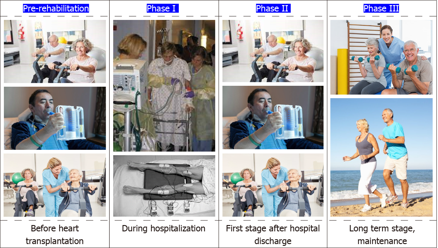
Rehabilitation is a complex process, individualized for each patient. Three main phases of rehabilitation can be differentiated according to the updated guidelines about preventive cardiology and rehabilitation of the ESC[75] (Figure 2): (1) Phase 1 is the phase of the in-hospital rehabilitation including early interventions and mobilization immediately after hospital admission[75]; (2) Phase 2 is probably the most critical part in patients with heart transplantation. It is being implemented just after the hospital discharge. It promotes and delivers in-patient and out-patient rehabilitative services for clinical stabilization[75]. In-patient cardiac rehabilitation is being performed to unstable patients in order to stabilize them before the longer-term cardiac rehabilitation program after hospital discharge. Clinically unstable patients after an acute event, with advanced HF under continuous medication or with implantable devices, heart transplant recipients and patients unable to attend a formal outpatient rehabilitation program for any personal reasons are considered as high risk[75]. On the other hand, early out-patient cardiac rehabilitation is being used for independent patients early after hospital discharge, usually within 3 to 6 mo after a cardiovascular event. The mean duration is 8 to 12 wk, most times continuing for one year after the event[75]. Finally, a home-based program is another form of rehabilitation assessed and supported by the rehabilitation group at patient’s home. It may include regular visits to the rehabilitation center and contacts with the team. The activities of a home-based program are similar to those of an early outpatient cardiac rehabilitation program[75]; and (3) Phase 3 is the long-term out-patient type of cardiac rehabilitation. The main aim of phase 3 rehabilitation is to promote long-term exercise and rehabilitation in patients out of hospital and the community. Moreover, it usually results in maintenance of the fitness level and better outcomes in heart transplant recipients[75].

Figure 2
Figure 2 Stages of a cardiac rehabilitation program.

Another important phase of rehabilitation is the “pre-rehabilitation” stage. Heart transplant recipients are doing regular aerobic or combined exercise before transplantation in order to maintain a higher fitness level and reduce complications afterwards like intensive care unit (ICU) acquired weakness or cardiac cachexia.

Significant components of a cardiac rehabilitation program for heart transplant patients

The initial step of the enrollment of a heart transplant recipient in a cardiac rehabilitation program is the risk assessment of the patient by the rehabilitation team. The risk assessment consists of clinical examination including sings such as examination of the wound healing or symptoms of the transplant’s rejection, imaging techniques such as chest X ray for infection, pleural effusion or diaphragm paralysis and echocardiography for RV and LV function or pericardial effusion[75]. Moreover, tests for exercise capacity including CPET 30 d after transplantation or bicycle ergometer and modified Bruce protocols and Naughton protocols on treadmill are recommended[75]. Patient education on the risk of acute rejection is also a significant variable of a rehabilitation program. Patients should be instructed to practice self-monitoring during their rehabilitation process. In the case of transplant rejection, usually presented with significant reduce in blood pressure, unexpected variations of heart rate, fever or fatigue, exercise training should be immediately stopped and appropriate interventions are needed[75]. As far as health care professionals are concerned, they need to be aware of all aspects of this condition. For example, physicians should have full knowledge regarding the possible reasons for patients’ limited exercise tolerance which could possibly be the immune-suppression therapy side effects, chronotropic incompetence or LV diastolic dysfunction[75]. They should also be aware of all necessary actions to prevent complications which could harm patients and avoid infections, and therefore transplant rejection[75].

The second step of a cardiac rehabilitation program is physical activity counselling. Most specifically, heart transplant recipients enrolling a rehabilitation program should perform chronic dynamic and resistance exercises in order to prevent the side-effects of immunosuppressive therapy. In addition, exercise intensity should be increased slowly over time so that patients could reach a score of 12-14 in the Borg scale[75].

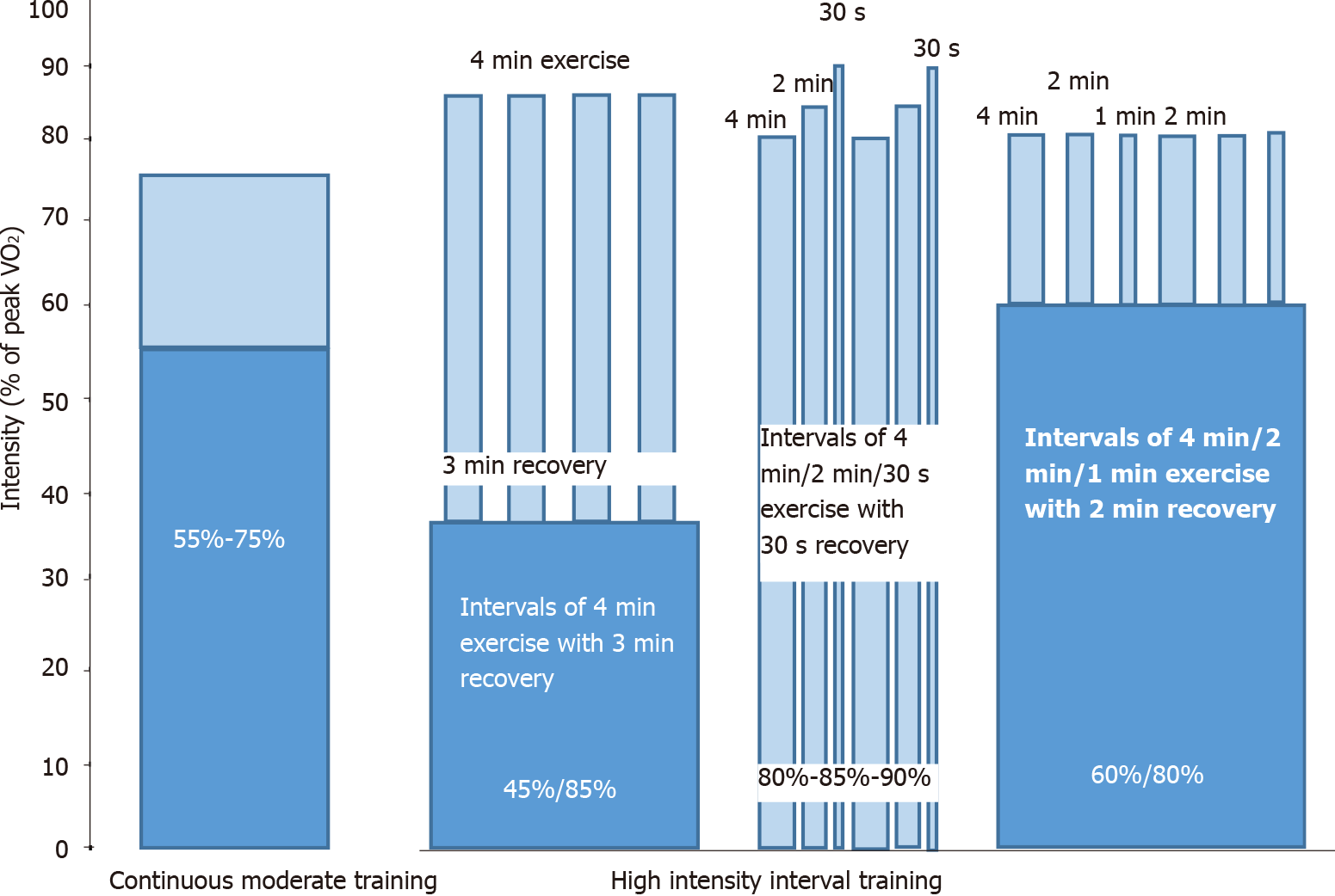
Exercise training is the most important aspect of a cardiac rehabilitation program. Early training program could be beneficial in the early and the long-term post-operative period. Early mobilization of heart transplant recipients could be achieved by implementing kinesiotherapy of the upper and lower limbs and prevention of respiratory infections could be achieved by performing respiratory physiotherapy[75]. Supervised exercise programs during the initial phase may be crucial to verify individual responses, tolerability and adaptability to exercise and clinical stability[75]. Aerobic exercise should be performed immediately after CPET for patients’ prescription. Specifically, regular aerobic exercise may start in the second or third week after transplantation while resistance exercise should be added after 6 to 8 wk. However, exercise should be discontinued during corticosteroid bolus therapy for rejection[75]. A duration of at least 30-40 min/d of combined aerobic and resistance training at moderate level, slowly progressing warm-up, closed-chain resistive activities and cycling in each exercise training session should be achieved[75]. The intensity of aerobic exercise could be calculated according to peak VO2 (< 50% or 10% below Ventilatory Anaerobic Threshold determined by CEPT) or peak work load (< 50%)[77]. Resistance training should consist of 2-3 sets with 10-12 repetitions per set at 40%-70% of the 1-repetition maximum (RM) test with > 1 min recovery between sets in order to achieve 5 sets of 10 repetitions at 70% of the 1-RM test[75]. Aerobic exercise could be either continuous moderate training (COMT) or high intensity interval training (HIIT) (Figure 3). COMT includes sessions consisted of aerobic exercise of 40 min with a continuous intensity of 55%-75% of peak VO2[75,78]. HIIT may varies between different rehabilitation centers. It could either consist of 40-min exercise of high intensity (blocks of 4 min-2 min-30 s according to 80%, 85% and 90% of peak VO2 with 1 or 2 min recovery)[79] or 16-min interval training (intervals of 4, 2 and 1-min duration at > 80% of peak VO2 with a 2-min active rest period of approximately 60% of peak VO2)[78]. Another very common HIIT protocol consists of 4 min × 4 min exercise bouts at 85%-95% of maximum heart rate, with 3 min recovery between them corresponding to 11-13 on the Borg scale[80]. Duration between COMT and HIIT sessions is similar and a 10-min pre-training warm up above 50% of peak VO2, as well as a 10-min post-training stretching and exercises are included in both protocols. HIIT is suggested for hemodynamic stable heart transplant recipients with beneficial effects for them[75,78-80].

Figure 3
Figure 3 Different protocols of continuous moderate training and high intensity interval training. VO2: Oxygen uptake.

Except for exercise training, there are other important parameters which contribute to the success level of a cardiac rehabilitation program. Patients should be guided by expert nutritionists in order to maintain a balanced diet without sudden weight gain that could increase the risk of cardiac allograft vasculopathy or other classical cardiovascular risk factors[75] and avoid food that could lead to infection such as raw meat or seafood, un-pasteurized milk or cheese and raw eggs. A healthy lifestyle should be adopted by patients in their daily program. Monitoring of blood pressure, lower sodium intake, avoidance of hyperlipidemia and tobacco smoking, and adherence to the suggested medication would increase the beneficial effect of rehabilitation and reduce drug side effects. Appropriate medication with diltiazem, amlodipine and angiotensin-converting enzyme inhibitors, usually completed by diuretics, is mandatory. Also, statins, daily exercise and healthy diet should be applied in patients with hyperlipidemia in order to reduce the possibility of cardiovascular disease and improve survival[75]. Finally, psychosocial management is being considered as an important element in each cardiac rehabilitation program. Patients usually present high levels of depression, apprehensiveness or anxiety, and therefore support coping strategies should be implemented by expert psychologists[75].

Effects of exercise training in heart transplant patients

As far as pre-rehabilitation stage is concerned, 2 clinical trials were recently conducted in patients awaiting heart transplantation. In the first study[81], 7 end-stage HF patients awaiting heart transplantation while on intravenous inotropic support performed exercise training on a cycle ergometer while 11 patients followed the conventional protocol. 6-MWT assessed exercise capacity and manovacuometry assessed inspiratory muscle strength before and after each protocol. The intergroup comparison revealed significant increase in 6-MWT and inspiratory muscle strength in the intervention group compared to the control (P < 0.01)[81]. In the second study[82], 24 HF patients with advanced symptoms awaiting heart transplantation performed HIIT during hospitalization. HIIT was shown to improve skeletal muscle strength, and most specifically knee extensor strength, and decrease brain natriuretic peptide levels in these patients, however, without having any effect on hand grip strength[82].

Exercise training, as early as possible after hospital discharge, is being considered beneficial for the acute and long-term outcomes of heart transplant patients[83-86]. It has been shown to improve endothelial function assessed by brachial artery flow-mediated dilatation (FMD)[79]. In addition, it reduces systolic blood pressure, pro-atrial natriuretic peptide and high sensitive C-reactive protein (CRP)[79]. Two clinical trials investigated the effects of exercise training within the first year of hospital discharge after transplantation. In Braith et al[85] study, 8 wk after transplantation, 10 heart transplant recipients performed COMT on a treadmill 3 d/wk for 12 wk and 10 recipients took standard medical care for the same time period. Patients performed warm-up for 5 min, treadmill walking for 30 min and cooldown for 5 min within the first month. After the first month, treadmill walking increased to 35-40 min with an intensity between 11 and 13 or 12 and 14 of the Borg scale. Brachial artery reactivity was assessed using flow-mediated dilation. This randomized clinical trial proved the benefit of aerobic exercise on peripheral artery function in the early period after heart transplantation, demonstrating increase in brachial artery FMD in contrast with the progressive decline in patients who did not undergo rehabilitation. In addition, resting norepinephrine decreased significantly (P < 0.05) after exercise in the training group compared to controls and peak VO2 increased 26% in the trained patients but remained unchanged in controls[85]. In another study of the same institution[42], 8 heart transplant recipients, 2 mo after transplant, underwent a 6-mo resistance training program (2 d/wk, 10-15 repetitions at 50% of 1-RM in the beginning and then increase by 5%-10% in resistance in each set) for upper and lower body while 7 recipients were used as a control group. The aim of the study was to show the shift of type II fibers to type I fibers through biopsy of the right vastus lateralis, and therefore the beneficial effect of resistance training in the reverse of skeletal muscle myopathy even within a few days after heart transplantation[42].

Several studies have shown the effects of exercise training in heart transplant patients who enrolled a cardiac rehabilitation program in more than one year after hospital discharge. Most of these studies examined the effect of HIIT protocol in different functional capacity and vascular endothelial function indices. Nytrøen et al[80], included 48 clinically stable heart transplant recipients 1-8 (mean time: 4.1 ± 2.2) years after transplantation. Maximal CPET on a treadmill was performed in both 12-mo HIIT patients (intervention group) and patients who received usual care for the same time period. The HIIT group performed warm up for 10 min, followed by four 4 min exercise bouts at 85% to 95% of their maximum heart rate, with 3 min recovery time between them (intensity 11-13 on the Borg scale). Exercise group presented higher mean peak VO2 and predicted peak VO2 compared to controls (P < 0.001). Muscular exercise capacity and general health were also improved. Hermann et al[79], examined the effect of HIIT on peak VO2 and FMD of the brachial artery in 14 patients after heart transplantation who performed an 8-wk HIIT program. Each session included a warm up above 50% of peak VO2 and then 42 min of HIIT divided in 4 min-2 min-30 s intervals at 80%, 85% and 90% of peak VO2 (intensity at 18-19 of the Borg scale) with 1- or 2-min recovery between the intervals. There was also a control group of 13 patients after heart transplantation who did not exercise. Blood pressure and several indices were also evaluated at baseline and 8 wk later. There was a significant increase in peak VO2 and FMD in patients performed HIIT compared to controls, but nitroglycerin-induced vasodilation remained unchanged. Moreover, HIIT reduced systolic blood pressure in heart transplant recipients while it remained unchanged in controls, indicating thus the benefits of HIIT in endothelial after transplantation. Monk-Hansen et al[87], did not observe improvement of LV function in heart transplant recipients after an 8-wk exercise training program, although an increase in peak VO2 was noticed.

A single study recently examined the effects of COMT on ambulatory blood pressure and arterial stiffness of heart transplant recipients. In this study[88], 40 patients either performed 40 min endurance exercise at 70% of peak VO2 (3 times per week) for 12 wk or did not perform any kind of exercise. All patients underwent CPET, 24-h ambulatory blood pressure monitoring, and carotid-femoral pulse wave velocity assessment in 2 time periods; at baseline and after 12 wk. COMT reduced ambulatory blood pressure but pulse wave velocity remained unchanged, suggesting thus that it could be beneficial for the treatment of hypertension in heart transplant recipients.

Comparing HIIT an COMT, Yardley et al[89] showed that heart transplant patients had similar beneficial effect in inflammatory indices such as CRP, blood platelets and angiogenesis, but indices of angiogenesis including VEGF and angiopoietin 2 after HIIT seemed to increase more than COMT.

Finally, combined exercise, including aerobic exercise and muscle strength training, is still under investigation.

Limitations and perspectives

In most studies there are gaps in methodology which could lead to bias. Inclusion criteria, different baseline exercise capacity and fitness level, differentiations in exercise training protocols and small number of samples are some variables that may lead to systemic bias and underpowered conclusions. Taking these factors into consideration, it would be safer to reach a conclusion that HIIT is effective and feasible in heart transplant patients rather than state that it is more beneficial than exercise with moderate intensity. Moreover, patients may drop out of the program either for logistic reasons or for complications of the transplantation caused by transplant rejection, infections and side effects of the immunosuppressive therapy.

Many cases of heart transplant patients could be inspiring examples of the remarkable human exercise capacity. A combination between the conventional post-heart transplantation multi-disciplinary medical therapy with the carefully monitored aerobic or combined endurance exercise training could be a real breakthrough in the field of medicine.

CONCLUSION

Exercise training improves exercise capacity, cardiac and vascular endothelial function in heart transplant recipients. Pre-rehabilitation regular aerobic or combined exercise is beneficial for patients with end-stage HF awaiting heart transplantation in order to maintain a higher fitness level and reduce complications afterwards like ICU acquired weakness or cardiac cachexia. All hospitalized patients after heart transplantation should be referred to early mobilization of skeletal muscles through kinesiotherapy of the upper and lower limbs and respiratory physiotherapy in order to prevent infections of the respiratory system prior to hospital discharge. Moreover, health care providers should suggest all heart transplant recipients to participate in a rehabilitation program after hospital discharge. Although HIIT seems to have more benefits than COMT especially in stable transplant patients, individualized training based on the abilities and needs of each patient still remains the most appropriate approach. Cardiac rehabilitation appears to be safe in heart transplant patients. However, long-term follow-up data is incomplete and, therefore, further high quality and adequately-powered studies are needed to demonstrate the long-term benefits of exercise training in this population.

Footnotes

Provenance and peer review: Invited article; Externally peer reviewed.

Specialty type: Transplantation

Country/Territory of origin: Greece

Peer-review report’s scientific quality classification

Grade A (Excellent): 0

Grade B (Very good): B

Grade C (Good): 0

Grade D (Fair): 0

Grade E (Poor): 0

P-Reviewer: Zhang ZX S-Editor: Wang JJ L-Editor: A P-Editor: Wang JJ

References
1.  Lund LH, Edwards LB, Dipchand AI, Goldfarb S, Kucheryavaya AY, Levvey BJ, Meiser B, Rossano JW, Yusen RD, Stehlik J. International Society for Heart and Lung Transplantation. The Registry of the International Society for Heart and Lung Transplantation: Thirty-third Adult Heart Transplantation Report-2016; Focus Theme: Primary Diagnostic Indications for Transplant. J Heart Lung Transplant. 2016;35:1158-1169.  [PubMed]  [DOI]  [Cited in This Article: ]  [Cited by in Crossref: 288]  [Cited by in F6Publishing: 295]  [Article Influence: 36.9]  [Reference Citation Analysis (0)]
2.  Potena L, Zuckermann A, Barberini F, Aliabadi-Zuckermann A. Complications of Cardiac Transplantation. Curr Cardiol Rep. 2018;20:73.  [PubMed]  [DOI]  [Cited in This Article: ]  [Cited by in Crossref: 19]  [Cited by in F6Publishing: 28]  [Article Influence: 4.7]  [Reference Citation Analysis (0)]
3.  McCartney SL, Patel C, Del Rio JM. Long-term outcomes and management of the heart transplant recipient. Best Pract Res Clin Anaesthesiol. 2017;31:237-248.  [PubMed]  [DOI]  [Cited in This Article: ]  [Cited by in Crossref: 42]  [Cited by in F6Publishing: 72]  [Article Influence: 10.3]  [Reference Citation Analysis (0)]
4.  Tucker WJ, Beaudry RI, Samuel TJ, Nelson MD, Halle M, Baggish AL, Haykowsky MJ. Performance Limitations in Heart Transplant Recipients. Exerc Sport Sci Rev. 2018;46:144-151.  [PubMed]  [DOI]  [Cited in This Article: ]  [Cited by in Crossref: 19]  [Cited by in F6Publishing: 19]  [Article Influence: 3.2]  [Reference Citation Analysis (0)]
5.  Habedank D, Ewert R, Hummel M, Wensel R, Hetzer R, Anker SD. Changes in exercise capacity, ventilation, and body weight following heart transplantation. Eur J Heart Fail. 2007;9:310-316.  [PubMed]  [DOI]  [Cited in This Article: ]  [Cited by in Crossref: 28]  [Cited by in F6Publishing: 28]  [Article Influence: 1.6]  [Reference Citation Analysis (0)]
6.  Carter R, Al-Rawas OA, Stevenson A, Mcdonagh T, Stevenson RD. Exercise responses following heart transplantation: 5 year follow-up. Scott Med J. 2006;51:6-14.  [PubMed]  [DOI]  [Cited in This Article: ]  [Cited by in Crossref: 27]  [Cited by in F6Publishing: 30]  [Article Influence: 1.7]  [Reference Citation Analysis (0)]
7.  Agapitou V, Tzanis G, Dimopoulos S, Karatzanos E, Karga H, Nanas S. Effect of combined endurance and resistance training on exercise capacity and serum anabolic steroid concentration in patients with chronic heart failure. Hellenic J Cardiol. 2018;59:179-181.  [PubMed]  [DOI]  [Cited in This Article: ]  [Cited by in Crossref: 11]  [Cited by in F6Publishing: 11]  [Article Influence: 1.8]  [Reference Citation Analysis (0)]
8.  Kourek C, Karatzanos E, Psarra K, Ntalianis A, Mitsiou G, Delis D, Linardatou V, Pittaras T, Vasileiadis I, Dimopoulos S, Nanas S. Endothelial progenitor cells mobilization after maximal exercise in patients with chronic heart failure. Hellenic J Cardiol. 2021;62:70-72.  [PubMed]  [DOI]  [Cited in This Article: ]  [Cited by in Crossref: 6]  [Cited by in F6Publishing: 7]  [Article Influence: 2.3]  [Reference Citation Analysis (0)]
9.  Kourek C, Karatzanos E, Psarra K, Georgiopoulos G, Delis D, Linardatou V, Gavrielatos G, Papadopoulos C, Nanas S, Dimopoulos S. Endothelial progenitor cells mobilization after maximal exercise according to heart failure severity. World J Cardiol. 2020;12:526-539.  [PubMed]  [DOI]  [Cited in This Article: ]  [Cited by in Crossref: 5]  [Cited by in F6Publishing: 7]  [Article Influence: 1.8]  [Reference Citation Analysis (0)]
10.  Kourek C, Alshamari M, Mitsiou G, Psarra K, Delis D, Linardatou V, Pittaras T, Ntalianis A, Papadopoulos C, Panagopoulou N, Vasileiadis I, Nanas S, Karatzanos E. The acute and long-term effects of a cardiac rehabilitation program on endothelial progenitor cells in chronic heart failure patients: Comparing two different exercise training protocols. Int J Cardiol Heart Vasc. 2021;32:100702.  [PubMed]  [DOI]  [Cited in This Article: ]  [Cited by in Crossref: 8]  [Cited by in F6Publishing: 10]  [Article Influence: 2.5]  [Reference Citation Analysis (0)]
11.  Koutroumpi M, Dimopoulos S, Psarra K, Kyprianou T, Nanas S. Circulating endothelial and progenitor cells: Evidence from acute and long-term exercise effects. World J Cardiol. 2012;4:312-326.  [PubMed]  [DOI]  [Cited in This Article: ]  [Cited by in Crossref: 44]  [Cited by in F6Publishing: 46]  [Article Influence: 3.8]  [Reference Citation Analysis (0)]
12.  Sawyer BJ, Tucker WJ, Bhammar DM, Ryder JR, Sweazea KL, Gaesser GA. Effects of high-intensity interval training and moderate-intensity continuous training on endothelial function and cardiometabolic risk markers in obese adults. J Appl Physiol (1985). 2016;121:279-288.  [PubMed]  [DOI]  [Cited in This Article: ]  [Cited by in Crossref: 88]  [Cited by in F6Publishing: 116]  [Article Influence: 14.5]  [Reference Citation Analysis (0)]
13.  Awad M, Czer LS, Hou M, Golshani SS, Goltche M, De Robertis M, Kittleson M, Patel J, Azarbal B, Kransdorf E, Esmailian F, Trento A, Kobashigawa JA. Early Denervation and Later Reinnervation of the Heart Following Cardiac Transplantation: A Review. J Am Heart Assoc. 2016;5.  [PubMed]  [DOI]  [Cited in This Article: ]  [Cited by in Crossref: 58]  [Cited by in F6Publishing: 69]  [Article Influence: 8.6]  [Reference Citation Analysis (0)]
14.  Idema RN, van den Meiracker AH, Balk AH, Bos E, Schalekamp MA, Man in 't Veld AJ. Abnormal diurnal variation of blood pressure, cardiac output, and vascular resistance in cardiac transplant recipients. Circulation. 1994;90:2797-2803.  [PubMed]  [DOI]  [Cited in This Article: ]  [Cited by in Crossref: 36]  [Cited by in F6Publishing: 37]  [Article Influence: 1.2]  [Reference Citation Analysis (0)]
15.  Bennett AL, Ventura HO. Hypertension in Patients with Cardiac Transplantation. Med Clin North Am. 2017;101:53-64.  [PubMed]  [DOI]  [Cited in This Article: ]  [Cited by in Crossref: 15]  [Cited by in F6Publishing: 14]  [Article Influence: 2.0]  [Reference Citation Analysis (0)]
16.  Goldstein D  The autonomic nervous system in health and disease. New York: Marcel Dekker Inc, 2001; 13: 263-264.  [PubMed]  [DOI]  [Cited in This Article: ]
17.  Goland S, Siegel RJ, Burton K, De Robertis MA, Rafique A, Schwarz E, Zivari K, Mirocha J, Trento A, Czer LS. Changes in left and right ventricular function of donor hearts during the first year after heart transplantation. Heart. 2011;97:1681-1686.  [PubMed]  [DOI]  [Cited in This Article: ]  [Cited by in Crossref: 43]  [Cited by in F6Publishing: 44]  [Article Influence: 3.4]  [Reference Citation Analysis (0)]
18.  Wilhelmi M, Pethig K, Wilhelmi M, Nguyen H, Strüber M, Haverich A. Heart transplantation: echocardiographic assessment of morphology and function after more than 10 years of follow-up. Ann Thorac Surg. 2002;74:1075-1079; discussion 1079.  [PubMed]  [DOI]  [Cited in This Article: ]  [Cited by in Crossref: 24]  [Cited by in F6Publishing: 26]  [Article Influence: 1.2]  [Reference Citation Analysis (0)]
19.  Badano LP, Miglioranza MH, Edvardsen T, Colafranceschi AS, Muraru D, Bacal F, Nieman K, Zoppellaro G, Marcondes Braga FG, Binder T, Habib G, Lancellotti P; Document reviewers. European Association of Cardiovascular Imaging/Cardiovascular Imaging Department of the Brazilian Society of Cardiology recommendations for the use of cardiac imaging to assess and follow patients after heart transplantation. Eur Heart J Cardiovasc Imaging. 2015;16:919-948.  [PubMed]  [DOI]  [Cited in This Article: ]  [Cited by in Crossref: 133]  [Cited by in F6Publishing: 141]  [Article Influence: 15.7]  [Reference Citation Analysis (0)]
20.  Nygaard S, Christensen AH, Rolid K, Nytrøen K, Gullestad L, Fiane A, Thaulow E, Døhlen G, Godang K, Saul JP, Wyller VBB. Autonomic cardiovascular control changes in recent heart transplant recipients lead to physiological limitations in response to orthostatic challenge and isometric exercise. Eur J Appl Physiol. 2019;119:2225-2236.  [PubMed]  [DOI]  [Cited in This Article: ]  [Cited by in Crossref: 12]  [Cited by in F6Publishing: 11]  [Article Influence: 2.2]  [Reference Citation Analysis (0)]
21.  Kao AC, Van Trigt P 3rd, Shaeffer-McCall GS, Shaw JP, Kuzil BB, Page RD, Higginbotham MB. Central and peripheral limitations to upright exercise in untrained cardiac transplant recipients. Circulation. 1994;89:2605-2615.  [PubMed]  [DOI]  [Cited in This Article: ]  [Cited by in Crossref: 119]  [Cited by in F6Publishing: 124]  [Article Influence: 4.1]  [Reference Citation Analysis (0)]
22.  Scott JM, Esch BT, Haykowsky MJ, Warburton DE, Toma M, Jelani A, Taylor D, Paterson I, Poppe D, Liang Y, Thompson R. Cardiovascular responses to incremental and sustained submaximal exercise in heart transplant recipients. Am J Physiol Heart Circ Physiol. 2009;296:H350-H358.  [PubMed]  [DOI]  [Cited in This Article: ]  [Cited by in Crossref: 21]  [Cited by in F6Publishing: 22]  [Article Influence: 1.4]  [Reference Citation Analysis (0)]
23.  Marconi C, Marzorati M. Exercise after heart transplantation. Eur J Appl Physiol. 2003;90:250-259.  [PubMed]  [DOI]  [Cited in This Article: ]  [Cited by in Crossref: 64]  [Cited by in F6Publishing: 64]  [Article Influence: 3.0]  [Reference Citation Analysis (0)]
24.  Braith RW, Edwards DG. Exercise following heart transplantation. Sports Med. 2000;30:171-192.  [PubMed]  [DOI]  [Cited in This Article: ]  [Cited by in Crossref: 70]  [Cited by in F6Publishing: 73]  [Article Influence: 3.0]  [Reference Citation Analysis (0)]
25.  Clemmensen TS, Eiskjaer H, Løgstrup BB, Mellemkjaer S, Andersen MJ, Tolbod LP, Harms HJ, Poulsen SH. Clinical features, exercise hemodynamics, and determinants of left ventricular elevated filling pressure in heart-transplanted patients. Transpl Int. 2016;29:196-206.  [PubMed]  [DOI]  [Cited in This Article: ]  [Cited by in Crossref: 10]  [Cited by in F6Publishing: 10]  [Article Influence: 1.1]  [Reference Citation Analysis (0)]
26.  Patel AR, Kuvin JT, Pandian NG, Smith JJ, Udelson JE, Mendelsohn ME, Konstam MA, Karas RH. Heart failure etiology affects peripheral vascular endothelial function after cardiac transplantation. J Am Coll Cardiol. 2001;37:195-200.  [PubMed]  [DOI]  [Cited in This Article: ]  [Cited by in Crossref: 45]  [Cited by in F6Publishing: 45]  [Article Influence: 2.0]  [Reference Citation Analysis (0)]
27.  Hognestad A, Holm T, Simonsen S, Kjekshus J, Andreassen AK. Serial measurements of peripheral vascular reactivity and exercise capacity in congestive heart failure and after heart transplantation. J Card Fail. 2005;11:447-454.  [PubMed]  [DOI]  [Cited in This Article: ]  [Cited by in Crossref: 8]  [Cited by in F6Publishing: 8]  [Article Influence: 0.4]  [Reference Citation Analysis (0)]
28.  Witman MA, Fjeldstad AS, McDaniel J, Ives SJ, Zhao J, Barrett-O'Keefe Z, Nativi JN, Stehlik J, Wray DW, Richardson RS. Vascular function and the role of oxidative stress in heart failure, heart transplant, and beyond. Hypertension. 2012;60:659-668.  [PubMed]  [DOI]  [Cited in This Article: ]  [Cited by in Crossref: 32]  [Cited by in F6Publishing: 31]  [Article Influence: 2.6]  [Reference Citation Analysis (0)]
29.  Tomczak CR, Jendzjowsky NG, Riess KJ, Tymchak W, Kim D, Haennel R, Haykowsky MJ. Relation of etiology of heart failure (ischemic versus nonischemic) before transplantation to delayed pulmonary oxygen uptake kinetics after heart transplantation. Am J Cardiol. 2007;99:1745-1749.  [PubMed]  [DOI]  [Cited in This Article: ]  [Cited by in Crossref: 4]  [Cited by in F6Publishing: 4]  [Article Influence: 0.2]  [Reference Citation Analysis (0)]
30.  Bussières LM, Pflugfelder PW, Taylor AW, Noble EG, Kostuk WJ. Changes in skeletal muscle morphology and biochemistry after cardiac transplantation. Am J Cardiol. 1997;79:630-634.  [PubMed]  [DOI]  [Cited in This Article: ]  [Cited by in Crossref: 22]  [Cited by in F6Publishing: 25]  [Article Influence: 0.9]  [Reference Citation Analysis (0)]
31.  Pierce GL, Magyari PM, Aranda JM Jr, Edwards DG, Hamlin SA, Hill JA, Braith RW. Effect of heart transplantation on skeletal muscle metabolic enzyme reserve and fiber type in end-stage heart failure patients. Clin Transplant. 2007;21:94-100.  [PubMed]  [DOI]  [Cited in This Article: ]  [Cited by in Crossref: 11]  [Cited by in F6Publishing: 11]  [Article Influence: 0.6]  [Reference Citation Analysis (1)]
32.  Schaufelberger M, Eriksson BO, Lönn L, Rundqvist B, Sunnerhagen KS, Swedberg K. Skeletal muscle characteristics, muscle strength and thigh muscle area in patients before and after cardiac transplantation. Eur J Heart Fail. 2001;3:59-67.  [PubMed]  [DOI]  [Cited in This Article: ]  [Cited by in Crossref: 51]  [Cited by in F6Publishing: 53]  [Article Influence: 2.3]  [Reference Citation Analysis (0)]
33.  Kennel PJ, Mancini DM, Schulze PC. Skeletal Muscle Changes in Chronic Cardiac Disease and Failure. Compr Physiol. 2015;5:1947-1969.  [PubMed]  [DOI]  [Cited in This Article: ]  [Cited by in Crossref: 11]  [Cited by in F6Publishing: 12]  [Article Influence: 1.3]  [Reference Citation Analysis (0)]
34.  Drexler H, Riede U, Münzel T, König H, Funke E, Just H. Alterations of skeletal muscle in chronic heart failure. Circulation. 1992;85:1751-1759.  [PubMed]  [DOI]  [Cited in This Article: ]  [Cited by in Crossref: 521]  [Cited by in F6Publishing: 507]  [Article Influence: 15.8]  [Reference Citation Analysis (0)]
35.  Minotti JR, Christoph I, Oka R, Weiner MW, Wells L, Massie BM. Impaired skeletal muscle function in patients with congestive heart failure. Relationship to systemic exercise performance. J Clin Invest. 1991;88:2077-2082.  [PubMed]  [DOI]  [Cited in This Article: ]  [Cited by in Crossref: 160]  [Cited by in F6Publishing: 167]  [Article Influence: 5.1]  [Reference Citation Analysis (0)]
36.  Mettauer B, Zoll J, Sanchez H, Lampert E, Ribera F, Veksler V, Bigard X, Mateo P, Epailly E, Lonsdorfer J, Ventura-Clapier R. Oxidative capacity of skeletal muscle in heart failure patients versus sedentary or active control subjects. J Am Coll Cardiol. 2001;38:947-954.  [PubMed]  [DOI]  [Cited in This Article: ]  [Cited by in Crossref: 100]  [Cited by in F6Publishing: 102]  [Article Influence: 4.4]  [Reference Citation Analysis (0)]
37.  Gumucio JP, Mendias CL. Atrogin-1, MuRF-1, and sarcopenia. Endocrine. 2013;43:12-21.  [PubMed]  [DOI]  [Cited in This Article: ]  [Cited by in Crossref: 198]  [Cited by in F6Publishing: 233]  [Article Influence: 21.2]  [Reference Citation Analysis (0)]
38.  Schmidt T, Bjarnason-Wehrens B, Predel HG, Reiss N. Exercise after Heart Transplantation: Typical Alterations, Diagnostics and Interventions. Int J Sports Med. 2021;42:103-111.  [PubMed]  [DOI]  [Cited in This Article: ]  [Cited by in Crossref: 2]  [Cited by in F6Publishing: 2]  [Article Influence: 0.5]  [Reference Citation Analysis (0)]
39.  Lampert E, Mettauer B, Hoppeler H, Charloux A, Charpentier A, Lonsdorfer J. Structure of skeletal muscle in heart transplant recipients. J Am Coll Cardiol. 1996;28:980-984.  [PubMed]  [DOI]  [Cited in This Article: ]  [Cited by in Crossref: 47]  [Cited by in F6Publishing: 50]  [Article Influence: 1.8]  [Reference Citation Analysis (0)]
40.  Hasselgren PO, Alamdari N, Aversa Z, Gonnella P, Smith IJ, Tizio S. Corticosteroids and muscle wasting: role of transcription factors, nuclear cofactors, and hyperacetylation. Curr Opin Clin Nutr Metab Care. 2010;13:423-428.  [PubMed]  [DOI]  [Cited in This Article: ]  [Cited by in Crossref: 60]  [Cited by in F6Publishing: 62]  [Article Influence: 4.4]  [Reference Citation Analysis (0)]
41.  Hokanson JF, Mercier JG, Brooks GA. Cyclosporine A decreases rat skeletal muscle mitochondrial respiration in vitro. Am J Respir Crit Care Med. 1995;151:1848-1851.  [PubMed]  [DOI]  [Cited in This Article: ]  [Cited by in Crossref: 68]  [Cited by in F6Publishing: 69]  [Article Influence: 2.4]  [Reference Citation Analysis (0)]
42.  Braith RW, Magyari PM, Pierce GL, Edwards DG, Hill JA, White LJ, Aranda JM Jr. Effect of resistance exercise on skeletal muscle myopathy in heart transplant recipients. Am J Cardiol. 2005;95:1192-1198.  [PubMed]  [DOI]  [Cited in This Article: ]  [Cited by in Crossref: 53]  [Cited by in F6Publishing: 54]  [Article Influence: 2.8]  [Reference Citation Analysis (0)]
43.  Henderson NK, Sambrook PN, Kelly PJ, Macdonald P, Keogh AM, Spratt P, Eisman JA. Bone mineral loss and recovery after cardiac transplantation. Lancet. 1995;346:905.  [PubMed]  [DOI]  [Cited in This Article: ]  [Cited by in Crossref: 43]  [Cited by in F6Publishing: 46]  [Article Influence: 1.6]  [Reference Citation Analysis (0)]
44.  Maillet M, van Berlo JH, Molkentin JD. Molecular basis of physiological heart growth: fundamental concepts and new players. Nat Rev Mol Cell Biol. 2013;14:38-48.  [PubMed]  [DOI]  [Cited in This Article: ]  [Cited by in Crossref: 398]  [Cited by in F6Publishing: 380]  [Article Influence: 34.5]  [Reference Citation Analysis (0)]
45.  Weiner RB, Baggish AL. Exercise-induced cardiac remodeling. Prog Cardiovasc Dis. 2012;54:380-386.  [PubMed]  [DOI]  [Cited in This Article: ]  [Cited by in Crossref: 91]  [Cited by in F6Publishing: 97]  [Article Influence: 8.1]  [Reference Citation Analysis (0)]
46.  Hickson RC, Galassi TM, Dougherty KA. Repeated development and regression of exercise-induced cardiac hypertrophy in rats. J Appl Physiol Respir Environ Exerc Physiol. 1983;54:794-797.  [PubMed]  [DOI]  [Cited in This Article: ]  [Cited by in Crossref: 18]  [Cited by in F6Publishing: 19]  [Article Influence: 0.5]  [Reference Citation Analysis (0)]
47.  Wisløff U, Loennechen JP, Currie S, Smith GL, Ellingsen Ø. Aerobic exercise reduces cardiomyocyte hypertrophy and increases contractility, Ca2+ sensitivity and SERCA-2 in rat after myocardial infarction. Cardiovasc Res. 2002;54:162-174.  [PubMed]  [DOI]  [Cited in This Article: ]  [Cited by in Crossref: 163]  [Cited by in F6Publishing: 165]  [Article Influence: 7.5]  [Reference Citation Analysis (1)]
48.  Spence AL, Naylor LH, Carter HH, Buck CL, Dembo L, Murray CP, Watson P, Oxborough D, George KP, Green DJ. A prospective randomised longitudinal MRI study of left ventricular adaptation to endurance and resistance exercise training in humans. J Physiol. 2011;589:5443-5452.  [PubMed]  [DOI]  [Cited in This Article: ]  [Cited by in Crossref: 141]  [Cited by in F6Publishing: 144]  [Article Influence: 11.1]  [Reference Citation Analysis (0)]
49.  Mitchell A, Fujisawa T, Newby D, Mills N, Cruden NL. Vascular injury and repair: a potential target for cell therapies. Future Cardiol. 2015;11:45-60.  [PubMed]  [DOI]  [Cited in This Article: ]  [Cited by in Crossref: 20]  [Cited by in F6Publishing: 20]  [Article Influence: 2.2]  [Reference Citation Analysis (0)]
50.  Broughton KM, Wang BJ, Firouzi F, Khalafalla F, Dimmeler S, Fernandez-Aviles F, Sussman MA. Mechanisms of Cardiac Repair and Regeneration. Circ Res. 2018;122:1151-1163.  [PubMed]  [DOI]  [Cited in This Article: ]  [Cited by in Crossref: 114]  [Cited by in F6Publishing: 123]  [Article Influence: 20.5]  [Reference Citation Analysis (0)]
51.  Wysoczynski M, Dassanayaka S, Zafir A, Ghafghazi S, Long BW, Noble C, DeMartino AM, Brittian KR, Bolli R, Jones SP. A New Method to Stabilize C-Kit Expression in Reparative Cardiac Mesenchymal Cells. Front Cell Dev Biol. 2016;4:78.  [PubMed]  [DOI]  [Cited in This Article: ]  [Cited by in Crossref: 29]  [Cited by in F6Publishing: 30]  [Article Influence: 3.8]  [Reference Citation Analysis (0)]
52.  Vujic A, Lerchenmüller C, Wu TD, Guillermier C, Rabolli CP, Gonzalez E, Senyo SE, Liu X, Guerquin-Kern JL, Steinhauser ML, Lee RT, Rosenzweig A. Exercise induces new cardiomyocyte generation in the adult mammalian heart. Nat Commun. 2018;9:1659.  [PubMed]  [DOI]  [Cited in This Article: ]  [Cited by in Crossref: 97]  [Cited by in F6Publishing: 118]  [Article Influence: 19.7]  [Reference Citation Analysis (0)]
53.  Fulghum K, Hill BG. Metabolic Mechanisms of Exercise-Induced Cardiac Remodeling. Front Cardiovasc Med. 2018;5:127.  [PubMed]  [DOI]  [Cited in This Article: ]  [Cited by in Crossref: 53]  [Cited by in F6Publishing: 51]  [Article Influence: 8.5]  [Reference Citation Analysis (0)]
54.  Olver TD, Ferguson BS, Laughlin MH. Molecular Mechanisms for Exercise Training-Induced Changes in Vascular Structure and Function: Skeletal Muscle, Cardiac Muscle, and the Brain. Prog Mol Biol Transl Sci. 2015;135:227-257.  [PubMed]  [DOI]  [Cited in This Article: ]  [Cited by in Crossref: 41]  [Cited by in F6Publishing: 41]  [Article Influence: 4.6]  [Reference Citation Analysis (0)]
55.  Goodwin GW, Taegtmeyer H. Improved energy homeostasis of the heart in the metabolic state of exercise. Am J Physiol Heart Circ Physiol. 2000;279:H1490-H1501.  [PubMed]  [DOI]  [Cited in This Article: ]  [Cited by in Crossref: 93]  [Cited by in F6Publishing: 96]  [Article Influence: 4.0]  [Reference Citation Analysis (0)]
56.  Lopaschuk GD. Metabolic Modulators in Heart Disease: Past, Present, and Future. Can J Cardiol. 2017;33:838-849.  [PubMed]  [DOI]  [Cited in This Article: ]  [Cited by in Crossref: 83]  [Cited by in F6Publishing: 98]  [Article Influence: 12.3]  [Reference Citation Analysis (0)]
57.  Foryst-Ludwig A, Kreissl MC, Benz V, Brix S, Smeir E, Ban Z, Januszewicz E, Salatzki J, Grune J, Schwanstecher AK, Blumrich A, Schirbel A, Klopfleisch R, Rothe M, Blume K, Halle M, Wolfarth B, Kershaw EE, Kintscher U. Adipose Tissue Lipolysis Promotes Exercise-induced Cardiac Hypertrophy Involving the Lipokine C16:1n7-Palmitoleate. J Biol Chem. 2015;290:23603-23615.  [PubMed]  [DOI]  [Cited in This Article: ]  [Cited by in Crossref: 37]  [Cited by in F6Publishing: 45]  [Article Influence: 5.0]  [Reference Citation Analysis (0)]
58.  Tao J, Yang Z, Wang JM, Tu C, Pan SR. Effects of fluid shear stress on eNOS mRNA expression and NO production in human endothelial progenitor cells. Cardiology. 2006;106:82-88.  [PubMed]  [DOI]  [Cited in This Article: ]  [Cited by in Crossref: 50]  [Cited by in F6Publishing: 50]  [Article Influence: 2.8]  [Reference Citation Analysis (0)]
59.  Yang Z, Wang JM, Chen L, Luo CF, Tang AL, Tao J. Acute exercise-induced nitric oxide production contributes to upregulation of circulating endothelial progenitor cells in healthy subjects. J Hum Hypertens. 2007;21:452-460.  [PubMed]  [DOI]  [Cited in This Article: ]  [Cited by in Crossref: 55]  [Cited by in F6Publishing: 65]  [Article Influence: 3.8]  [Reference Citation Analysis (0)]
60.  Tzanis G, Manetos C, Dimopoulos S, Vasileiadis I, Malliaras K, Kaldara E, Karatzanos E, Nanas S. Attenuated Microcirculatory Response to Maximal Exercise in Patients With Chronic Heart Failure. J Cardiopulm Rehabil Prev. 2016;36:33-37.  [PubMed]  [DOI]  [Cited in This Article: ]  [Cited by in Crossref: 10]  [Cited by in F6Publishing: 8]  [Article Influence: 1.0]  [Reference Citation Analysis (0)]
61.  Ross MD, Wekesa AL, Phelan JP, Harrison M. Resistance exercise increases endothelial progenitor cells and angiogenic factors. Med Sci Sports Exerc. 2014;46:16-23.  [PubMed]  [DOI]  [Cited in This Article: ]  [Cited by in Crossref: 65]  [Cited by in F6Publishing: 67]  [Article Influence: 6.7]  [Reference Citation Analysis (0)]
62.  Van Craenenbroeck EM, Beckers PJ, Possemiers NM, Wuyts K, Frederix G, Hoymans VY, Wuyts F, Paelinck BP, Vrints CJ, Conraads VM. Exercise acutely reverses dysfunction of circulating angiogenic cells in chronic heart failure. Eur Heart J. 2010;31:1924-1934.  [PubMed]  [DOI]  [Cited in This Article: ]  [Cited by in Crossref: 61]  [Cited by in F6Publishing: 62]  [Article Influence: 4.4]  [Reference Citation Analysis (0)]
63.  Sandri M, Beck EB, Adams V, Gielen S, Lenk K, Höllriegel R, Mangner N, Linke A, Erbs S, Möbius-Winkler S, Scheinert D, Hambrecht R, Schuler G. Maximal exercise, limb ischemia, and endothelial progenitor cells. Eur J Cardiovasc Prev Rehabil. 2011;18:55-64.  [PubMed]  [DOI]  [Cited in This Article: ]  [Cited by in Crossref: 33]  [Cited by in F6Publishing: 34]  [Article Influence: 2.6]  [Reference Citation Analysis (0)]
64.  Van Guilder GP, Westby CM, Greiner JJ, Stauffer BL, DeSouza CA. Endothelin-1 vasoconstrictor tone increases with age in healthy men but can be reduced by regular aerobic exercise. Hypertension. 2007;50:403-409.  [PubMed]  [DOI]  [Cited in This Article: ]  [Cited by in Crossref: 118]  [Cited by in F6Publishing: 118]  [Article Influence: 6.9]  [Reference Citation Analysis (0)]
65.  Tinken TM, Thijssen DH, Black MA, Cable NT, Green DJ. Time course of change in vasodilator function and capacity in response to exercise training in humans. J Physiol. 2008;586:5003-5012.  [PubMed]  [DOI]  [Cited in This Article: ]  [Cited by in Crossref: 185]  [Cited by in F6Publishing: 196]  [Article Influence: 12.3]  [Reference Citation Analysis (0)]
66.  Chan KH, Simpson PJ, Yong AS, Dunn LL, Chawantanpipat C, Hsu C, Yu Y, Keech AC, Celermajer DS, Ng MK. The relationship between endothelial progenitor cell populations and epicardial and microvascular coronary disease-a cellular, angiographic and physiologic study. PLoS One. 2014;9:e93980.  [PubMed]  [DOI]  [Cited in This Article: ]  [Cited by in Crossref: 8]  [Cited by in F6Publishing: 11]  [Article Influence: 1.1]  [Reference Citation Analysis (0)]
67.  Fujisue K, Sugiyama S, Matsuzawa Y, Akiyama E, Sugamura K, Matsubara J, Kurokawa H, Maeda H, Hirata Y, Kusaka H, Yamamoto E, Iwashita S, Sumida H, Sakamoto K, Tsujita K, Kaikita K, Hokimoto S, Matsui K, Ogawa H. Prognostic Significance of Peripheral Microvascular Endothelial Dysfunction in Heart Failure With Reduced Left Ventricular Ejection Fraction. Circ J. 2015;79:2623-2631.  [PubMed]  [DOI]  [Cited in This Article: ]  [Cited by in Crossref: 35]  [Cited by in F6Publishing: 35]  [Article Influence: 3.9]  [Reference Citation Analysis (0)]
68.  Werner N, Kosiol S, Schiegl T, Ahlers P, Walenta K, Link A, Böhm M, Nickenig G. Circulating endothelial progenitor cells and cardiovascular outcomes. N Engl J Med. 2005;353:999-1007.  [PubMed]  [DOI]  [Cited in This Article: ]  [Cited by in Crossref: 1596]  [Cited by in F6Publishing: 1571]  [Article Influence: 82.7]  [Reference Citation Analysis (0)]
69.  Samman Tahhan A, Hammadah M, Sandesara PB, Hayek SS, Kalogeropoulos AP, Alkhoder A, Mohamed Kelli H, Topel M, Ghasemzadeh N, Chivukula K, Ko YA, Aida H, Hesaroieh I, Mahar E, Kim JH, Wilson P, Shaw L, Vaccarino V, Waller EK, Quyyumi AA. Progenitor Cells and Clinical Outcomes in Patients With Heart Failure. Circ Heart Fail. 2017;10.  [PubMed]  [DOI]  [Cited in This Article: ]  [Cited by in Crossref: 31]  [Cited by in F6Publishing: 33]  [Article Influence: 4.7]  [Reference Citation Analysis (0)]
70.  Ferraro E, Giammarioli AM, Chiandotto S, Spoletini I, Rosano G. Exercise-induced skeletal muscle remodeling and metabolic adaptation: redox signaling and role of autophagy. Antioxid Redox Signal. 2014;21:154-176.  [PubMed]  [DOI]  [Cited in This Article: ]  [Cited by in Crossref: 124]  [Cited by in F6Publishing: 143]  [Article Influence: 14.3]  [Reference Citation Analysis (0)]
71.  Leblanc PJ, Howarth KR, Gibala MJ, Heigenhauser GJ. Effects of 7 wk of endurance training on human skeletal muscle metabolism during submaximal exercise. J Appl Physiol (1985). 2004;97:2148-2153.  [PubMed]  [DOI]  [Cited in This Article: ]  [Cited by in Crossref: 34]  [Cited by in F6Publishing: 31]  [Article Influence: 1.6]  [Reference Citation Analysis (0)]
72.  McGlory C, Devries MC, Phillips SM. Skeletal muscle and resistance exercise training; the role of protein synthesis in recovery and remodeling. J Appl Physiol (1985). 2017;122:541-548.  [PubMed]  [DOI]  [Cited in This Article: ]  [Cited by in Crossref: 64]  [Cited by in F6Publishing: 79]  [Article Influence: 9.9]  [Reference Citation Analysis (0)]
73.  Puigserver P, Spiegelman BM. Peroxisome proliferator-activated receptor-gamma coactivator 1 alpha (PGC-1 alpha): transcriptional coactivator and metabolic regulator. Endocr Rev. 2003;24:78-90.  [PubMed]  [DOI]  [Cited in This Article: ]  [Cited by in Crossref: 1537]  [Cited by in F6Publishing: 1571]  [Article Influence: 74.8]  [Reference Citation Analysis (0)]
74.  Sandri M. Protein breakdown in muscle wasting: role of autophagy-lysosome and ubiquitin-proteasome. Int J Biochem Cell Biol. 2013;45:2121-2129.  [PubMed]  [DOI]  [Cited in This Article: ]  [Cited by in Crossref: 408]  [Cited by in F6Publishing: 477]  [Article Influence: 43.4]  [Reference Citation Analysis (0)]
75.  Piepoli MF, Corrà U, Adamopoulos S, Benzer W, Bjarnason-Wehrens B, Cupples M, Dendale P, Doherty P, Gaita D, Höfer S, McGee H, Mendes M, Niebauer J, Pogosova N, Garcia-Porrero E, Rauch B, Schmid JP, Giannuzzi P. Secondary prevention in the clinical management of patients with cardiovascular diseases. Core components, standards and outcome measures for referral and delivery: a policy statement from the cardiac rehabilitation section of the European Association for Cardiovascular Prevention & Rehabilitation. Endorsed by the Committee for Practice Guidelines of the European Society of Cardiology. Eur J Prev Cardiol. 2014;21:664-681.  [PubMed]  [DOI]  [Cited in This Article: ]  [Cited by in Crossref: 371]  [Cited by in F6Publishing: 395]  [Article Influence: 32.9]  [Reference Citation Analysis (0)]
76.  Rehabilitation after cardiovascular diseases, with special emphasis on developing countries. Report of a WHO Expert Committee. World Health Organ Tech Rep Ser. 1993;831:1-122.  [PubMed]  [DOI]  [Cited in This Article: ]
77.  Hansen JE, Sue DY, Wasserman K. Predicted values for clinical exercise testing. Am Rev Respir Dis. 1984;129:S49-S55.  [PubMed]  [DOI]  [Cited in This Article: ]  [Cited by in Crossref: 587]  [Cited by in F6Publishing: 630]  [Article Influence: 15.8]  [Reference Citation Analysis (0)]
78.  Dall CH, Snoer M, Christensen S, Monk-Hansen T, Frederiksen M, Gustafsson F, Langberg H, Prescott E. Effect of high-intensity training versus moderate training on peak oxygen uptake and chronotropic response in heart transplant recipients: a randomized crossover trial. Am J Transplant. 2014;14:2391-2399.  [PubMed]  [DOI]  [Cited in This Article: ]  [Cited by in Crossref: 37]  [Cited by in F6Publishing: 41]  [Article Influence: 4.1]  [Reference Citation Analysis (0)]
79.  Hermann TS, Dall CH, Christensen SB, Goetze JP, Prescott E, Gustafsson F. Effect of high intensity exercise on peak oxygen uptake and endothelial function in long-term heart transplant recipients. Am J Transplant. 2011;11:536-541.  [PubMed]  [DOI]  [Cited in This Article: ]  [Cited by in Crossref: 69]  [Cited by in F6Publishing: 75]  [Article Influence: 5.8]  [Reference Citation Analysis (0)]
80.  Nytrøen K, Rustad LA, Aukrust P, Ueland T, Hallén J, Holm I, Rolid K, Lekva T, Fiane AE, Amlie JP, Aakhus S, Gullestad L. High-intensity interval training improves peak oxygen uptake and muscular exercise capacity in heart transplant recipients. Am J Transplant. 2012;12:3134-3142.  [PubMed]  [DOI]  [Cited in This Article: ]  [Cited by in Crossref: 66]  [Cited by in F6Publishing: 75]  [Article Influence: 6.3]  [Reference Citation Analysis (0)]
81.  Forestieri P, Guizilini S, Peres M, Bublitz C, Bolzan DW, Rocco IS, Santos VB, Moreira RS, Breda JR, de Almeida DR, Carvalho AC, Arena R, Gomes WJ. A Cycle Ergometer Exercise Program Improves Exercise Capacity and Inspiratory Muscle Function in Hospitalized Patients Awaiting Heart Transplantation: a Pilot Study. Braz J Cardiovasc Surg. 2016;31:389-395.  [PubMed]  [DOI]  [Cited in This Article: ]  [Cited by in Crossref: 4]  [Cited by in F6Publishing: 5]  [Article Influence: 0.7]  [Reference Citation Analysis (0)]
82.  Taya M, Amiya E, Hatano M, Maki H, Nitta D, Saito A, Tsuji M, Hosoya Y, Minatsuki S, Nakayama A, Fujiwara T, Konishi Y, Yokota K, Watanabe M, Morita H, Haga N, Komuro I. High-intensity aerobic interval training can lead to improvement in skeletal muscle power among in-hospital patients with advanced heart failure. Heart Vessels. 2018;33:752-759.  [PubMed]  [DOI]  [Cited in This Article: ]  [Cited by in Crossref: 9]  [Cited by in F6Publishing: 11]  [Article Influence: 1.8]  [Reference Citation Analysis (0)]
83.  de Souza JAF, Araújo BTS, de Lima GHC, Dornelas de Andrade A, Campos SL, de Aguiar MIR, Carneiro RMD, Brandão DC. Effect of exercise on endothelial function in heart transplant recipients: systematic review and meta-analysis. Heart Fail Rev. 2020;25:487-494.  [PubMed]  [DOI]  [Cited in This Article: ]  [Cited by in Crossref: 2]  [Cited by in F6Publishing: 2]  [Article Influence: 0.7]  [Reference Citation Analysis (0)]
84.  Pierce GL, Schofield RS, Casey DP, Hamlin SA, Hill JA, Braith RW. Effects of exercise training on forearm and calf vasodilation and proinflammatory markers in recent heart transplant recipients: a pilot study. Eur J Cardiovasc Prev Rehabil. 2008;15:10-18.  [PubMed]  [DOI]  [Cited in This Article: ]  [Cited by in Crossref: 27]  [Cited by in F6Publishing: 29]  [Article Influence: 1.8]  [Reference Citation Analysis (0)]
85.  Braith RW, Schofield RS, Hill JA, Casey DP, Pierce GL. Exercise training attenuates progressive decline in brachial artery reactivity in heart transplant recipients. J Heart Lung Transplant. 2008;27:52-59.  [PubMed]  [DOI]  [Cited in This Article: ]  [Cited by in Crossref: 38]  [Cited by in F6Publishing: 35]  [Article Influence: 2.2]  [Reference Citation Analysis (0)]
86.  Schmidt A, Pleiner J, Bayerle-Eder M, Wiesinger GF, Rödler S, Quittan M, Mayer G, Wolzt M. Regular physical exercise improves endothelial function in heart transplant recipients. Clin Transplant. 2002;16:137-143.  [PubMed]  [DOI]  [Cited in This Article: ]  [Cited by in Crossref: 58]  [Cited by in F6Publishing: 56]  [Article Influence: 2.5]  [Reference Citation Analysis (0)]
87.  Monk-Hansen T, Dall CH, Christensen SB, Snoer M, Gustafsson F, Rasmusen H, Prescott E. Interval training does not modulate diastolic function in heart transplant recipients. Scand Cardiovasc J. 2014;48:91-98.  [PubMed]  [DOI]  [Cited in This Article: ]  [Cited by in Crossref: 16]  [Cited by in F6Publishing: 17]  [Article Influence: 1.7]  [Reference Citation Analysis (0)]
88.  Pascoalino LN, Ciolac EG, Tavares AC, Castro RE, Ayub-Ferreira SM, Bacal F, Issa VS, Bocchi EA, Guimarães GV. Exercise training improves ambulatory blood pressure but not arterial stiffness in heart transplant recipients. J Heart Lung Transplant. 2015;34:693-700.  [PubMed]  [DOI]  [Cited in This Article: ]  [Cited by in Crossref: 24]  [Cited by in F6Publishing: 29]  [Article Influence: 2.9]  [Reference Citation Analysis (0)]
89.  Yardley M, Gullestad L, Nytrøen K. Importance of physical capacity and the effects of exercise in heart transplant recipients. World J Transplant. 2018;8:1-12.  [PubMed]  [DOI]  [Cited in This Article: ]  [Cited by in CrossRef: 19]  [Cited by in F6Publishing: 15]  [Article Influence: 2.5]  [Reference Citation Analysis (0)]