
















Copyright: ©Author(s) 2026.
World J Transplant. Sep 18, 2026; 16(3): 122203
Published online Sep 18, 2026. doi: 10.5500/wjt.122203
Published online Sep 18, 2026. doi: 10.5500/wjt.122203
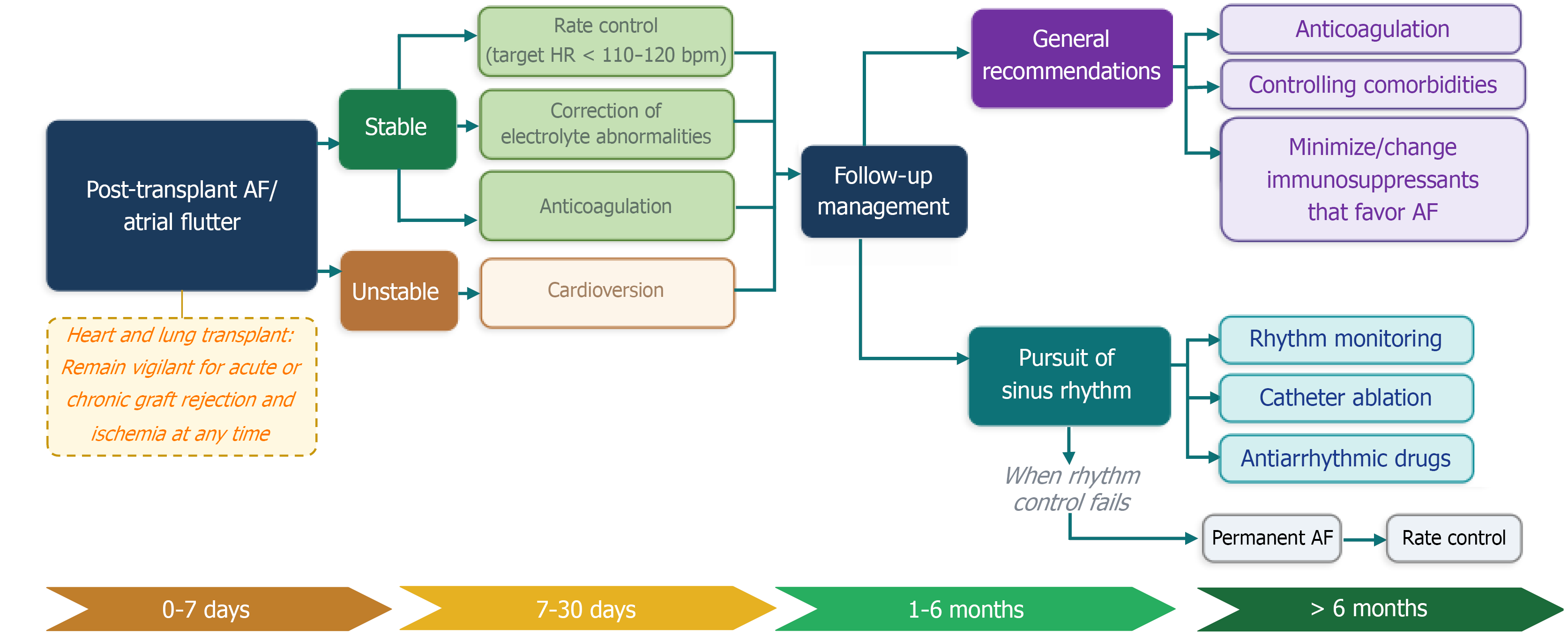
Figure 1 Early and follow-up management of post-transplant atrial tachyarrhythmias.
The timeline separates very early postoperative management (0-7 days), early reassessment (7-30 days), intermediate follow-up (1-6 months), and late arrhythmia evaluation (> 6 months). AF: Atrial fibrillation; HR: Heart rate.
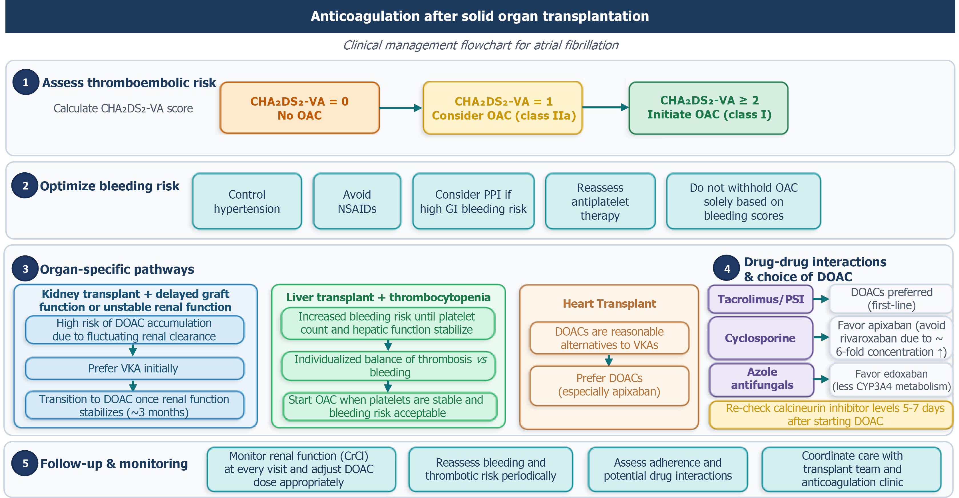
Figure 2 Clinical management flowchart for anticoagulation after solid-organ transplantation.
The figure integrates general CHA2DS2-VA-based thromboembolic risk assessment with organ-specific bleeding and pharmacokinetic considerations, including delayed graft function or unstable renal clearance after kidney transplantation, thrombocytopenia or unstable hepatic synthetic function after liver transplantation, and drug-drug interactions with calcineurin inhibitors, proliferation signal inhibitors, azole antifungals, and protease inhibitors. OAC: Oral anticoagulant; VKA: Vitamin K antagonist; NSAIDs: Non-steroidal anti-inflammatory drugs; PPI: Proton pump inhibitor; GI: Gastrointestinal; DOAC: Direct oral anticoagulant; PSI: Proliferation signal inhibitor; CYP3A4: Cytochrome P450 3A4; CrCl: Creatinine clearance.
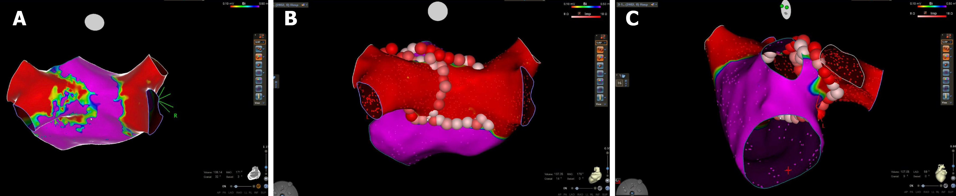
Figure 3 Representative original case from our center showing electroanatomical mapping and radiofrequency catheter ablation in a patient with atrial fibrillation following orthotopic heart transplantation, demonstrating recipient-donor atrial conduction across the left inferior pulmonary vein region.
A: Posterior view of the left atrium obtained using the CARTO 3 system (Biosense Webster, CA, United States). The bipolar voltage map uses a 0.10-0.50 mV display scale and demonstrates electrical continuity between recipient and donor atrial tissue in the area adjacent to the left inferior pulmonary vein, suggesting a recipient-graft atrioatrial conduction pathway and posterior wall scar that served as a trigger for atrial fibrillation. This finding guided the ablation strategy, which targeted isolation of the left pulmonary veins and posterior left atrial wall; B: Final electroanatomical map confirming complete electrical isolation of the posterior left atrial wall and left pulmonary veins following ablation, with no residual conduction across the previously identified recipient-donor atrial connection; C: Three-dimensional reconstruction showing radiofrequency ablation lesions placed to achieve circumferential isolation of the left pulmonary veins and posterior wall.
- Citation: Brigido ARD, Falcon HCS, Faria VS, Bernardi HGB, Sousa JCV, Belfort DSP, Carvalho GD, Lins PRG. Post-transplant atrial tachyarrhythmias: Epidemiology, mechanisms, outcomes, and management across solid organs. World J Transplant 2026; 16(3): 122203
- URL: https://www.wjgnet.com/2220-3230/full/v16/i3/122203.htm
- DOI: https://dx.doi.org/10.5500/wjt.122203