
















Copyright: ©Author(s) 2026.
World J Transplant. Sep 18, 2026; 16(3): 120920
Published online Sep 18, 2026. doi: 10.5500/wjt.120920
Published online Sep 18, 2026. doi: 10.5500/wjt.120920
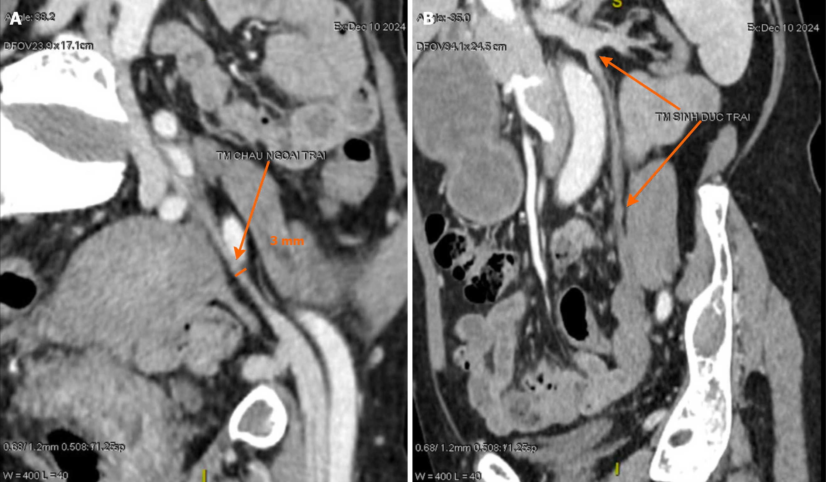
Figure 1 Preoperative computed tomography venography.
A: Markedly atrophic left external iliac vein measuring approximately 3 mm in diameter (arrow). This small caliber was insufficient for adequate renal allograft venous drainage; B: Left ovarian (gonadal) vein measuring 3.7 mm in diameter at its maximal segment (arrow), identified as a potential alternative venous outflow pathway within the operative field.
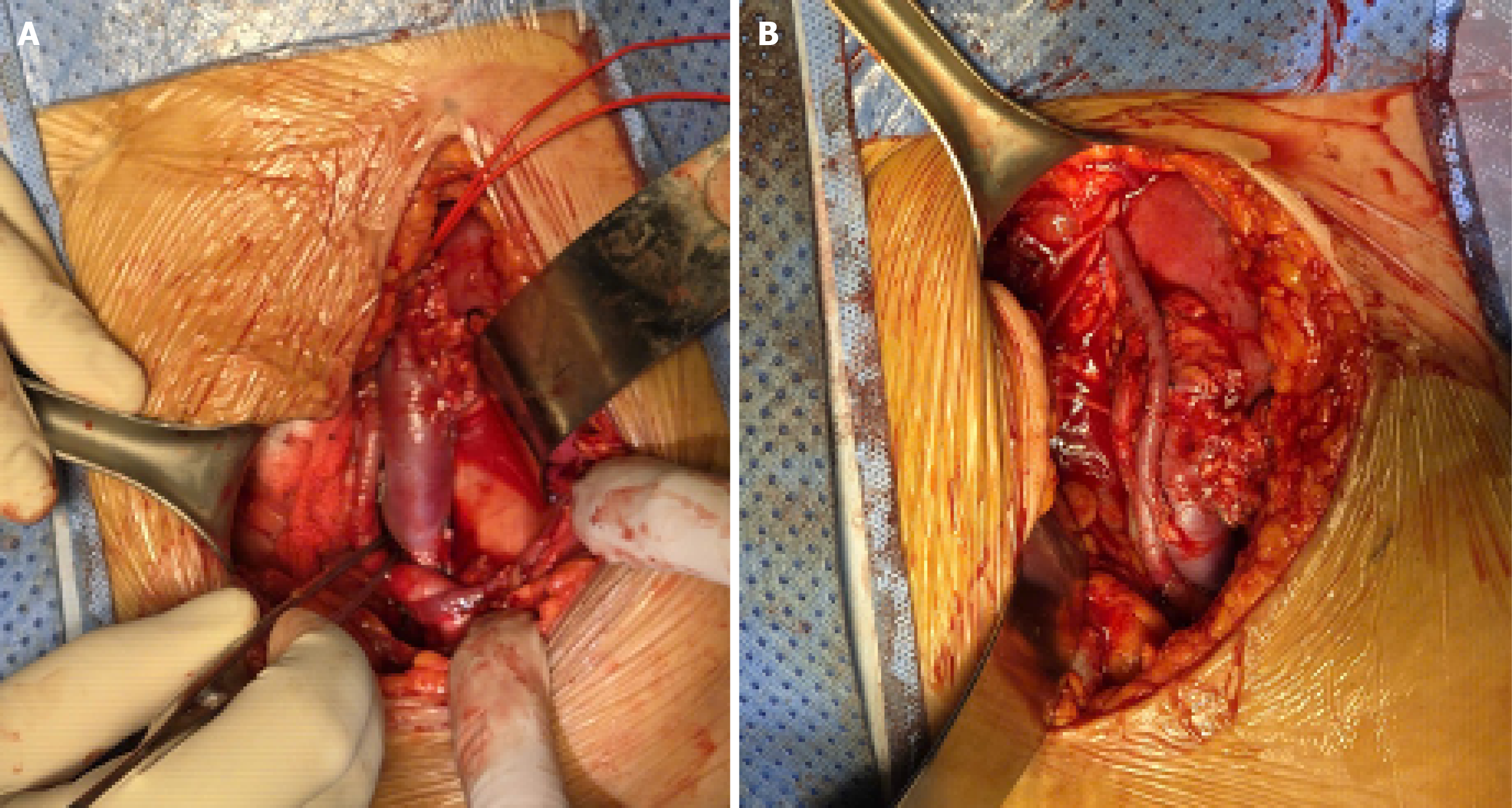
Figure 2 Intraoperative findings.
A: After clamp release, the left renal vein became markedly distended and the graft appeared congested and tense, consistent with inadequate outflow; B: Salvage bypass using an end-to-side great saphenous vein interposition graft from the renal vein to the left ovarian vein, creating an additional venous drainage pathway.
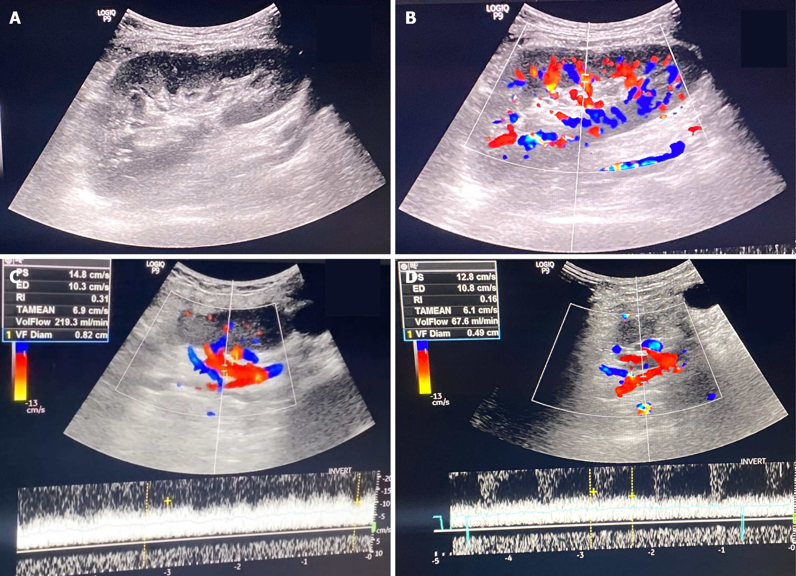
Figure 3 Follow-up ultrasonography at 8 months.
A: Gray-scale imaging of the renal graft showing preserved corticomedullary differentiation without hydronephrosis; B: Color Doppler demonstrating homogeneous parenchymal perfusion; C: Spectral Doppler measurement at the renal vein showing an outflow volume of 219.3 mL/minute; D: Spectral Doppler measurement at the great saphenous vein conduit showing a flow of 67.6 mL/minute.
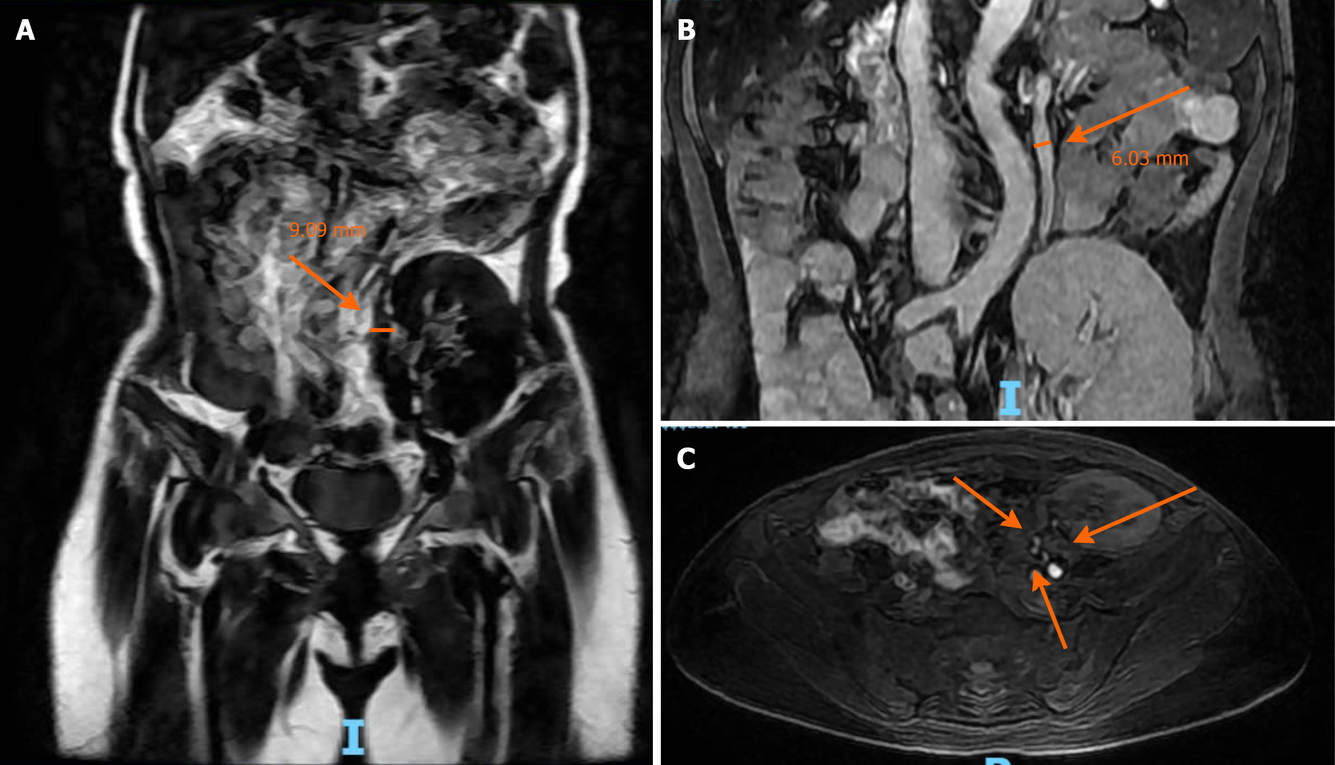
Figure 4 Follow-up magnetic resonance imaging at 8 months.
A: Great saphenous vein conduit dilated to 9.09 mm (arrow); B: Left ovarian vein enlarged to 6.03 mm (arrow); C: Patent triple arterial anastomoses supplying the renal graft (arrows).
- Citation: Nguyen DD, Pham HN, Nguyen HK, Bui KCL, Vo TM, Pham HN. Ovarian vein bypass for renal allograft salvage in iliac vein atrophy: A case report and review of literature. World J Transplant 2026; 16(3): 120920
- URL: https://www.wjgnet.com/2220-3230/full/v16/i3/120920.htm
- DOI: https://dx.doi.org/10.5500/wjt.120920