Copyright
©The Author(s) 2025.
World J Transplant. Dec 18, 2025; 15(4): 105597
Published online Dec 18, 2025. doi: 10.5500/wjt.v15.i4.105597
Published online Dec 18, 2025. doi: 10.5500/wjt.v15.i4.105597
Figure 1 Urgent abdominal contrast-enhanced computed tomography scan.
A: Large, retro-peritoneal hematoma (orange arrow); B: Rupture in the upper pole of the transplanted kidney (orange arrow); C: Area of parenchymal hypoperfusion just below the fracture (orange arrow).
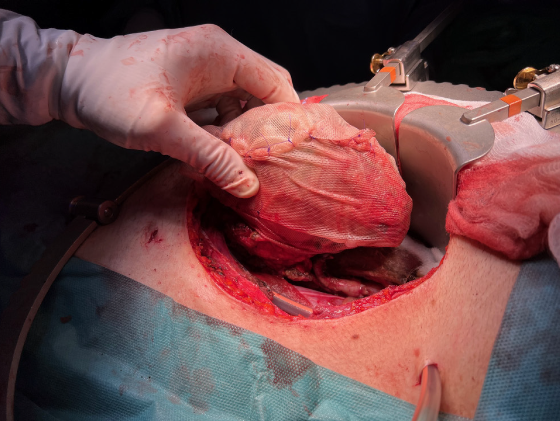
Figure 2
Intra-operative photograph showing the kidney graft covered by a fibrin sealant and wrapped with a tailored polyglactin 910 knitted mesh.
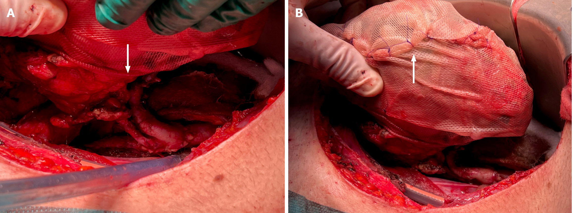
Figure 3 Intra-operative photographs.
A: Tailored polyglactin 910 knitted mesh cut in the center and trimmed to let the renal graft vessels to perfectly fit in the gap; B: Extremities of the mesh tightened and tied together with absorbable polydioxanone sutures.
Figure 4 Serial post-operative ultrasound scans of the kidney graft showing progressive resorption of the polyglactin 910 knitted mesh.
A: After seven days (white arrow); B: After three months (white arrow); C: After six months.
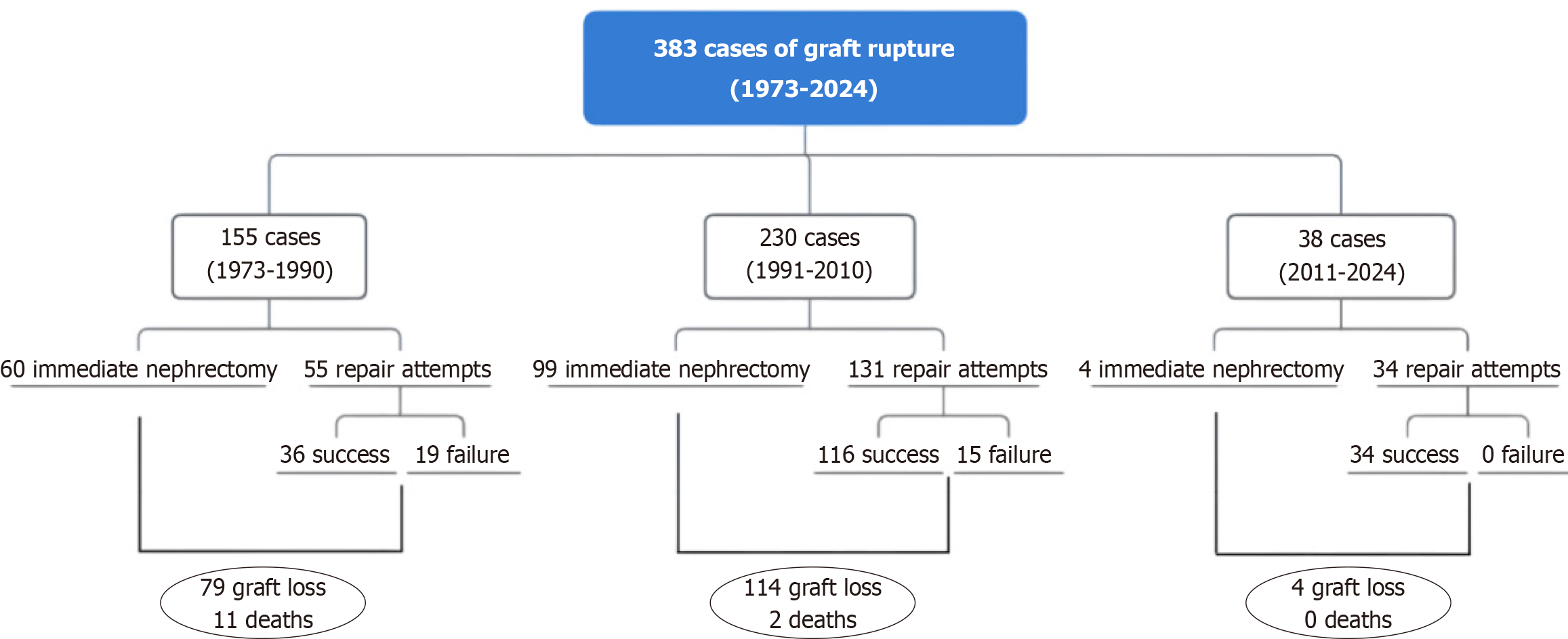
Figure 5
Flow diagrams summarizing and comparing the main outcomes of the episodes of spontaneous kidney graft rupture reported in the literature and sorted in different publication eras: 1973-1990, 1991-2010, and 2011-2024.
- Citation: Campioli E, Sikharulidze A, Cenzi LM, Spagnoletti G, Ferraresso M, Favi E. Suture-free polyglactin 910 mesh repair of kidney graft rupture: A case report and review of literature. World J Transplant 2025; 15(4): 105597
- URL: https://www.wjgnet.com/2220-3230/full/v15/i4/105597.htm
- DOI: https://dx.doi.org/10.5500/wjt.v15.i4.105597


