Copyright
©The Author(s) 2025.
World J Transplant. Mar 18, 2025; 15(1): 96696
Published online Mar 18, 2025. doi: 10.5500/wjt.v15.i1.96696
Published online Mar 18, 2025. doi: 10.5500/wjt.v15.i1.96696
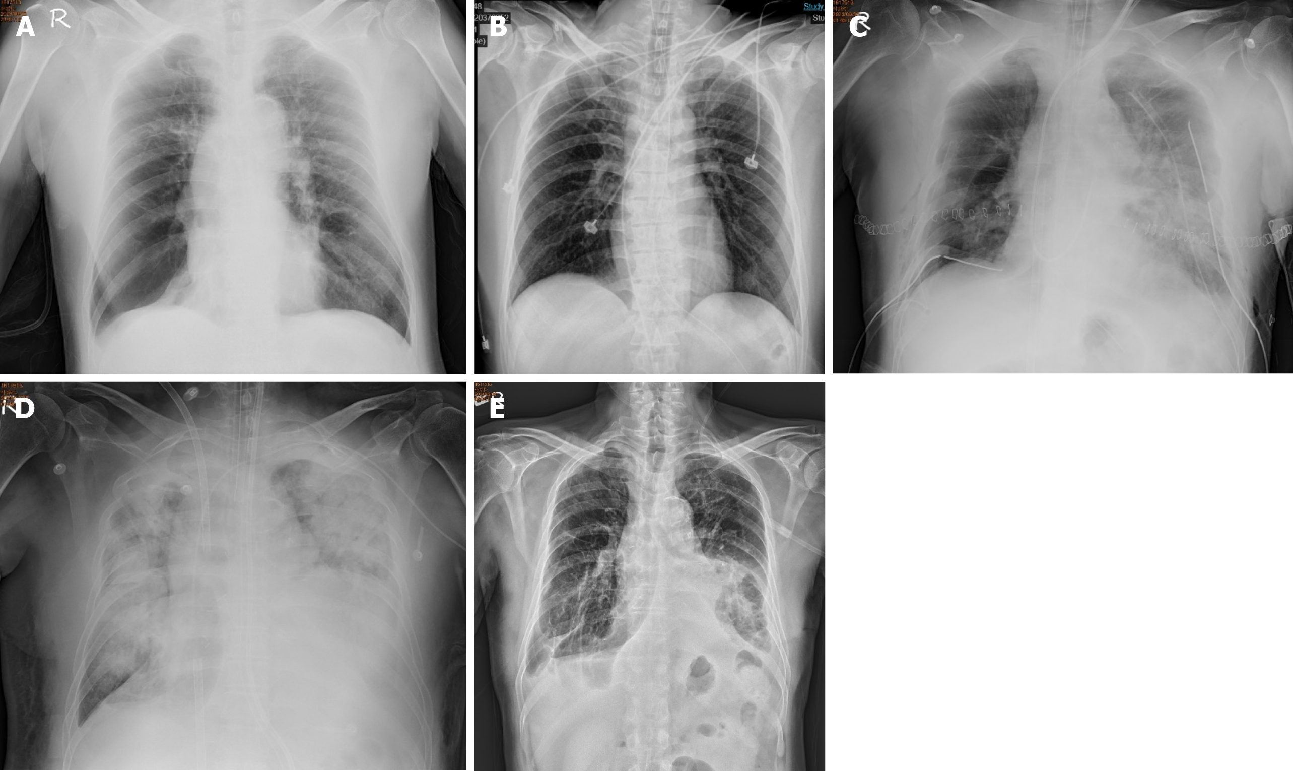
Figure 1 Chest plain film.
A: The pretransplant chest plain film showed hyperinflation and increased opacification of both lung fields except for the right lower lung field which showed hypertranslucency; B: Donor chest plain film showing clear bilateral lung fields without opacity; C: Postoperative chest plain film showing increased infiltration in the left lung, with bilateral chest tube retention in the pleural cavities and surgical stitches over the chest wall; D: Chest plain film on postoperative day 8 showing diffuse patchy opacities over both lungs with veno-venous extracorporeal membrane oxygenation support; E: Chest plain film on postoperative day 121 showing a stable condition after the removal of the tracheostomy tube with full expansion of both lungs.
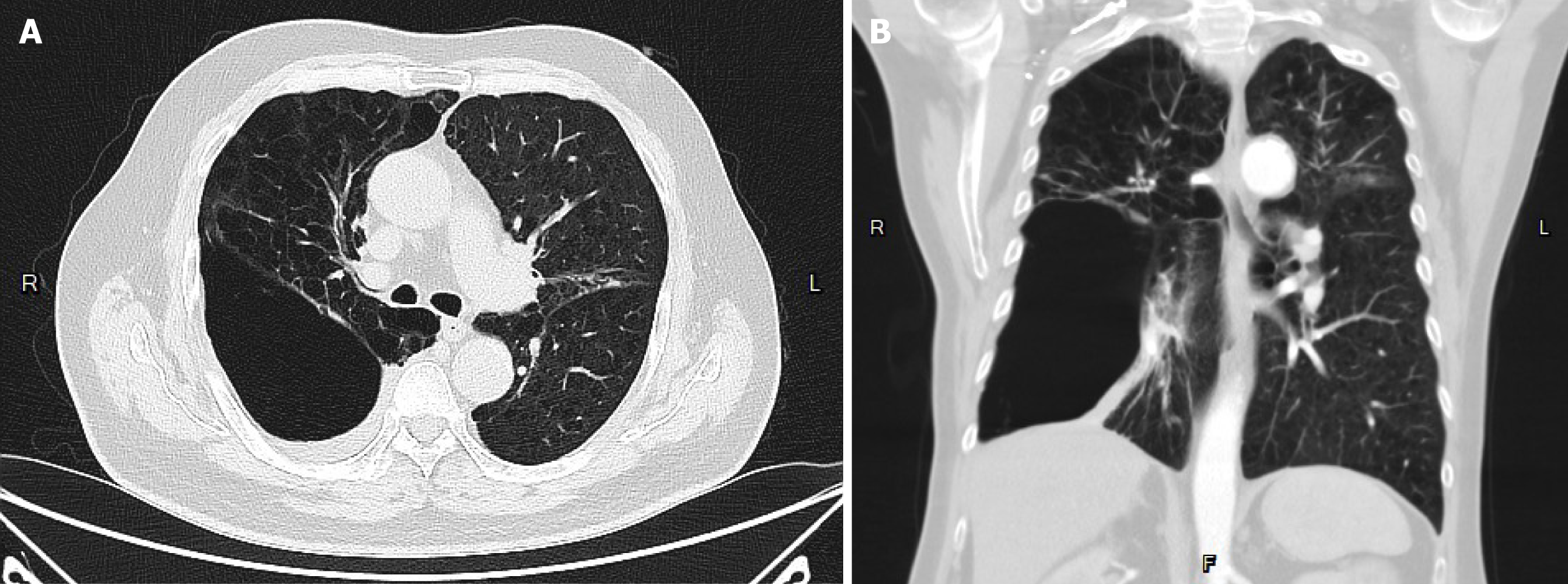
Figure 2 Recipient’s pretransplant chest computed tomography.
A: Axial view; B: Coronal view. Pretransplant chest computed tomography showed diffuse emphysematous change of bilateral lungs, with marked destruction of the right lung.
Figure 3 Post-operative flexible bronchoscopy.
A: Right bronchial anastomosis; B: Left bronchial anastomosis. Bilateral bronchial anastomoses showing no dehiscence or stricture under flexible bronchoscopy.
- Citation: Kuo YS, Lin KH, Chen YY, Tsai YM, Wu TH, Huang HK, Huang TW. Success of intravenous immunoglobulin and steroids in managing severe COVID-19 following lung transplantation: A case report. World J Transplant 2025; 15(1): 96696
- URL: https://www.wjgnet.com/2220-3230/full/v15/i1/96696.htm
- DOI: https://dx.doi.org/10.5500/wjt.v15.i1.96696


