©The Author(s) 2024.
World J Transplant. Jun 18, 2024; 14(2): 95009
Published online Jun 18, 2024. doi: 10.5500/wjt.v14.i2.95009
Published online Jun 18, 2024. doi: 10.5500/wjt.v14.i2.95009
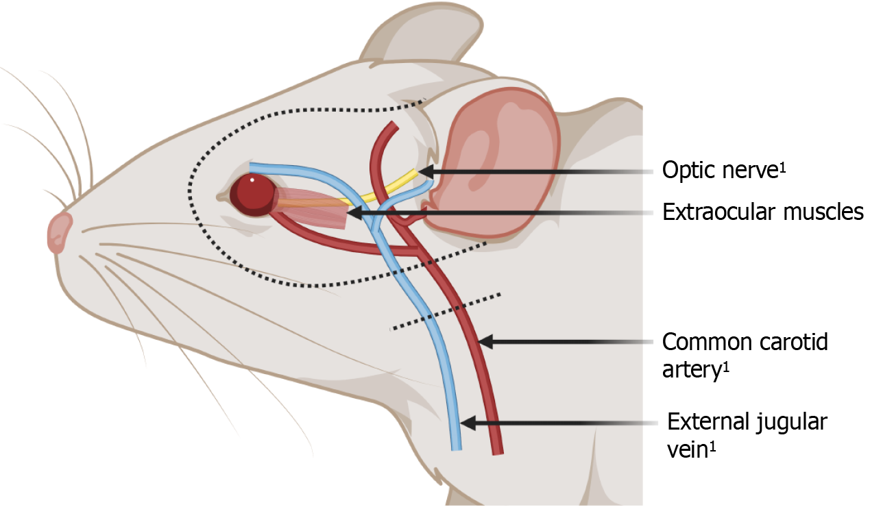
Figure 1 Anatomical illustration of a rodent model for whole-eye transplantation.
1Anastamosis with the recipient. Created with BioRender.com (Supplementary material).
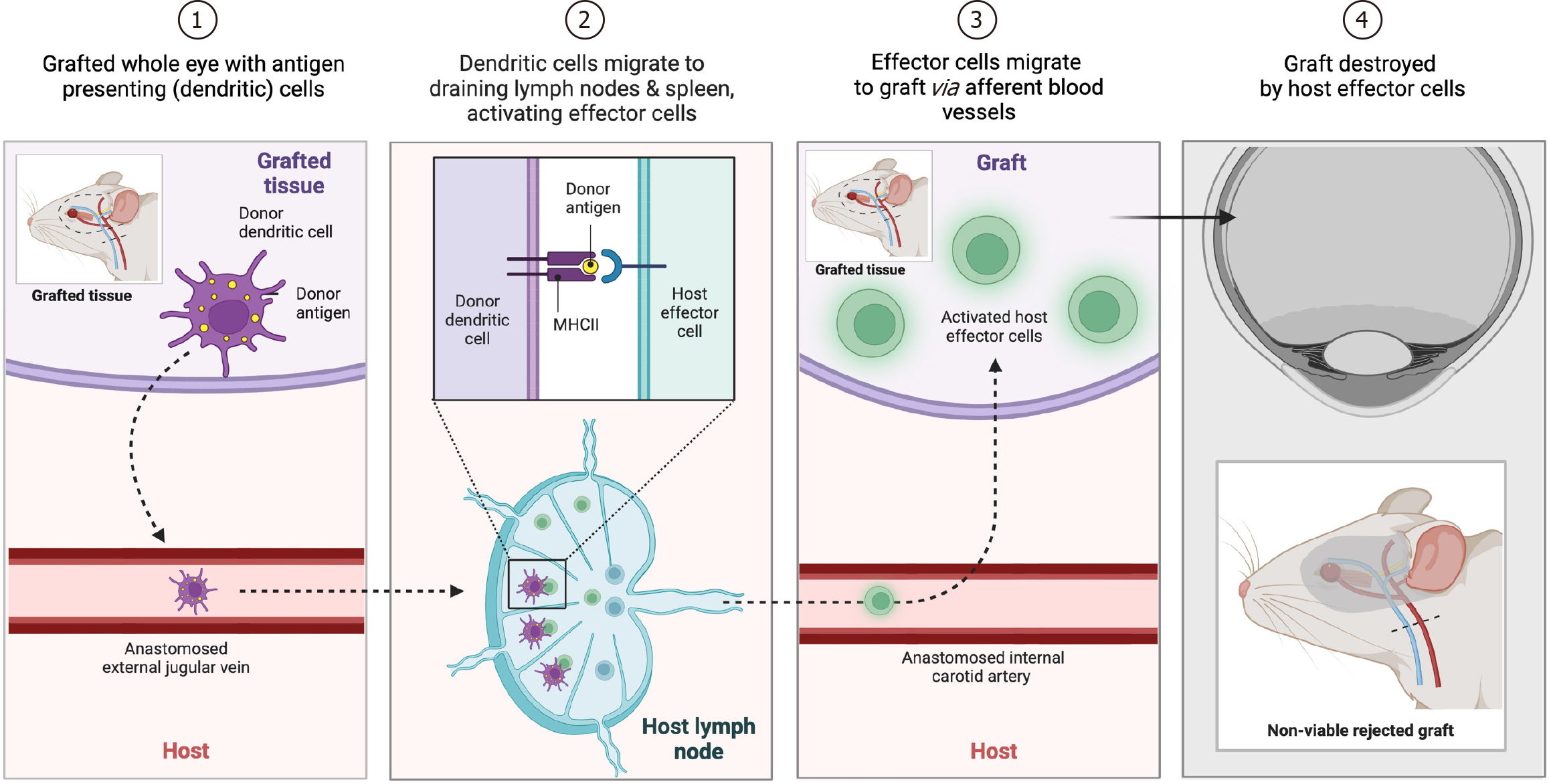
Figure 2 Schematic figure of different phases of the rejection process, mediated by antigen-presenting cells and activated effector cells, following whole-eye transplantation in a representative rodent model.
Created with BioRender.com (Supplementary material).
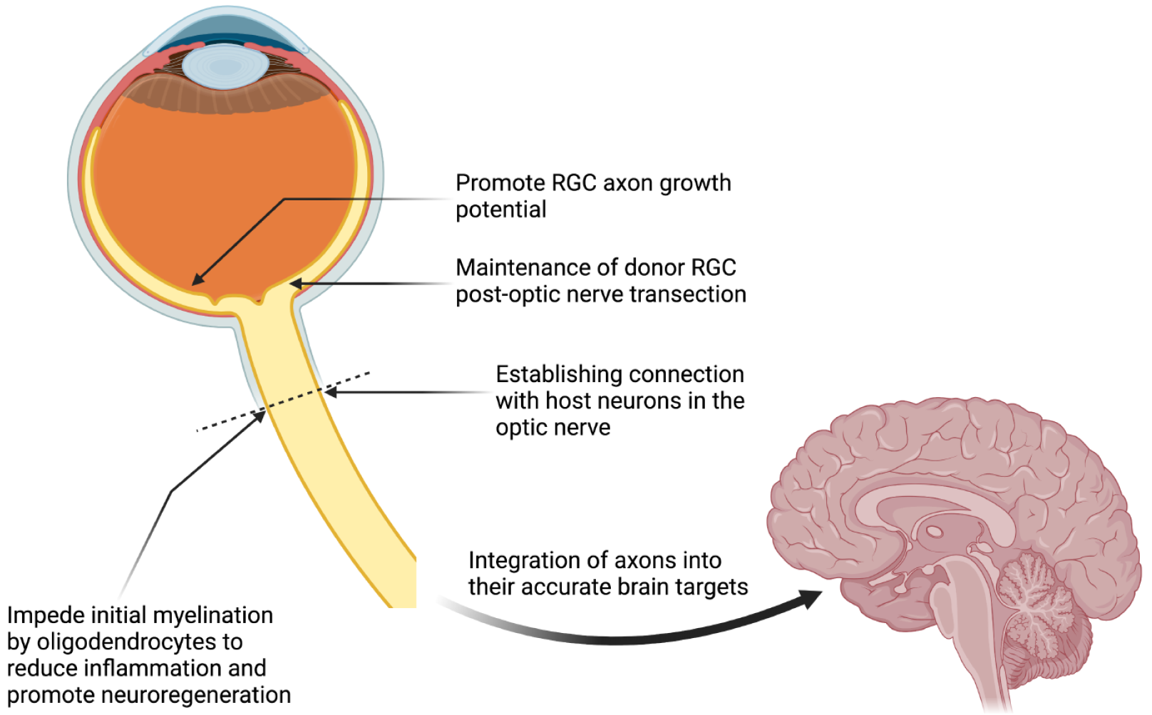
Figure 3 Challenges in neural pathway regeneration for restoring visual function following whole-eye transplantation.
RGC: Retinal ganglion cell. Created with BioRender.com (Supplementary material).
- Citation: Scarabosio A, Surico PL, Tereshenko V, Singh RB, Salati C, Spadea L, Caputo G, Parodi PC, Gagliano C, Winograd JM, Zeppieri M. Whole-eye transplantation: Current challenges and future perspectives. World J Transplant 2024; 14(2): 95009
- URL: https://www.wjgnet.com/2220-3230/full/v14/i2/95009.htm
- DOI: https://dx.doi.org/10.5500/wjt.v14.i2.95009


