Copyright
©The Author(s) 2023.
World J Transplant. Dec 18, 2023; 13(6): 290-298
Published online Dec 18, 2023. doi: 10.5500/wjt.v13.i6.290
Published online Dec 18, 2023. doi: 10.5500/wjt.v13.i6.290
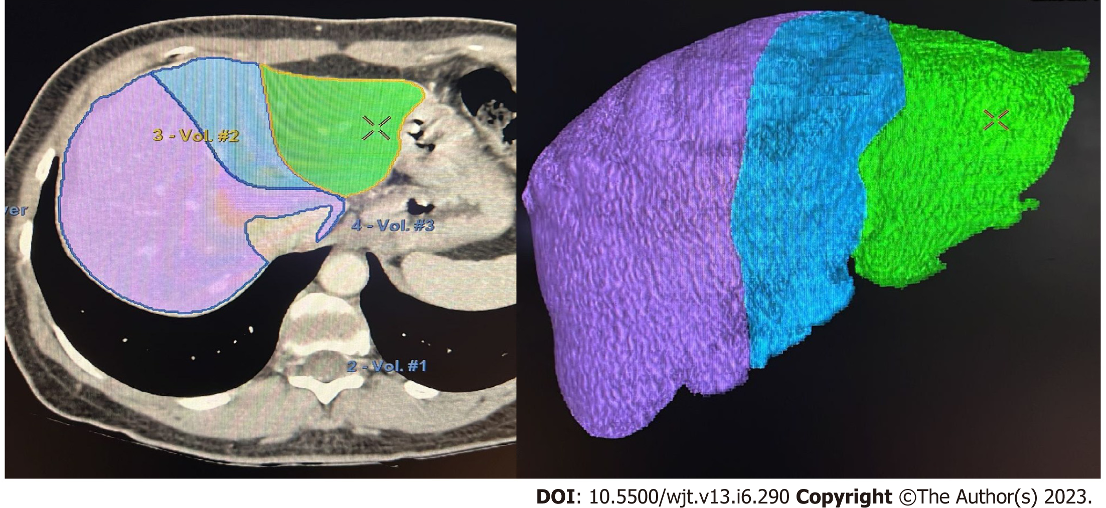
Figure 1 Manual volumetric study performed in our institution for pre-operative living-donor evaluation (Hepatic VCAR-GE Healthcare).
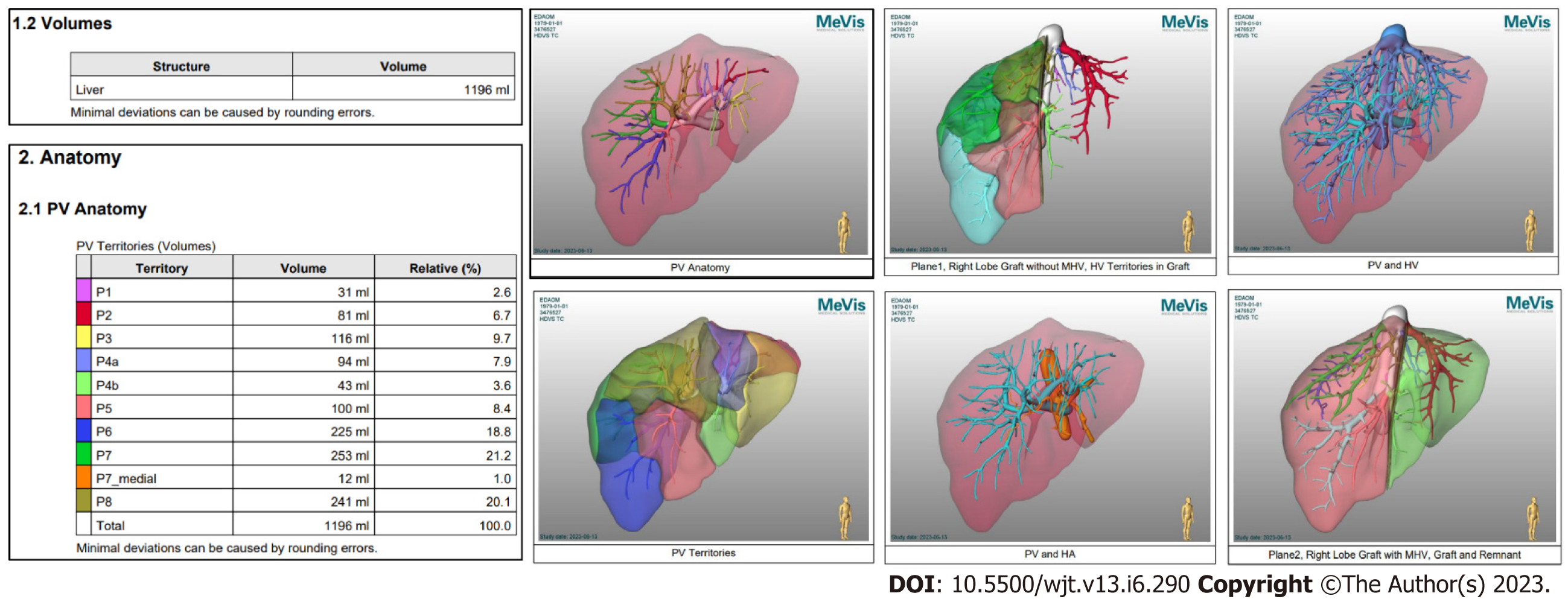
Figure 2 MeVis software images and tables output: The software returns multiple images and tables.
PV: Peripheral vein; MHV: Middle hepatic vein; HA: Hepatic artery.
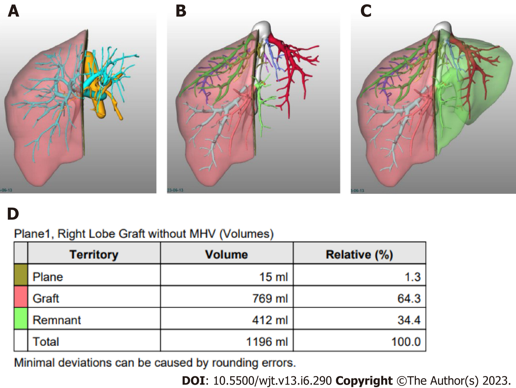
Figure 3 Resection planes volumetric estimation using MeVis.
A: Right Lobe Graft without middle hepatic vein (MHV), peripheral vein and hepatic artery; B: Right Lobe Graft without MHV, HV; C: Right Lobe Graft without MHV, Graft and Remnant; D: Table showing total, plane, graft and remnant liver volumes. MHV: Middle hepatic vein.
- Citation: Machry M, Ferreira LF, Lucchese AM, Kalil AN, Feier FH. Liver volumetric and anatomic assessment in living donor liver transplantation: The role of modern imaging and artificial intelligence. World J Transplant 2023; 13(6): 290-298
- URL: https://www.wjgnet.com/2220-3230/full/v13/i6/290.htm
- DOI: https://dx.doi.org/10.5500/wjt.v13.i6.290


