©The Author(s) 2017.
World J Clin Infect Dis. Feb 25, 2018; 8(1): 1-3
Published online Feb 25, 2018. doi: 10.5495/wjcid.v8.i1.1
Published online Feb 25, 2018. doi: 10.5495/wjcid.v8.i1.1
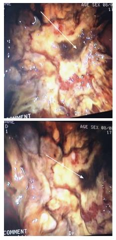
Figure 1 Gastroscopy image showing mocormycosis indicated by arrows.
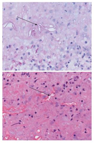
Figure 2 Histology of gastric mucormycosis shown by arrows.
- Citation: Kgomo MK, Elnagar AA, Mashoshoe K, Thomas P, Van Hougenhouck-Tulleken WG. Gastric mucormycosis: A case report. World J Clin Infect Dis 2018; 8(1): 1-3
- URL: https://www.wjgnet.com/2220-3176/full/v8/i1/1.htm
- DOI: https://dx.doi.org/10.5495/wjcid.v8.i1.1


