©2014 Baishideng Publishing Group Inc.
World J Clin Infect Dis. May 25, 2014; 4(2): 5-8
Published online May 25, 2014. doi: 10.5495/wjcid.v4.i2.5
Published online May 25, 2014. doi: 10.5495/wjcid.v4.i2.5
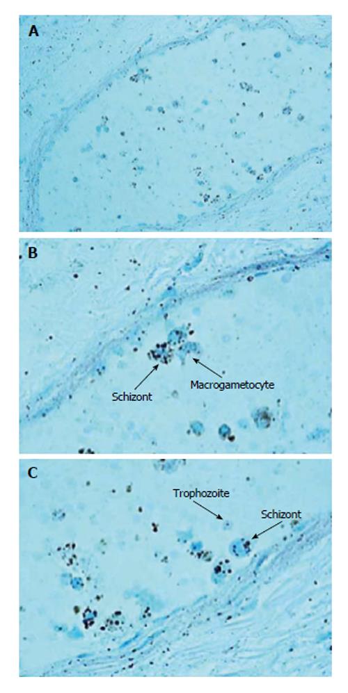
Figure 1 Different stages of the hematic schizogonic cycle of malarial parasite (schizont, trophozoite and crescent-shape macrogametocyte) in a splenic vessel.
Giemsa stain, magnification A: 20 ×; B: 100 ×; C: 100 ×.
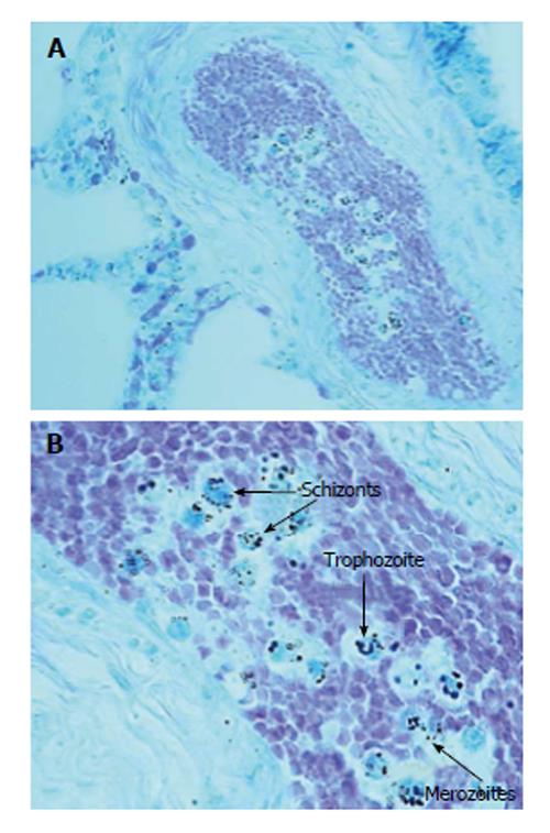
Figure 2 Malarial parasites in a pulmonary vessel.
Giemsa stain showing merozoite, schizont and trophozoite stages; magnification A: 20 ×; B: 40 ×.
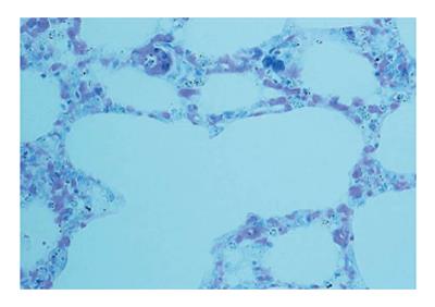
Figure 3 Numerous schizonts in the capillaries of pulmonary alveolar septa.
Giemsa stain, 20 × magnification.
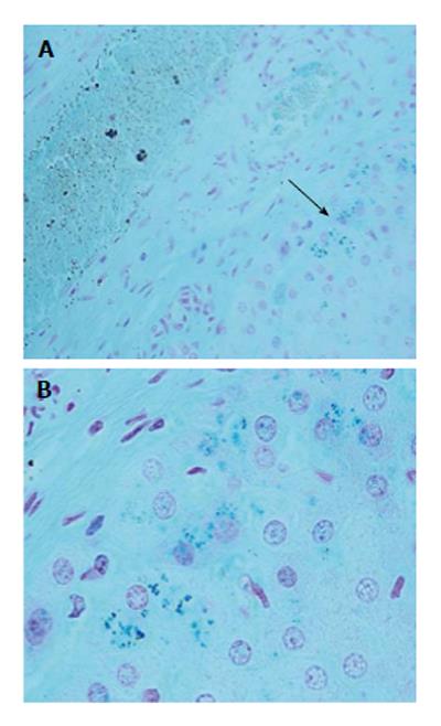
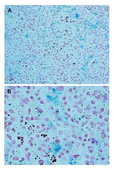
Figure 4 Malarial pigment grain accumulation in the splenic reticuloendothelial system.
Perls stain showing hemozoin and hemosiderin; magnification A: 40 ×; B: 100 ×.
Figure 5 Malarial pigment grain accumulation in the splenic reticuloendothelial system.
Perls staining method for iron showing hemozoin and hemosiderin; magnification A: 20 ×; B: 100 ×.
- Citation: Pusiol T, Lavezzi AM, Radice F, Alfonsi G, Matturri L. Unsuspected imported malaria in a case of sudden infant death. World J Clin Infect Dis 2014; 4(2): 5-8
- URL: https://www.wjgnet.com/2220-3176/full/v4/i2/5.htm
- DOI: https://dx.doi.org/10.5495/wjcid.v4.i2.5


