©The Author(s) 2023.
World J Clin Infect Dis. Feb 28, 2023; 13(1): 1-10
Published online Feb 28, 2023. doi: 10.5495/wjcid.v13.i1.1
Published online Feb 28, 2023. doi: 10.5495/wjcid.v13.i1.1
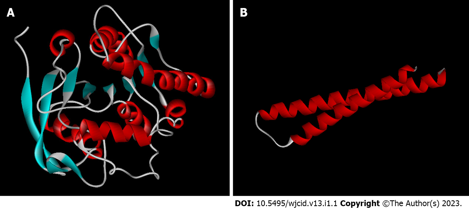
Figure 1 Three-dimensional structure models of Mycobacterium leprae antigens predicted in silico.
A: 85B antigen; B: ML0050. Blue: β-sheets; red: α-helices; white: Loops.
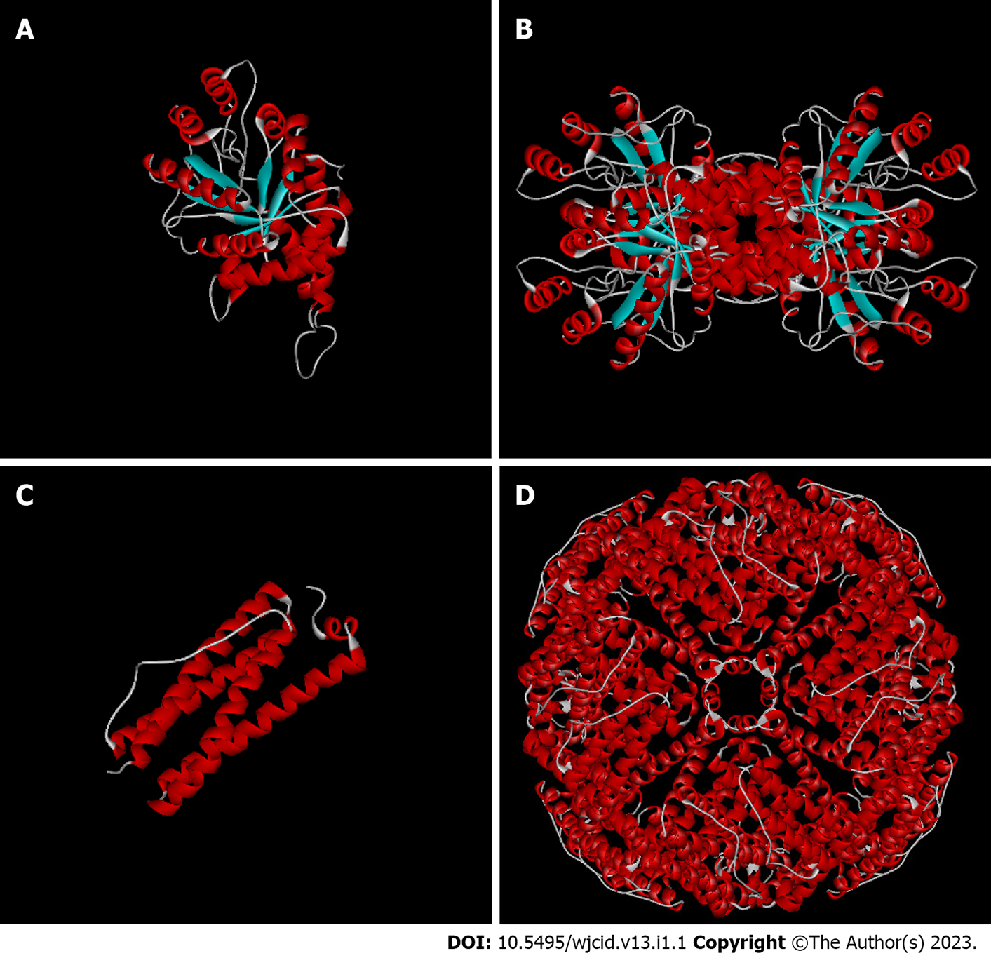
Figure 2 Three-dimensional models of the monomers and quaternary structures of Mycobacterium leprae antigens predicted in silico.
A: ML0286 monomer; B: ML 0286 quaternary structure (tetramer); C: ML2038 monomer; D: ML2038 quaternary structure (24 subunits). Blue: β-sheets; red: α-helices; white: Loops.
Figure 3 Z-score plot for three-dimensional structure models of the monomers of Mycobacterium leprae antigens predicted in silico.
A: 85B antigen; B: ML0050; C: ML0286; D: ML2038. The red star indicates the position of the model among the non-redundant structures deposited in the Protein Data Bank. Z-score < 1 indicates higher quality models. Arrow points the star position. PDB: Protein Data Bank.
- Citation: Melo de Assis BL, Viana Vieira R, Rudenco Gomes Palma IT, Bertolini Coutinho M, de Moura J, Peiter GC, Teixeira KN. Three-dimensional models of antigens with serodiagnostic potential for leprosy: An in silico study. World J Clin Infect Dis 2023; 13(1): 1-10
- URL: https://www.wjgnet.com/2220-3176/full/v13/i1/1.htm
- DOI: https://dx.doi.org/10.5495/wjcid.v13.i1.1


