Copyright
©The Author(s) 2025.
World J Exp Med. Jun 20, 2025; 15(2): 100166
Published online Jun 20, 2025. doi: 10.5493/wjem.v15.i2.100166
Published online Jun 20, 2025. doi: 10.5493/wjem.v15.i2.100166
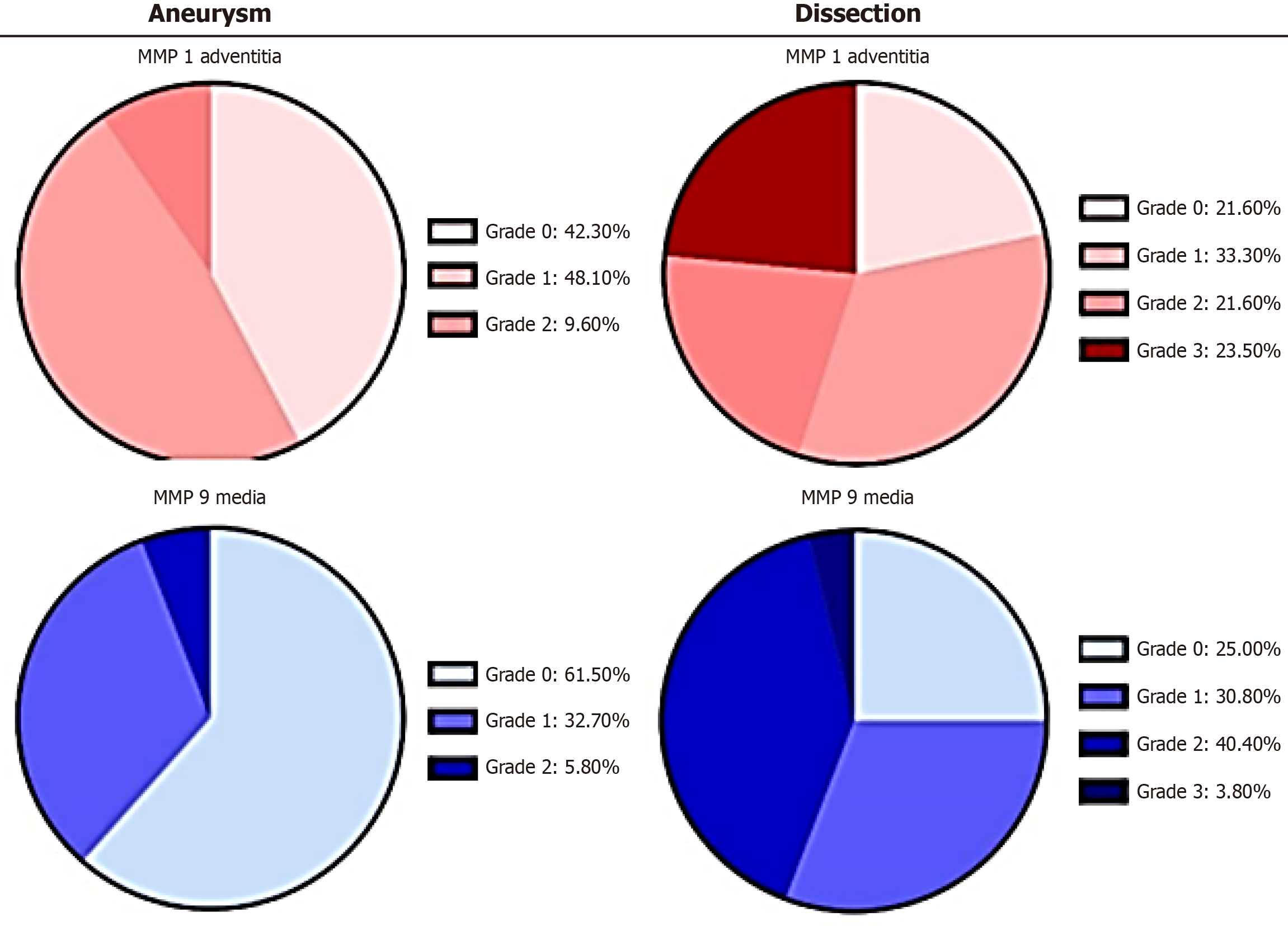
Figure 1 An example of the distribution differences of matrix metalloproteinas-1 and matrix metalloproteinas-9 between aneurysm and dissection.
The charts illustrate the absolute mean values (%) of immunohistological detection of the corresponding target antigens in the adventitia and media for the groups “aneurysm” and “dissection” (n = 52 each). Minimal staining was observed in the intima. Grading evaluations were conducted independently by two observers/pathologists. MMPs: Matrix metalloproteinases.
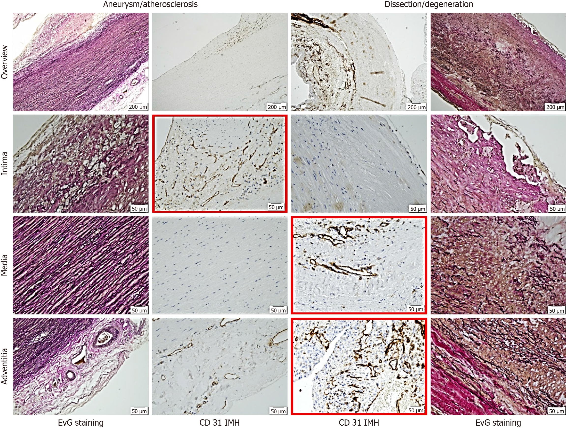
Figure 2 Representative histological and immunohistological findings in aneurysm and dissection.
Note the differing staining patterns of vessels: In the intima for aneurysmal changes and in the media and adventitia for dissection (red frames). The distinct pattern of small vessels reflects vascularization or neovascularization, suggesting the primarily degenerative nature of dissection pathogenesis. Staining methods included hematoxylin (S3301, Dako), Elastica van Gieson, and immunohistochemistry using a primary antibody (monoclonal Mouse Anti-human CD31, Endothelial Cell Clone JC70A, Dako). EvG: Elastica van Gieson.
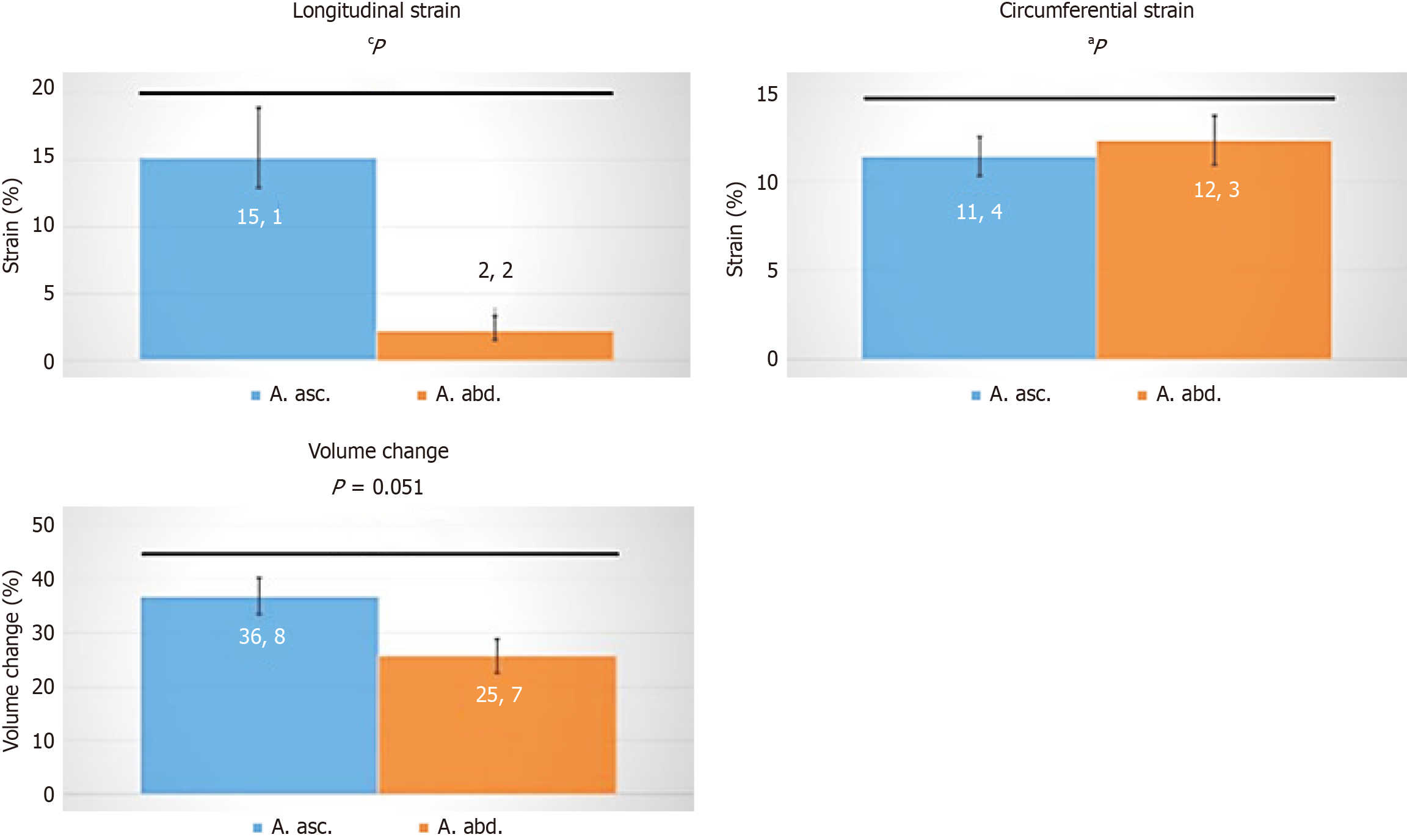
Figure 3 Differences in strain between the ascending (blue) and descending (orange) aortic root, representing varying volume charges.
Statistical analysis was performed using Wolfram MATHEMATICA 9. Data were tested for normal distribution with the function Distribution Fit test. Normal distribution was rejected for P < 0.05. For normally distributed data, values are presented as mean ± SEM, and an unpaired two-tailed t-test was conducted. For non-normally distributed data, values are given as median, and a Mann-Whitney U-test was performed. aP < 0.01; cP < 0.005.
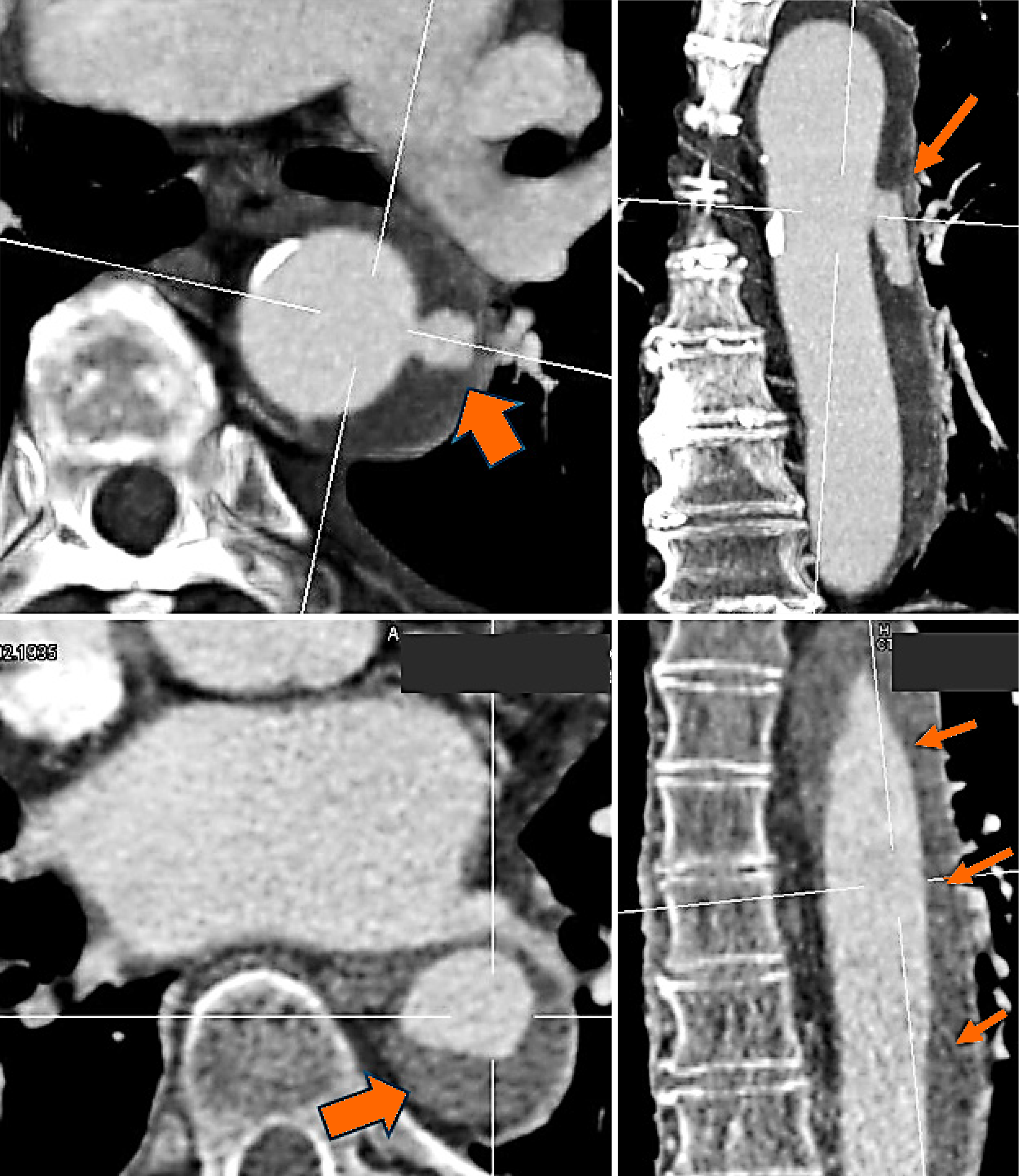
Figure 4 A clinical radiologic image (angio-computed tomography) of a current dissection leading to the urgent admission of a patient to the emergency unit at our hospital.
A small lesion in the inner layer of the aorta is clearly visible. Penetrating blood spreads into the already damaged medial areas (arrows), resulting in hemorrhage throughout the vascular tube.
- Citation: Irqsusi M, Rodepeter FR, Günther M, Kirschbaum A, Vogt S. Matrix metalloproteinases and their tissue inhibitors as indicators of aortic aneurysm and dissection development in extracellular matrix remodeling. World J Exp Med 2025; 15(2): 100166
- URL: https://www.wjgnet.com/2220-315x/full/v15/i2/100166.htm
- DOI: https://dx.doi.org/10.5493/wjem.v15.i2.100166


