©The Author(s) 2024.
World J Exp Med. Dec 20, 2024; 14(4): 98005
Published online Dec 20, 2024. doi: 10.5493/wjem.v14.i4.98005
Published online Dec 20, 2024. doi: 10.5493/wjem.v14.i4.98005
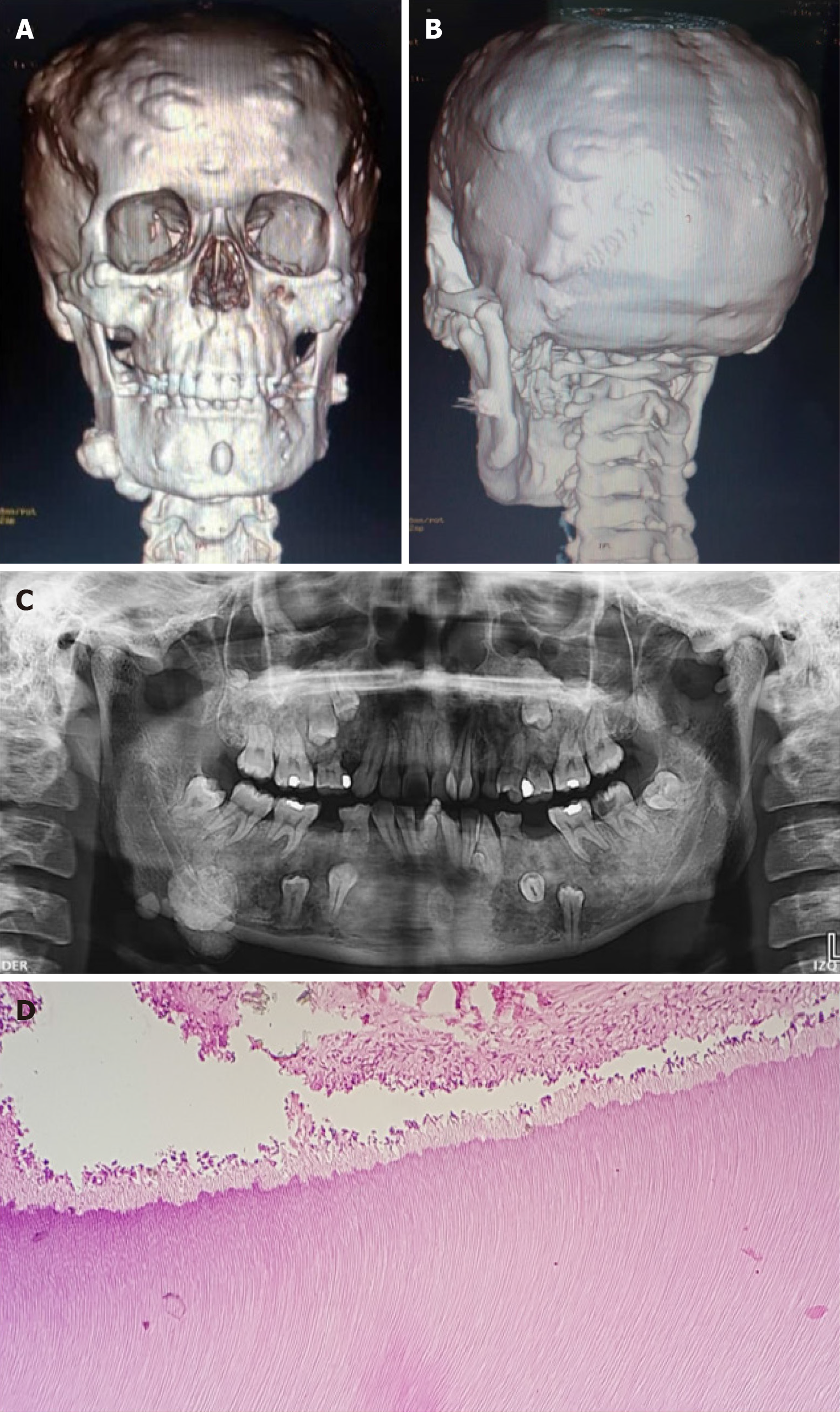
Figure 1 Gardner syndrome in a 22-years-female patient.
A and B: Three-dimensional reconstruction images show multiple variable sized osteomas in craniofacial bones; C: Panoramic radiograph shows radiopacity in the right posterior region of the mandible diagnostic as odontoma; D: Haematoxylin and eosin staining section of the odontoma characterized by the presence of dentinal tubules, odontoblasts, and dental pulp.
- Citation: Schuch LF, Silveira FM, Pereira-Prado V, Sicco E, Pandiar D, Villarroel-Dorrego M, Bologna-Molina R. Clinicopathological and molecular insights into odontogenic tumors associated with syndromes: A comprehensive review. World J Exp Med 2024; 14(4): 98005
- URL: https://www.wjgnet.com/2220-315x/full/v14/i4/98005.htm
- DOI: https://dx.doi.org/10.5493/wjem.v14.i4.98005


