Copyright
©The Author(s) 2024.
World J Exp Med. Mar 20, 2024; 14(1): 87256
Published online Mar 20, 2024. doi: 10.5493/wjem.v14.i1.87256
Published online Mar 20, 2024. doi: 10.5493/wjem.v14.i1.87256
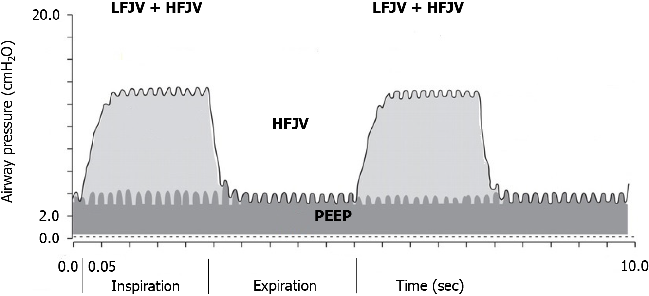
Figure 1 Illustration of superimposed high-frequency jet ventilation with typical pressure curves of continuous high-frequency jet ventilation and two cycles of simultaneous low-frequency jet ventilation.
The low-frequency jet ventilation provides the upper pressure level, and the high-frequency jet ventilation generates the lower pressure level (i.e., the positive end-expiratory pressure). PEEP: Positive end-expiratory pressure; HFJV: High-frequency jet ventilation; LFJV: Low-frequency jet ventilation.
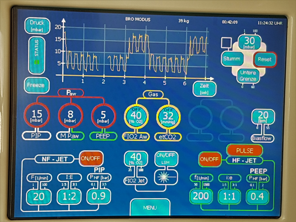
Figure 2 The TwinStreamTM ICU monitor displays the selected superimposed high-frequency jet ventilation setting.
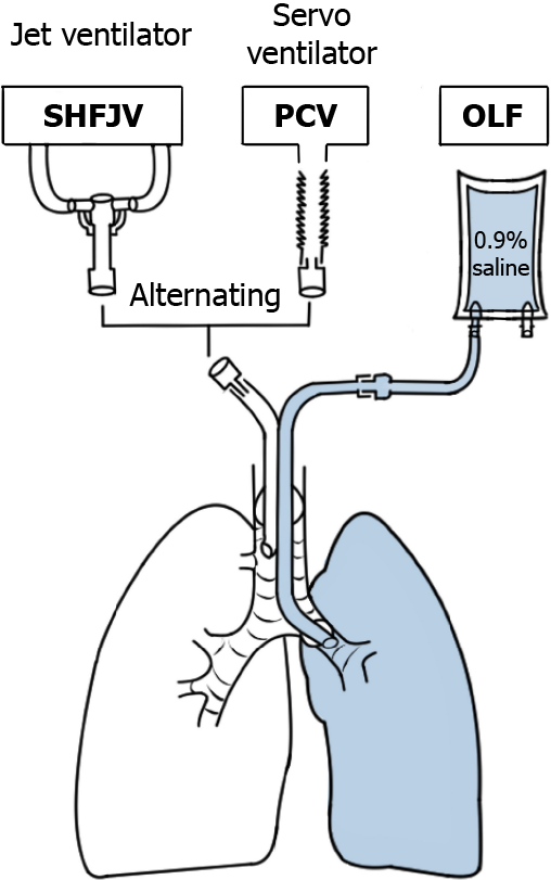
Figure 3 Experimental set-up.
SHFJV: Superimposed high-frequency jet ventilation; PCV: Pressure-controlled ventilation; OLF: One-lung flooding.
Figure 4 Flow scheme of the experimental protocol.
SHFJV: Superimposed high-frequency jet ventilation. In the control group (n = 5) both lungs were ventilated with pressure-controlled ventilation (two-lung ventilation) throughout the experimental time without lung flooding, but the position change was the same. OLF: One-lung flooding (left lung); OLV: One-lung ventilation; TLV: Two-lung ventilation; PCV: Pressure-controlled ventilation.
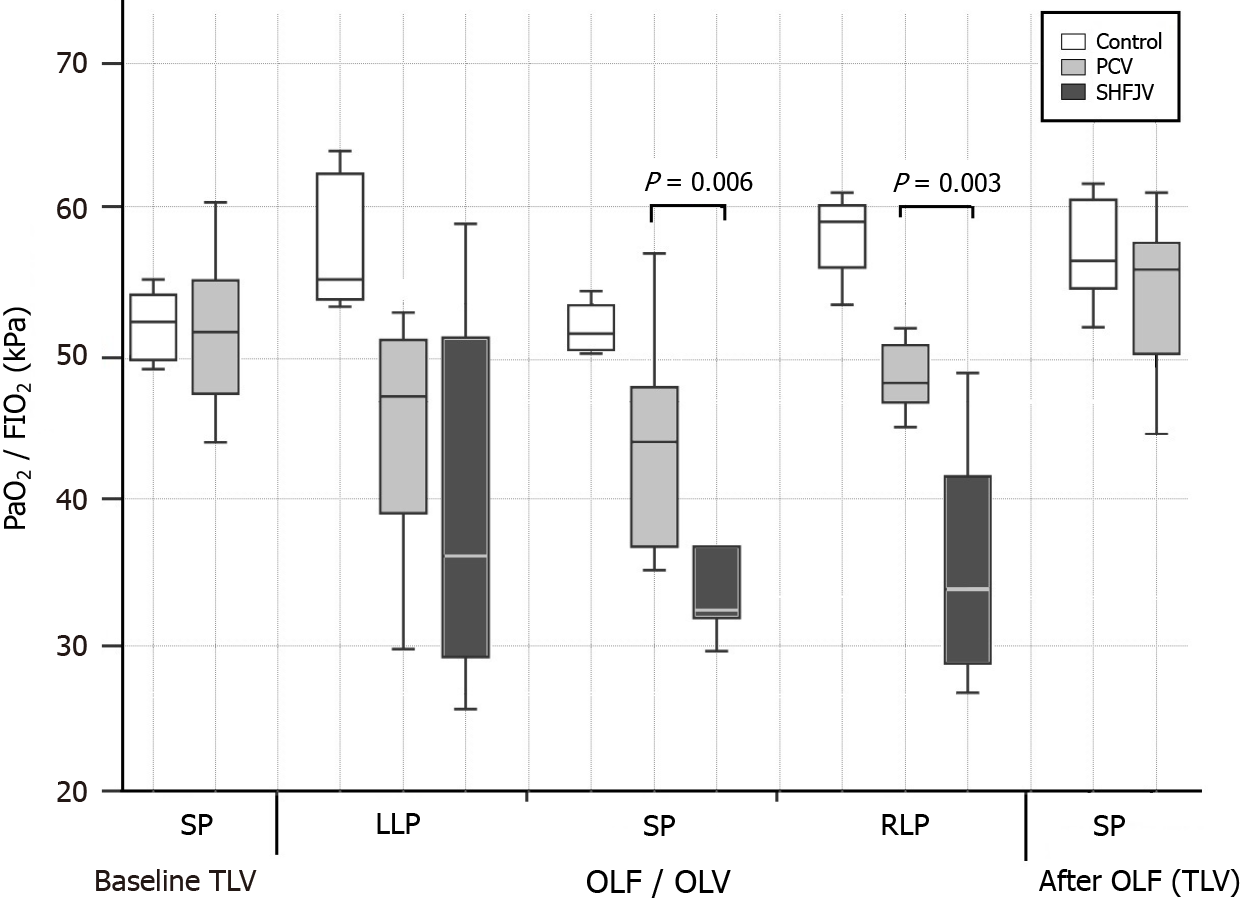
Figure 5 Boxplot of arterial partial pressure of oxygen/ fraction of inspired oxygen ratio for controls, pressure-controlled ventilation and superimposed high-frequency jet ventilation at baseline, one-lung flooding/one-lung ventilation in different animal positions and after one-lung flooding.
In the supine and right lateral decubitus position after one-lung flooding, PaO2/FiO2 ratio during superimposed high-frequency jet ventilation was significantly lower than during PCV. SHFJV: Superimposed high-frequency jet ventilation; PCV: Pressure-controlled ventilation; OLF: One-lung flooding; OLV: One-lung ventilation; TLV: Two-lung ventilation; LLP: Left lateral position; SP: Supine position; RLP: Right lateral position; PaO2/FiO2: Arterial partial pressure of oxygen/fraction of inspired oxygen (Horowitz index).
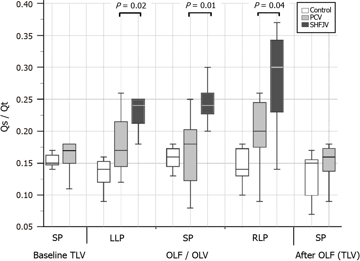
Figure 6 Boxplot of shunt fraction for controls, pressure-controlled ventilation and superimposed high-frequency jet ventilation at baseline, one-lung flooding/one-lung ventilation in different animal positions and after one-lung flooding.
In all positions after one-lung flooding, Qs/Qt during superimposed high-frequency jet ventilation was significantly higher than during pressure-controlled ventilation. SHFJV: Superimposed high-frequency jet ventilation; PCV: Pressure-controlled ventilation; OLF: One-lung flooding; OLV: One-lung ventilation; TLV: Two-lung ventilation; LLP: Left lateral position; SP: Supine position; RLP: Right lateral position; Qs/Qt: Right-to-left shunt fraction.
Figure 7 Chest X-ray.
A: Before one-lung flooding. Correct position of the left-sided double-lumen endobronchial tube was confirmed using a fiber optic bronchoscope. Pulmonary artery catheter was placed in the left pulmonary artery; B: after one-lung flooding of the left lung wing. A pulmonary artery catheter placed in the left pulmonary artery. An endobronchial catheter placed in the left lower bronchus and inserted through the left lumen of the tube; C: 30 min after fluid drainage and conventional ventilation of both lungs. The X-ray shows normally aerated lung wings.
- Citation: Lesser T, Wolfram F, Braun C, Gottschall R. Effects of unilateral superimposed high-frequency jet ventilation on porcine hemodynamics and gas exchange during one-lung flooding. World J Exp Med 2024; 14(1): 87256
- URL: https://www.wjgnet.com/2220-315x/full/v14/i1/87256.htm
- DOI: https://dx.doi.org/10.5493/wjem.v14.i1.87256


