Copyright
©2013 Baishideng Publishing Group Co.
World J Exp Med. Nov 20, 2013; 3(4): 100-107
Published online Nov 20, 2013. doi: 10.5493/wjem.v3.i4.100
Published online Nov 20, 2013. doi: 10.5493/wjem.v3.i4.100
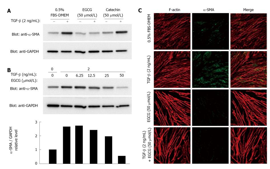
Figure 1 Effects of (-)-epigallocatechin-3-gallate on expression of α-smooth muscle actin.
A: Lysates of MRC-5 cells were obtained from cells treated with 0.5% FBS in DMEM alone, (-)-epigallocatechin-3-gallate (EGCG) (50 μmol/L), or catechin (50 μmol/L) for 24 h. After SDS-PAGE, proteins were blotted onto Immobilon, and probed with anti-α-smooth muscle actin (α-SMA) antibody. Glyceraldehyde 3-phosphate dehydrogenase (GAPDH) was used as an internal control; B: MRC-5 cells were treated with the indicated amounts of EGCG. α-SMA was detected in the same manner as in (A). The expression levels of α-SMA were normalized to those of GAPDH; C: Expression of α-SMA in cells treated with EGCG (50 μmol/L) in the absence (-) or presence (+) of transforming growth factor-β (TGF-β) (2 ng/mL) were examined by confocal microscopy. Green: α-SMA (fluorescein isothiocyanate conjugated goat anti-mouse IgG); Red: Actin stress fiber (rhodamine-labeled phalloidin); blue: Nuclei (DAPI). FBS: fetal bovine serum.
Figure 2 Effects of scavenging compounds on expression of α-smooth muscle actin.
MRC-5 cells were treated with edaravone (100 μmol/L) (A) or N-acetylcysteine (NAC) (2.5 mmol/L) (B) for 1 h, and then stimulated with transforming growth factor-β (TGF-β) for 24 h. Cell lysates were electrophoresed, blotted onto Immobilon, and probed with anti-α-smooth muscle actin (α-SMA). GAPDH: Glyceraldehyde 3-phosphate dehydrogenase. FBS: Fetal bovine serum.
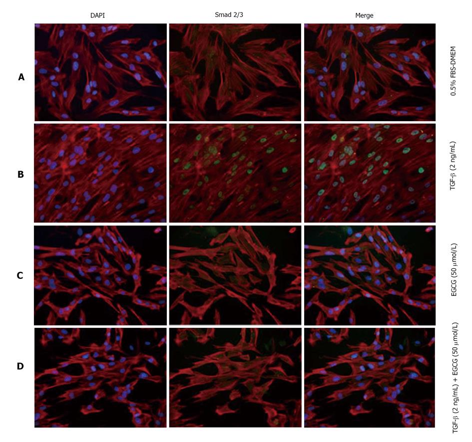
Figure 3 Effects of (-)-epigallocatechin-3-gallate on activation and localization of Smad2/3.
MRC-5 cells were treated with transforming growth factor-β (TGF-β) and/or (-)-epigallocatechin-3-gallate (EGCG). Cells were examined by confocal microscopy. Subcellular localization of Smad2/3 (green) and actin stress fibers (red) are shown. Nuclei were stained by DAPI (blue). A: Control; B: Treated with TGF-β; C: Treated with EGCG; D: Treated with TGF-β and EGCG. DAPI: 4',6-diamidino-2-phenylindole.
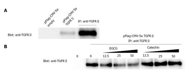
Figure 4 (-)-Epigallocatechin-3-gallate interferes with binding between transforming growth factor-β and its type II receptor.
A: Positive control of the immunoprecipitation experiment. Cell lysates from transfected COS-7 cells were treated with anti-transforming growth factor-β type II receptor (TGFRII). TGFRII was recovered in the immunoprecipitation product of the lysate; B: Effects of (-)-epigallocatechin-3-gallate (EGCG) and catechin on the antigen-antibody interaction. After cells were treated with EGCG or catechin, anti-TGFRII bound to Protein G was added to each lysate. Western blotting was performed using anti-TGFRII.
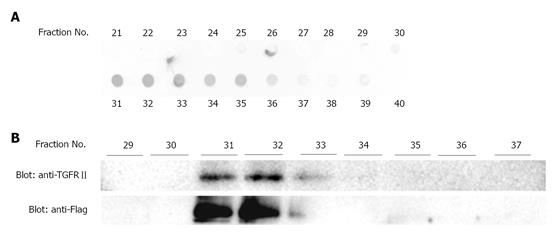
Figure 5 Transforming growth factor-β type II receptor binds to (-)-epigallocatechin-3-gallate.
A: Proteins in the fractions were detected by staining with Coomassie Brilliant Blue. An aliquot of each fraction was blotted on polyvinylidene difluoride membrane and stained; B: The protein-containing fractions revealed in (A) were electrophoresed and Western blotting was performed using anti-Flag antibody. TGFRII: Transforming growth factor-β type II receptor.
- Citation: Tabuchi M, Hayakawa S, Honda E, Ooshima K, Itoh T, Yoshida K, Park AM, Higashino H, Isemura M, Munakata H. Epigallocatechin-3-gallate suppresses transforming growth factor-beta signaling by interacting with the transforming growth factor-beta type II receptor. World J Exp Med 2013; 3(4): 100-107
- URL: https://www.wjgnet.com/2220-315X/full/v3/i4/100.htm
- DOI: https://dx.doi.org/10.5493/wjem.v3.i4.100


