Pravda J. Dissipation of the mitochondrial proton motive force drives sepsis pathogenesis and explains hyperlactatemia’s predictive value in sepsis mortality. World J Crit Care Med 2026; 15(3): 120314 [PMID: 42707451 DOI: 10.5492/wjccm.120314]
Corresponding Author of This Article
Jay Pravda, MD, Senior Scientist, Department of Disease Pathogenesis, Inflammatory Disease Research Centre, 4371 Northlake Blvd No. 247, Palm Beach Gardens, FL 33410, United States. jay.pravda@protonmail.com
Research Domain of This Article
Medicine, Research & Experimental
Article-Type of This Article
review-article
Open-Access Policy of This Article
This article is an open-access article which was selected by an in-house editor and fully peer-reviewed by external reviewers. It is distributed in accordance with the Creative Commons Attribution Non Commercial (CC BY-NC 4.0) license, which permits others to distribute, remix, adapt, build upon this work non-commercially, and license their derivative works on different terms, provided the original work is properly cited and the use is non-commercial. See: http://creativecommons.org/licenses/by-nc/4.0/
Baishideng Publishing Group Inc, 7041 Koll Center Parkway, Suite 160, Pleasanton, CA 94566, USA
Share the Article
Pravda J. Dissipation of the mitochondrial proton motive force drives sepsis pathogenesis and explains hyperlactatemia’s predictive value in sepsis mortality. World J Crit Care Med 2026; 15(3): 120314 [PMID: 42707451 DOI: 10.5492/wjccm.120314]
Author contributions: Pravda J is the sole author of this manuscript and solely responsible for its content; Pravda J performed all the research, collected, analyzed, and interpreted all the data; Pravda J conceived of and developed the hydrogen peroxide-based pathogenesis of sepsis; Pravda J prepared and wrote the manuscript and performed all critical revisions; Pravda J certifies that this manuscript is the product of his original research; and Pravda J has overall responsibility for this manuscript.
Conflict-of-interest statement: The corresponding author states that there is no conflict of interest.
Corresponding author: Jay Pravda, MD, Senior Scientist, Department of Disease Pathogenesis, Inflammatory Disease Research Centre, 4371 Northlake Blvd No. 247, Palm Beach Gardens, FL 33410, United States. jay.pravda@protonmail.com
Received: February 24, 2026 Revised: March 12, 2026 Accepted: April 21, 2026 Published online: September 9, 2026 Processing time: 185 Days and 11.8 Hours
Abstract
Sepsis remains a major cause of global mortality, yet its underlying pathogenesis is still incompletely understood. The current Sepsis-3 definition describes sepsis as a “life-threatening organ dysfunction caused by a dysregulated host response to infection”, but offers no mechanistic explanation for how infection or inflammation lead to metabolic collapse and organ failure. This work proposes a unifying biochemical model in which sepsis originates as an intramitochondrial disturbance of redox homeostasis triggered by an early hypermetabolic surge in mitochondrial hydrogen peroxide. According to this framework, excess hydrogen peroxide overwhelms mitochondrial reductive buffering systems, leading to aconitase (Krebs cycle) inhibition, impaired NADH and FADH2 generation, dissipation of the proton motive force, and subsequent failure of oxidative phosphorylation. This sequence provides a coherent explanation for hallmark features of sepsis, including hyperlactatemia, metabolic acidosis, hypothermia, ATP depletion, bioenergetic failure and increased mortality. The model also accounts for interindividual variability in sepsis susceptibility through differences in mitochondrial reductive capacity and offers insight into why animal models fail to translate to humans. Additionally, toxic systemic hydrogen peroxide elevation may contribute independently to sepsis heterogeneity by oxidatively inhibiting multiple enzyme systems and inducing lymphocyte apoptosis, providing a mechanistic basis for immunosuppression and post-sepsis syndrome. Confirming a role for hydrogen peroxide in initiating and perpetuating these events positions impaired mitochondrial redox homeostasis as a central driver of sepsis pathogenesis and generates testable predictions regarding specific therapy and future research.
Core Tip: Despite extensive research spanning several decades and numerous unsuccessful clinical trials, the underlying cause of sepsis remains elusive. However, the characteristic metabolic disturbances observed in sepsis—such as hyperlactatemia, bioenergetic failure, metabolic acidosis, hypothermia, and oxidative stress—can be explained by dissipation of the mitochondrial proton motive force. Emerging evidence suggests that hydrogen peroxide–mediated disruption of this proton gradient represents the proximal mechanism driving the pathogenesis of sepsis.
Citation: Pravda J. Dissipation of the mitochondrial proton motive force drives sepsis pathogenesis and explains hyperlactatemia’s predictive value in sepsis mortality. World J Crit Care Med 2026; 15(3): 120314
Sepsis has been a scourge on humanity since antiquity and continues to exact a heavy toll in terms of morbidity and mortality, representing 20% of all global deaths[1]. In 2017, global sepsis cases approached 50 million, resulting in 11 million deaths, with approximately half occurring in children[1]. With over 38 million survivors annually, sepsis survivorship is acknowledged to be an ongoing major loss of health globally[2]. Tragically, one-third of in-hospital deaths involve patients who had sepsis during their stay[3]. The cumulative mortality rate for severe sepsis survivors one year after discharge is reported to be over 48%, with a significantly increased risk of dying up to 5 years post-discharge[4,5]. Moreover, nearly 50% of sepsis survivors admitted to the intensive care unit (ICU) did not return to work two years after discharge while other studies report that almost all sepsis survivors developed one or more new physical, cognitive or psychological impairments with 40% suffering from all three[6,7]. In the United States, the estimated annual cost of sepsis care is $62 billion—a figure widely considered to be a conservative estimate[8]. Research has failed to yield specific effective therapeutic interventions targeting the root cause of this often-fatal condition, as evidenced by the hundreds of failed clinical trials since the 1970s[9]. The urgent need for effective strategies to treat or prevent sepsis is evident in light of its devastating impact on global health and national healthcare systems.
A consistent but underexplained feature of sepsis is the profound metabolic disturbance that emerges during the course of the syndrome. Hyperlactatemia, metabolic acidosis, hypothermia, ATP depletion, and bioenergetic failure are well-documented hallmarks of severe sepsis, yet their origin remains incompletely understood. Mitochondrial dysfunction has long been implicated, but existing models do not specify the biochemical trigger that initiates mitochondrial failure or explain why organ dysfunction can progress even after pathogen clearance and appropriate antimicrobial therapy. Moreover, current paradigms do not account for why some individuals exposed to similar infectious or inflammatory stimuli develop sepsis while others do not, nor do they explain why animal models fail to reproduce human outcomes, and none have yielded a unifying biochemical model capable of explaining the full spectrum of sepsis phenotypes or guiding effective therapies. The persistent failure of clinical trials targeting inflammatory or immune pathways underscores the need for a mechanistic framework that identifies the initiating events driving metabolic collapse and organ dysfunction.
The present work proposes a mechanistic model in which sepsis originates as an intramitochondrial disturbance of redox homeostasis driven by an early surge in mitochondrial hydrogen peroxide (H2O2) during the hypermetabolic response to a systemic insult. According to this framework, excess H2O2 overwhelms mitochondrial reductive buffering systems, leading to aconitase (critical Krebs cycle enzyme) inhibition, impaired NADH and FADH2 generation, dissipation of the proton motive force (PMF), and subsequent failure of oxidative phosphorylation. As described below, this sequence provides a coherent explanation for the metabolic abnormalities observed in sepsis and offers a rationale for the strong association between hyperlactatemia and mortality since dissipation of the PMF leads to elevated lactate and eventual death.
The model also predicts that individuals with diminished mitochondrial reductive capacity—such as reduced glutathione (GSH) reserves or impaired antioxidant recycling—are more susceptible to H2O2 accumulation and therefore more likely to progress to sepsis. This concept provides a mechanistic basis for interindividual and interspecies variability in sepsis susceptibility and may help explain why many animal models fail to translate to human disease. Together, these considerations suggest that impaired mitochondrial redox homeostasis may represent a central, unifying mechanism underlying the metabolic, immunologic, and clinical manifestations of sepsis.
The goal of this work is to articulate this mechanistic framework, integrate it with existing biochemical and clinical observations, and outline the testable predictions it generates. By providing a specific biochemical pathway linking early hypermetabolism to organ dysfunction, this model aims to outline a specific therapeutic intervention to restore mitochondrial redox homeostasis, complement and extend current definitions of sepsis and guide future research toward the initiating events that drive the syndrome.
NORMAL CELLULAR BIOENERGETICS
Cellular bioenergetics is primarily centered in mitochondria. Pyruvate, the final product of glycolysis, is transported into the mitochondrial matrix, where it undergoes enzymatic oxidative decarboxylation via pyruvate dehydrogenase (PD) to produce acetyl CoA. This acetyl CoA is subsequently oxidized through the Krebs cycle, yielding electron carriers NADH and FADH2. These carriers facilitate the transfer of high-energy electrons to the electron transport chain (ETC), initiating the translocation of protons from the matrix into the mitochondrial intermembrane space (IMS). The accumulation of protons within the IMS establishes an electrochemical proton gradient known as the PMF.
The PMF is harnessed to drive the mitochondrial pyruvate carrier (MPC), nicotinamide nucleotide transhydrogenase (NNT), and ATP synthase, which provides nearly all ATP fueling for cellular function. These essential components, embedded within the mitochondrial inner membrane (MIM), play a crucial role in sustaining cellular energy metabolism (Figure 1A).
Figure 1 Normal and sepsis bioenergetics.
A: Normal cellular bioenergetics. During normal cellular bioenergetics, cytoplasmic pyruvate (a), the final product of glycolysis, is transported into the mitochondrial matrix via the mitochondrial pyruvate carrier (MPC) (b). There, it undergoes oxidation, beginning with pyruvate dehydrogenase (PD), to generate acetyl CoA, which fuels the Krebs cycle. This process yields NADH and FADH2, which transfer high-energy electrons to the electron transport chain (ETC). The series of sequential enzymatic reactions starting with PD to terminal electron transfer in the ETC complex IV can be visualized as the mitochondrial energy flux (MEF) (c). The ETC utilizes the high-energy electrons to translocate (pump) protons from the mitochondrial matrix into the intermembrane space, establishing the electrochemical proton gradient known as the proton motive force (PMF) (d), whose protons power ATP synthase to synthesize ATP (e). ATP is transported into the cytoplasm by adenine nucleotide translocase (ANT) (F). The MEF is essential for sustaining both ETC function and the PMF. At the terminal step, electrons are transferred to molecular oxygen via ETC complex IV (cytochrome c oxidase), generating water (g). However, up to 4% of electrons prematurely escape into the matrix (electron leak) (h), reducing molecular oxygen to superoxide (i), which is subsequently converted to H2O2 (k) by superoxide dismutase (J). H2O2 is highly toxic and must be immediately degraded. H2O2 is neutralized by glutathione peroxidase (GPx) and glutathione (GSH) (L). The oxidized cofactor GSH disulfide (GSSG) is regenerated to its reduced form (GSH) by GSSG reductase (EC #1.8.1.7) (m) with reducing equivalents provided by NADPH (n). In the process, NADPH is oxidized to NADP+, which is regenerated to NADPH by nicotinamide nucleotide transhydrogenase (NNT) (o) in the inner mitochondrial membrane. NNT is powered by the PMF. Peroxiredoxin and reduced thioredoxin (not shown) are also critical to mitochondrial antioxidant defense (Figure 2). Uncoupling protein (p) dissipates the proton gradient as heat instead of making ATP, reducing energy efficiency to regulate temperature; B: Sepsis bioenergetics: The same as panel A, with the addition of orange dashed arrows to indicate molecular targets inhibited by elevated levels of mitochondrial H2O2, specifically PD and Krebs cycle enzymes. These inhibitory effects compromise mitochondrial energy metabolism and diminish MEF. Consequently, proton translocation into the intermembrane space is impaired, leading to dissipation of the PMF. The loss of PMF disrupts the function of key mitochondrial components, including the MPC, ATP synthase, NNT, and ANT. The terminal metabolic abnormalities associated with each component dysfunction are highlighted in orange text and include elevated lactate levels, increased mitochondrial H2O2, hypothermia, bioenergetic failure, and metabolic acidemia. Each of these parameters serves as a biomarker for the dissipation of the PMF, a critical bioenergetic gradient essential for sustaining life. Their individual associations with increased mortality underscore the fundamental role of PMF integrity in survival. Deficiency of vitamin B1 (thiamine) and magnesium (Mg2+) compromises PD functionality, predisposing to dissipation of the PMF and sepsis. Importantly, correction of these abnormalities in isolation does not improve outcomes in sepsis, as it fails to normalize mitochondrial H2O2 and restore the PMF. Collectively, these findings strongly support the conclusion that dissipation of the PMF represents the proximal and unifying cause of sepsis pathophysiology. Additionally, the figure also illustrates direct inhibition of ATP synthase, ANT, and GPx mediated by elevated levels of H2O2, further exacerbating the previously described metabolic disturbances. H2O2-induced oxidative damage to mitochondrial DNA may play a contributory role in the development of post-sepsis syndrome. Question marks (?) indicate absence of electrons. UCP: Uncoupling protein; MPC: Pyruvate carrier; PD: Pyruvate dehydrogenase; ETC: Electron transport chain; MEF: Mitochondrial energy flux; PMF: Proton motive force; ANT: Adenine nucleotide translocase; GPx: Glutathione peroxidase; GSH: Glutathione; GSSG: Glutathione disulfide; GDR: Glutathione disulfide reductase; OMM: Outer mitochondrial membrane; SOD: Superoxide dismutase; IMM: Inner mitochondrial membrane; NNT: Nucleotide transhydrogenase; Prx: Peroxiredoxin; Trx-r: Thioredoxin; mtDNA: Mitochondrial DNA; H2O2: Hydrogen peroxide.
Mitochondrial energy metabolism operates as a self-sustaining biological engine, driving its own function by utilizing the PMF to power the MPC transport of pyruvate into the matrix. Pyruvate is then used to generate the high energy electrons needed to maintain the PMF, which simultaneously supplies the energy for other essential components embedded within the IMM, such as ATP synthase and NNT. Any disruption in the components driving the directional flow of energy—collectively the mitochondrial energy flux (MEF) — (Figure 1A) can impair the translocation of protons into the IMS and compromise the PMF, which in turn disrupts the function of the MPC, ATP synthase and NNT.
Mitochondrial bioenergetic integrity is critically dependent on available matrix reductive capacity—primarily mediated by GSH and thioredoxin (Trx-r) systems—to neutralize H2O2 produced through electron leakage during oxidative phosphorylation (Figure 1A, bottom left). Excessive accumulation of H2O2 can impair the MEF and compromise energy metabolism. The following section explores the role of H2O2 in the pathogenetic chain of events leading to the development of sepsis.
CELLULAR BIOENERGETICS DURING SEPSIS
Excess H2O2 production occurs during the early hypermetabolic phase of sepsis. Blood H2O2 levels in sepsis have been recorded as high as 558 μmol/L (µM), which is over 110 times the normal upper limit of 5 μmol/L and nearly 12 × the cytotoxic threshold of 50 µM[10-12]. This concentration is also nearly 280000 times higher than the average intracellular H2O2 concentration of 2 nM[13]. Studies report that sepsis survivors exhibit an early and robust antioxidant defense, characterized by significantly elevated GSH levels compared to non-survivors, who experience a progressive decline in GSH as sepsis advances[14-16]. As GSH is essential for the degradation of H2O2, its progressive depletion aligns with the markedly elevated H2O2 levels observed in patients with sepsis. An association of H2O2 with sepsis is underscored by reports of fatal sepsis in a young man following intravenous infusion of H2O2[17].
Under physiological conditions, most intracellular H2O2 is produced inside mitochondria as a result of electron leakage from the ETC[18-21]. H2O2 is highly toxic and is kept at low nanomolar levels by conversion to water via GSH peroxidase (GPx) (EC 1.11.1.9) and peroxiredoxin (PRx) (EC 1.11.1.24) that collectively degrade 99% of mitochondrial H2O2[22,23]. NADPH supplies the reducing equivalents that allow mitochondrial enzymatic antioxidant systems to effectively degrade H2O2 (Figure 1B)[22,24]. NADPH is generated by NNT embedded in the MIM, which is powered by the PMF. The highly elevated blood levels of H2O2 in sepsis indicate that its production exceeds the capacity of mitochondrial antioxidant systems to effectively neutralize it. But why have the antioxidant systems failed?
Early in its development, sepsis is characterized by a hypermetabolic response with increased consumption of oxygen, 90% of which is utilized by cytochrome C oxidase or complex IV (EC 7.1.1.9) in the mitochondrial ETC to generate the supraphysiological amounts of ATP needed during this period of heightened energy demands[25-29]. Up to 4% of electrons are leaked from the ETC[29]. These leaked electrons reduce molecular oxygen prematurely in the matrix to form superoxide anion radical, which is dismutated by superoxide dismutase to form H2O2.
A hypermetabolic response leads to increased electron leakage, resulting in a significant increase in H2O2 production. Experimental data from murine models demonstrate a 15-fold rise in H2O2 generation during state 3 respiration[30], a phenomenon that closely parallels the early hypermetabolic phase of sepsis. Under these conditions, the rapid surge in H2O2 production can exceed the degradation capacity of critical reducing equivalents, such as GSH and reduced Trx-r, which are essential for preventing mitochondrial H2O2 accumulation[30]. The antioxidant role of GSH and Trx-r as a first line of antioxidant defense is especially critical during a hypermetabolic response such as sepsis[30].
Notably, mitochondria contain only 10% of the total cellular GSH and lack the capability to synthesize it within the matrix[31]. Instead, GSH must be imported from the cytoplasm, a process that can take several hours after experimental depletion of mitochondrial GSH (mGSH)[32,33]. Consequently, a sustained hypermetabolic response can deplete key H2O2-reducing equivalents, leading to excessive intracellular accumulation of H2O2[34,35]. Experimentally, increased H2O2 formation at elevated ETC activity is observable when redox buffering systems become overwhelmed[36].
Empirical evidence supporting an early hypermetabolic response in sepsis is demonstrated by a significant elevation in mitochondrial enzyme activity in healthy human volunteers within two hours of intravenous endotoxin administration, simulating the initial hypermetabolic phase of sepsis[37]. Comparable hypermetabolic responses have been observed in laboratory animal models, such as rats and rabbits, following IV endotoxin infusion[38]. In humans, the hypermetabolic response to surgical trauma is reported to reduce serum and muscle GSH by 20% and 40%, respectively, compared to preoperative values[39].
Moreover, in critically ill ICU patients, blood GSH levels are reported to be 57% of those observed in control groups, while muscle glutamine—the storage form of glutamate essential for GSH synthesis—is 72% lower than in healthy controls[40]. Furthermore, studies have identified a reduction in Trx-r during both the acute and convalescent phases of pediatric sepsis[41]. Taken together, this implies a sustained elevation in the production of mitochondrial H2O2, which has overwhelmed mitochondrial antioxidant capacity, starting from the early hypermetabolic phase of sepsis through the recovery period. This is significantly magnified by the H2O2-induced inhibition of almost all mitochondrial antioxidant defenses as described in the next section.
Antioxidant enzyme inhibition
Compounding the hypermetabolic generation of H2O2 during the early phase of sepsis is the oxidative inhibition of critical antioxidant enzymes. As the hypermetabolic production of H2O2 increases, excess H2O2 interacts with the peroxidatic active sites on PRx and GPx, leading to irreversible oxidative modifications of cysteine and selenocysteine residues, respectively. This oxidative damage results in enzyme inactivation (hyperoxidation), impairing their ability to degrade mitochondrial H2O2[42-44] (Figure 2).
Figure 2 Antioxidant enzyme inhibition: The mitochondrial proton motive force maintains cellular redox homeostasis.
The electron transport chain (ETC) is a major source of hydrogen peroxide (H2O2). Up to 4% of electrons “leak” out prematurely from the ETC into the mitochondrial matrix forming superoxide anion radical (O2•ˉ), which is dismutated to H2O2 by superoxide dismutase. H2O2 is highly toxic to cells and must be disposed of. This detoxification process relies on two key regenerative redox enzyme systems—glutathione peroxidase (GPx) and peroxiredoxin (PRx)—which facilitate the reduction of H2O2 to water using glutathione (GSH) and reduced thioredoxin (TRX-r), respectively. During this reaction, GSH is oxidized to glutathione disulfide (GSSG), while TRX-r is converted to oxidized thioredoxin (TRx-o). To regenerate these reducing agents, glutathione disulfide reductase (EC 1.8.1.7) and thioredoxin disulfide reductase (EC 1.8.1.9) catalyze the reduction of GSSG and TRx-o, respectively, using NADPH as an electron donor. NADPH is replenished by nicotinamide nucleotide transhydrogenase (NNT; EC 7.1.1.1), which facilitates electron transfer from NADH to NADP+. This reaction is driven by the proton motive force (detailed below), linking mitochondrial energy metabolism to redox homeostasis. Thus, mitochondrial enzymatic redox buffering systems critically depend on the proton motive force (PMF) to sustain H2O2 detoxification and maintain cellular redox homeostasis. However, the functionality of this tightly interdependent antioxidant system can be compromised by excessive H2O2 production during the early hypermetabolic stage of sepsis. An acute H2O2 load can deplete GSH and Trx-r, leading to the accumulation of H2O2 followed by hyperoxidation of GPx and PRx, resulting in their conversion to dehydroalanine (DHA) and sulfinic acid derivatives, respectively (orange dashed arrows). These oxidative modifications deactivate both enzymes, which are responsible for eliminating approximately 99% of mitochondrial H2O2. Deactivation of these enzymes results in significantly increased free H2O2 accumulation within the mitochondrion, which disrupts mitochondrial energy metabolism initiating dissipation of the PMF (Figure 1B). Dissipation of the PMF compromises NNT functionality, further reducing mitochondrial capacity for H2O2 detoxification leading to toxic systemic levels of H2O2. NNT: Nucleotide transhydrogenase; GDR: Glutathione disulfide reductase; TDR: Thioredoxin disulfide reductase; GPx: Glutathione peroxidase; PRx: Peroxiredoxin; TRX-r: Reduced thioredoxin; TRx-o: Oxidized thioredoxin; GSH: Glutathione; GSSG: Glutathione disulfide; ETC: Electron transport chain; SOD: Superoxide dismutase; H2O2: Hydrogen peroxide; DHA: Dehydroalanine.
In support of this interpretation, studies have reported reduced GPx activity in sepsis patients compared to healthy individuals[45]. Similarly, PRx is extremely sensitive to H2O2-mediated oxidative inactivation under conditions of excessive H2O2[23,46]. While the role of the mitochondrial PRx 3 isoform remains largely unexplored in sepsis[47], research indicates that PRx 3 undergoes rapid oxidation and inactivation by H2O2 during the early stages of receptor-mediated apoptosis, leading to increased mitochondrial H2O2 levels[46,48]. This implies a similar H2O2-induced antioxidant enzyme inactivation during the early hypermetabolic phase of sepsis. Additionally, lowering PRx3 levels with siRNA is reported to increase cellular H2O2, illustrating the importance of PRX 3 for maintaining normal H2O2 levels and redox homeostasis[49].
Collectively, these findings suggest that the initial hypermetabolic response characteristic of early-stage sepsis results in the generation of substantial quantities of H2O2. The excessive accumulation of H2O2 can overwhelm and deplete key reducing cofactors—namely GSH and reduced Trx-r—both essential for mitochondrial antioxidant defense, leading to an initial buildup of mitochondrial H2O2. This is subsequently compounded by H2O2-mediated oxidative inhibition of GPx and PRx, further impairing the cell’s antioxidant defenses. The resulting unchecked diffusion of H2O2 throughout the mitochondrial matrix can inhibit critical redox-sensitive enzymes, disrupting mitochondrial energy metabolism (MEF) and contributing to dissipation of the PMF, thereby promoting the development of sepsis. These mechanisms are explored in detail starting with the following section.
INHIBITION OF MITOCHONDRIAL ENERGY METABOLISM
As mitochondrial H2O2 levels continue to rise, there is a pronounced suppression of cellular bioenergetics. Data derived from studies on rat heart mitochondria demonstrate that the addition of H2O2 leads to a marked reduction in overall mitochondrial respiration. Specifically, key Krebs cycle enzymes—aconitase, α-ketoglutarate dehydrogenase, and succinate dehydrogenase—were inhibited by 96% (P ≤ 10-8), 40% (P ≤ 0.0004), and 40% (P ≤ 0.0002) respectively, after 7.5 minutes of exposure to 50 μmol/L H2O2, well within the upper range reported in human sepsis blood of 558 µM[10,50].
Under these circumstances, aconitase inhibition by 96% is the main bottleneck. With only approximately 4% of citrate being converted to isocitrate, most of the Krebs cycle is starved of substrate. With the small amount of isocitrate available and α-Ketoglutarate dehydrogenase inhibited by 40%, the conversion to succinyl-CoA is limited. This step generates NADH but it would be severely reduced based only on a 4% substrate input. Finally, with succinate dehydrogenase inhibited by 40%, the resulting FADH2 yield is also cut to approximately 60% of baseline but the substrate scarcity drastically reduces that amount. With this degree of enzyme inhibition, NADH levels would plummet since the NADH generating enzymes—isocitrate dehydrogenase, α-ketoglutarate dehydrogenase, and malate dehydrogenase—are deprived of substrate or directly impaired by H2O2. With such limiting conditions, the reduction in electron carriers, NADH and FADH2, could surpass 90%. This would de-energize mitochondrial energy metabolism (MEF) (Figure 1A) with a corresponding dissipation of the PMF. Offsetting mechanisms such as a compensatory increase in ATP via glycolysis, anapleurotic metabolism of glutamate and reverse function of ATP synthase to sustain the PMF are not long-term solutions for cell survival. In other words, without the MEF—the biological engine that maintains the PMF, life is not possible.
In support of the above, studies have shown that exposure of rat heart mitochondria to 12.5 μmol/L H2O2 for 5 minutes almost completely abolished oxygen utilization[50]. The decline in mitochondrial respiration after exposure to 12.5μmol/L H2O2 was predominantly due to diminished levels of reducing equivalents (NADH, FADH2) available for electron transport. Other studies have documented significant decline in NADH levels after exposure of isolated guinea pig nerve terminal mitochondria to 500 μmol/L H2O2 for 7 minutes[51]. These H2O2 exposure concentrations are within the upper range of 558 μmol/L for blood H2O2 recorded in sepsis patients[10].
Thus, H2O2 inhibits both the synthesis of electron carriers by the Krebs cycle and the utilization of these carriers by the ETC. The former decreases the amount of electron carriers entering the ETC, while the latter decreases electron transport through the ETC. Without these high-energy electrons passing through the ETC, cytochrome C oxidase (complex 4 of the ETC) does not have sufficient electrons to combine with oxygen to form water. Under these conditions, the gradient or driving force for the passive diffusion of oxygen into the mitochondrial matrix (the “oxygen sink”) cannot be maintained. Clinically, this manifests as the inability of cells to utilize (extract) oxygen during sepsis despite adequate tissue oxygen delivery, which has been described as cytopathic hypoxia[52]. Due to its high sensitivity to H2O2-induced inhibition, the loss of aconitase activity is considered a reliable marker of oxidative damage[53].
Upstream from the Krebs cycle, at the start of the MEF, H2O2 has been documented to inhibit PD; a pivotal enzyme responsible for converting newly imported mitochondrial pyruvate into acetyl-CoA[54]. This inhibition has profound downstream effects, as it reduces the amount of PD-generated acetyl-CoA entering the Krebs cycle, thereby decreasing the synthesis of electron carriers NADH and FADH2. A reduction in these electron carriers will de-energize the MEF and impair the ETC’s ability to maintain the mitochondrial electrochemical proton gradient, which leads to dissipation of the PMF. Furthermore, the diminished availability of electron carriers impairs mitochondrial thermogenesis, a critical process for maintaining body temperature through heat generation during the oxidation of NADH and FADH2. This disruption contributes to sepsis-induced hypothermia, as detailed further below.
Other enzymes involved in supplying acetyl CoA to the Krebs cycle such as succinyl-CoA: 3-oxoacid CoA transferase, a critical enzyme for generating acetyl CoA from ketone bodies, and mitochondrial acetoacetyl CoA thiolase, an essential enzyme involved in the production of acetyl CoA from beta-oxidation of short-chain fatty acids are both inhibited by H2O2[54,55]. Thus, H2O2 significantly reduces the entry of acetyl-CoA into the Krebs cycle, in addition to directly inhibiting the Krebs cycle and complex II of the ETC.
In essence, excess H2O2 is a metabolic poison that de-energizes the entire MEF. Although the inhibitory action of H2O2 serves a physiological intracellular signaling function, providing feedback control to modulate bioenergetic output as H2O2 diffuses within the mitochondrial matrix[56], it is critically dependent on an intact cellular redox buffering (antioxidant) system to prevent the accumulation of H2O2. However, during a hypermetabolic response to a systemic insult, mitochondrial H2O2 levels can rise beyond the buffering capacity of the local antioxidant system, leading to inhibition of the MEF and dissipation of the PMF, as discussed in the following section.
DISSIPATION OF THE PMF
The PMF plays an essential and irreplaceable role in maintaining cellular metabolism. It drives the activity of key MIM proteins—including the MPC, ATP synthase, and nicotinamide NNT—all of which rely exclusively on the proton gradient generated by the PMF for their function (Figure 1A). Accordingly, disruption of the PMF impairs the operation of these components, resulting in a cascade of metabolic disturbances such as hyperlactatemia, bioenergetic organ failure, metabolic acidosis, systemic oxidative stress (characterized by elevated H2O2 levels), and hypothermia. These metabolic abnormalities mirror the clinical features commonly observed in sepsis (Figure 1B).
The PMF is an electrochemical gradient that is maintained across the inner mitochondrial membrane. The PMF consists of two interrelated components: An electrical potential gradient (ΔΨ) of approximately 190 millivolts and a chemical gradient (ΔpH) of roughly 30 millivolts[57]. These gradients are both mediated by the proton, a positively charged subatomic particle that simultaneously establishes the Δψ through its charge and the ΔpH through its concentration differential across the inner mitochondrial membrane. Notably, ΔΨ accounts for approximately 85% of the total PMF[57], corresponding to an electric field strength of nearly 30 million volts per meter—comparable to the energy released by a lightning strike[36,58,59]. A reduction in ΔΨ can result in mitochondrial depolarization and loss of membrane potential. Given its predominant contribution to the PMF, a substantial decline in Δψ can lead to a complete dissipation of the PMF.
Experimental evidence demonstrates that exposure of guinea pig brain synaptosomes to 500 μmol/L H2O2 for 15 minutes induces significant mitochondrial depolarization, primarily due to a reduction in NADH levels and a consequent decline in the PMF[60]. Similar findings have been reported in rat endothelial cells, where treatment with 100 μmol/L H2O2 for 40 minutes significantly diminished mitochondrial membrane potential[61]. Additional studies involving various cell types—including mouse myoblasts (250 μmol/L H2O2 for 30 minutes) and human umbilical vein endothelial cells (220 μmol/L H2O2 for 2 hours)—also revealed substantial reductions in Δψ[62,63]. Notably, a 50% decrease in ΔΨ was observed in a human endothelial cell line following exposure to 300 μmol/L H2O2 for 2 hours[64]. Sepsis patients, however, are subjected to sustained elevations in H2O2 concentrations reaching up to 558 μmol/L over prolonged periods ranging from days to weeks. This chronic oxidative burden significantly amplifies the depolarizing impact of H2O2 on the ΔΨ, exacerbating mitochondrial dysfunction.
Further investigations involving peripheral blood mononuclear cells from patients in the early stages of sepsis—absent of shock or multiple organ failure—have demonstrated a reduction in mitochondrial membrane potential (ΔΨ) and oxygen consumption[65]. These findings are consistent with an accumulation of intramitochondrial hydrogen peroxide (H2O2, as previously described), and underscore the critical role of the PMF in the development of sepsis. This is corroborated by studies reporting that a reduction in mitochondrial membrane potential—the major power source for the PMF—serves as a biomarker for sepsis severity and clinical outcome, with non-survivors exhibiting significantly reduced membrane potential compared to survivors[66,67]. Other studies have shown that sepsis survivors successfully normalize mitochondrial membrane potential, whereas non-survivors are unable to do so, with membrane depolarization (i.e., PMF dissipation) correlating with sepsis severity[67].
Additional support comes from research indicating a significant correlation between mitochondrial membrane (ΔΨm) depolarization—reflecting dissipation of the PMF—and severity of sepsis, as indicated by the Acute Physiology and Chronic Health Evaluation II score and Sequential Organ Failure Assessment score (all, P < 0.05)[68]. A similar association was also found in other conditions that present with systemic inflammatory response syndrome (SIRS), which can progress to sepsis, such as penetrating trauma, peritonitis, pneumonia, necrotizing fasciitis, bloodstream infections, post-cardiac arrest states, and cerebrovascular disorders[68]. Platelet apoptosis was enhanced in sepsis and the above-mentioned conditions, all of which present with SIRS.
SIRS represents a hypermetabolic state characterized by elevated energy expenditure and as noted above, increased production of H2O2. Prior studies have shown that exposure to H2O2 can induce platelet apoptosis[69]. Collectively, these findings support a model in which the onset of a SIRS hypermetabolic state, regardless of cause, leads to increased H2O2 generation, subsequent dissipation of the PMF, and progression to sepsis, whose severity parallels the degree of PMF dissipation. It also indicates that infection is but one of many oxidative stressors that can lead to sepsis.
Thus, the above data supports dissipation of the PMF as the final common pathway resulting in bioenergetic organ failure, metabolic acidosis, hyperlactatemia, hypothermia, and widespread oxidative stress—which as detailed in Figure 1B—are the key metabolic abnormalities characteristic of sepsis. Under appropriate clinical conditions, each of these metabolic abnormalities serves as an independent risk factor for increased mortality in sepsis and concurrently functions as a biomarker indicative of a collapsing PMF. This dual prognostic role reflects the essential function of an intact PMF for sustaining cellular viability and life, indicating a proximal causal role for dissipation of the PMF in the development of sepsis. The following section will detail how dissipation of the PMF leads to each of these metabolic disturbances, beginning with bioenergetic failure.
BIOENERGETIC ORGAN FAILURE
H2O2-driven disruption of the PMF during sepsis impairs ATP synthase activity, leading to bioenergetic organ failure.
Bioenergetic failure occurs when cells or organs face an energy deficit that compromises their capacity to maintain essential physiological functions. Cellular processes are critically reliant on ATP, which supplies approximately 95% of the energy required for normal function[70]. In the context of sepsis, multiple studies have documented a reduction in intracellular ATP levels, accompanied by impaired activity of ATP synthase[71-73].
Several studies have implicated bioenergetic failure, characterized by reduced ATP levels, as a contributing factor in the pathogenesis of multi-organ dysfunction associated with sepsis[37,74]. Experimental evidence further supports this association: When peripheral blood mononuclear cells from control subjects were suspended in plasma derived from patients with septic shock, they exhibited impaired mitochondrial respiration, reduced oxygen consumption, and diminished ATP synthase activity[75]. These findings imply the presence of a water-soluble, membrane-permeable factor in sepsis plasma capable of inducing mitochondrial dysfunction. H2O2 meets these criteria. Prior research has demonstrated that disturbances in redox homeostasis, marked by elevated H2O2 levels, precede mitochondrial bioenergetic impairment[54]. Increased intracellular H2O2 has been shown to correlate with a reduction in mitochondrial membrane potential and a subsequent decline in ATP production[54]. Consistent with these observations, H2O2 concentrations are reported to be significantly elevated in sepsis[10].
These findings reinforce the conclusion that multi-organ failure in sepsis is driven by bioenergetic collapse, with H2O2 playing a causative role. The mechanistic basis for this involves H2O2-mediated inhibition of key mitochondrial processes, including PD, the tricarboxylic acid (Krebs) cycle, and the ETC, as illustrated in Figure 1B. Collectively, these pathways constitute the MEF (Figure 1). Disruption of the MEF reduces the generation of critical reducing equivalents—namely NADH and FADH2—which are required to sustain proton translocation across the inner mitochondrial membrane via the ETC. This impairment diminishes the electrochemical proton gradient and mitochondrial membrane potential, also referred to as the PMF. Loss of PMF compromises ATP synthase function, leading to reduced ATP production and, ultimately, bioenergetic failure at the organ level.
These findings indicate that, in the context of sufficient oxygen delivery and absence of other confounding factors, organ failure serves as a surrogate marker for the dissipation of the PMF in critical illness, particularly sepsis. Elevated systemic concentrations of H2O2 are capable of inducing multi-organ dysfunction through its inhibitory effects on mitochondrial metabolism (MEF) across diverse tissues.
Taken together, the evidence supports a central role for H2O2 in mediating PMF collapse, thereby driving the bioenergetic failure that underlies organ dysfunction in sepsis. The subsequent section will explore how dissipation of the PMF contributes to the development of metabolic acidemia.
Metabolic acidosis
H2O2-mediated PMF Dissipation in Sepsis Impairs ATP Synthase Function and Promotes Metabolic Acidemia.
During sepsis, the simultaneous development of hyperlactatemia and metabolic acidosis is commonly referred to as lactic acidosis. This implies that lactic acid enters the bloodstream, followed by dissociation into its conjugate base, the lactate anion, accompanied by the release of a proton, leading to elevated serum lactate and metabolic acidemia, respectively. However, the enzymatic process responsible for lactate production is facilitated by lactate dehydrogenase, which specifically generates lactate—not lactic acid—through the following reaction:
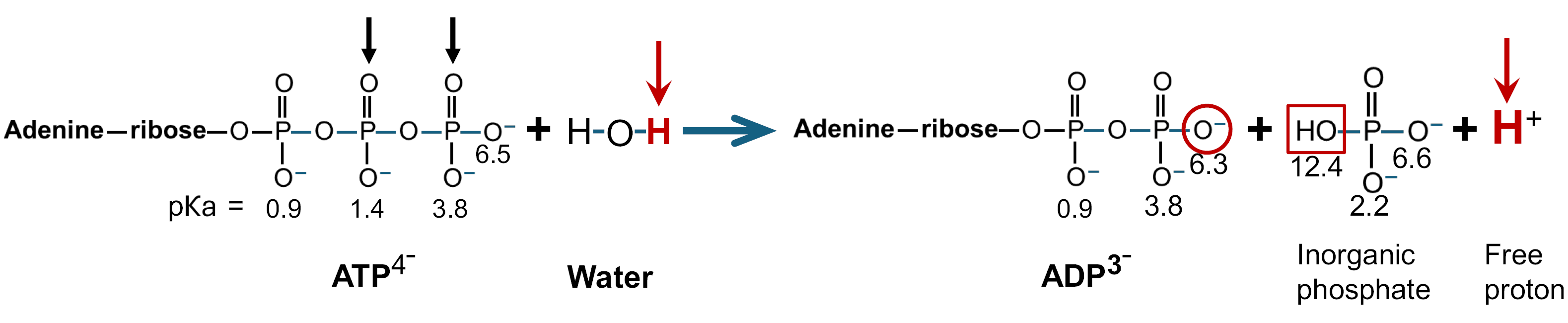
This reaction is not acidifying, as it does not generate protons; rather, it consumes a proton[76,77]. Consequently, the protons leading to metabolic acidosis during sepsis do not originate from lactic acid production. However, at physiological pH, the hydrolysis of cytoplasmic ATP will generate protons. The products of this reaction will differ depending on the ambient pH[78]. At pH > 7, ATP hydrolysis in the cytoplasm can be described by the following reaction[77,78]:
During ATP hydrolysis, water is inserted asymmetrically between the last two (beta and gamma) phosphate groups (black arrows). In this reaction, water donates a hydrogen atom to one phosphate group (red circle) and a hydroxyl moiety to the other (red square), resulting in the formation of ADP and inorganic phosphate, respectively[77]. However, ADP’s terminal hydroxy group (on the beta phosphate-red circle) has a pƘa of 6.3, leading to its ionization (deprotonation) at physiological pH. This generates a free proton in the cytoplasm, which originates from water (red arrows)[77,78]. At physiological PH (approximately 7.4), the hydroxy group on the inorganic phosphate (red square) remains un-ionized due to its significantly higher pƘa of 12.4[78].
Given that ATP is not stored in significant quantities for fuel, human cells—depending on their type—synthesize between 100 million and 5 billion ATP molecules per second in real time to replenish the ATP consumed through hydrolysis[77,79-81]. This process results in a significant number of protons being continuously released into the cytoplasm. While the protons generated by ATP hydrolysis have been implicated in sepsis-related metabolic acidosis[76,81], this raises a critical question: What is the normal fate of these protons, and what role do they play in the development of sepsis-associated metabolic acidemia?
In vitro studies have demonstrated that isolated rabbit heart mitochondria possess the ability to buffer (internalize) extramitochondrial protons, a process that is dependent on oxidative phosphorylation[82]. The inhibition of ATP synthase with oligomycin effectively abolished this mitochondrial proton buffering capacity[82]. Furthermore, at the conclusion of state 3 respiration—when the added ADP was fully consumed and mitochondrial respiration had ceased—the extramitochondrial proton concentration remained unchanged.
This suggests that the internalized protons were either utilized in other metabolic reactions within the matrix or were unable to exit due to the impermeability of the inner mitochondrial membrane. In other words, rather than remaining confined to the IMS, where they could diffuse through the permeable outer mitochondrial membrane into the surrounding culture medium, the protons were actively transported into the mitochondrial matrix.
The enzymatic activity of ATP synthase provides a mechanistic explanation for these observations. ATP synthase, embedded within the MIM, harnesses protons from the IMS, which are powered by the PMF, to enable ATP synthesis. As protons flow through ATP synthase into the mitochondrial matrix, they generate the energy required to catalyze the condensation reaction that combines ADP and inorganic phosphate into ATP. This reaction simultaneously incorporates a vicinal proton from the matrix into the formation of water. This process represents the reverse of ATP hydrolysis, as illustrated above. The interrelated reactions of ATP hydrolysis and synthesis can be summarized as follows[82,83]:
Thus, the continuous activity of ATP synthase during oxidative phosphorylation enables the internalization (buffering) of cytoplasmic protons into the mitochondrial matrix, where they are utilized for ATP (and water) synthesis. The newly synthesized ATP is subsequently exported to the cytoplasm, where it undergoes hydrolysis to fuel cellular processes. In the absence of ATP synthase, protons released during ATP hydrolysis accumulate in the cytoplasm and are transported out of the cell via membrane transporters[83], eventually reaching the bloodstream and contributing to acidemia. Since ATP synthase is powered by the PMF, dissipation of the PMF in sepsis will inhibit cytoplasmic proton buffering, resulting in metabolic acidosis. This implies that metabolic acidosis in sepsis serves as a biomarker for PMF dissipation.
This explains why the loss of acid-base buffering capacity portends a fatal outcome (independent predictor of mortality) in patients with sepsis, as it indicates dissipation of the PMF, without which life is not possible[84]. The internalization of cytoplasmic protons into the mitochondrial matrix not only prevents cytoplasmic acidosis and acidemia but may also contribute to the continued influx of cytoplasmic protons into the mitochondrial IMS. This continuous one-way proton flux into the matrix may prevent protons already present in the IMS from diffusing through the permeable outer mitochondrial membrane into the cytoplasm. Consequently, the mitochondrial matrix serves as a sink for cytoplasmic protons. In other words, as long as ATP synthase is functional, mitochondria can act as an active sink for cytoplasmic protons. This plays a crucial role in maintaining acid-base homeostasis, as well as contributing to the PMF and the essential metabolic processes it supports.
In summary, the evidence indicates that metabolic acidosis in sepsis is a direct consequence of ATP synthase dysfunction, driven by the dissipation of the PMF. This explains why attempts to correct metabolic acidosis in sepsis do not improve mortality[85], as addressing acidemia alone does not restore the PMF. Additionally, this relationship clarifies why bioenergetic organ failure occurs simultaneously with metabolic acidosis, as both conditions originate from ATP synthase impairment due to PMF dissipation. The following section will explain how PMF dissipation leads to hyperlactatemia and why it arises in parallel with metabolic acidosis and bioenergetic organ failure.
Hyperlactatemia
H2O2-mediated PMF dissipation in sepsis leads to dysfunction of the MPC followed by hyperlactatemia.
Elevated serum lactate levels are frequently observed in sepsis patients and are linked to considerable morbidity and mortality[71]. Research indicates that approximately two-thirds of individuals with sepsis exhibit increased serum lactate concentrations[71]. The in-hospital mortality for sepsis patients with hyperlactatemia ( ≥ 10 mmol/L) is reported to be 79% with a specificity of 99%[86]. Consequently, in the absence of a definitive causal definition to inform targeted therapeutic interventions, patients with sepsis are likely to continue experiencing high serum lactate levels, which serve as a strong predictive indicator of morbidity and mortality. However, lactate is neither the cause of sepsis nor a pathological entity itself[86]. This is corroborated by studies showing that interventions aimed at reducing serum lactate in lactic acidosis do not enhance survival rates, nor is lactate-level-guided resuscitation in early septic shock superior in reducing mortality compared with intervention guided by peripheral perfusion[87,88].
This raises the question of why elevated lactate is so prevalent and so strongly associated with morbidity and mortality in sepsis.
Lactate is synthesized from cytoplasmic pyruvate, the terminal product of glycolysis, via the enzyme lactate dehydrogenase. Concurrently, a portion of cytoplasmic pyruvate is translocated into the mitochondrial matrix through the MPC. The MPC is a transmembrane protein situated within the inner mitochondrial membrane that is the sole point of entry for pyruvate into the mitochondrial matrix[89]. The MPC translocates pyruvate into the mitochondrial matrix via a symport mechanism with a proton, a high number of which are transported into the mitochondrial IMS as a result of ETC activity. These protons constitute the electrochemical proton gradient, which creates the PMF (L in Figure 1A)[89,90].
The MPC cannot transport pyruvate into the mitochondrial matrix without the protons in the mitochondrial IMS. Thus, protons in the IMS generate the electrochemical gradient necessary to establish the PMF, which powers the MPC-mediated translocation of pyruvate into the mitochondrial matrix[91]. Consequently, dissipation of the PMF inhibits the MPC’s ability to transport pyruvate into the mitochondrial matrix. Under these conditions, pyruvate accumulates in the cytoplasm, where it is converted by lactate dehydrogenase into lactate. This lactate then exits the cell and enters the bloodstream, manifesting as hyperlactatemia.
Studies have shown that depolarization of the mitochondrial membrane potential (ΔΨm), reflecting dissipation of PMF, is significantly correlated with blood lactate concentration in sepsis and other conditions presenting with the SIRS[68]. In other words, greater dissipation of the PMF is associated with higher serum lactate concentrations. Because the PMF is the sole energy source driving the MPC, these findings indicate that impaired MPC function is contributing to the rise in circulating lactate.
Given that blood concentration of H2O2 reported in sepsis exceeds the threshold for Krebs cycle enzyme inhibition and H2O2 is permeable through cell membranes, it can be anticipated that the resulting excess free mitochondrial matrix H2O2 will suppress Krebs cycle activity and restrict the supply of NADH and FADH2 entering the ETC. This is followed by depletion of the electrochemical proton gradient and dissipation of the PMF. The end result is dysfunction of the MPC leading to hyperlactatemia, which is often described as lactic acidosis due to the accompanying metabolic acidosis, as noted above.
Thus, dissipation of the PMF by H2O2 accounts for the emergence of hyperlactatemia in sepsis. It also elucidates why hyperlactatemia is independently associated with a high risk of mortality since the PMF is the common sole power source for both the MPC and ATP synthase. Accordingly, dissipation of the PMF by H2O2 leads to simultaneous inhibition of the MPC and ATP synthase. This results in hyperlactatemia and impaired ATP synthesis with the latter culminating in bioenergetic organ failure and death. Because the inhibition of Krebs cycle activity by H2O2 is upstream of oxygen utilization by cytochrome C oxidase (complex IV), hyperlactatemia occurs when oxygen is available. This has been called cytopathic hypoxia.
This indicates that hyperlactatemia is an early biomarker for dissipation of the PMF in sepsis. This also explains why lowering serum lactate has no effect on sepsis mortality because doing so does not restore a dissipating PMF. It also clarifies the inverse relationship between elevated serum lactate and diminished serum bicarbonate in sepsis since hyperlactatemia and metabolic acidosis are both driven by a dissipating PMF[92]. In conclusion, the onset of hyperlactatemia in sepsis can be attributed to dissipation of the PMF and subsequent dysfunction of the MPC. Furthermore, PMF dissipation accounts for the strong association between hyperlactatemia and mortality in sepsis, as the PMF is essential for life, powering not only the MPC but also ATP synthase, which is crucial for ATP production.
Hypothermia
H2O2-mediated suppression of oxidative phosphorylation leads to impaired thermogenesis and hypothermia.
Hypothermia occurs in up to 35% of sepsis patients and is independently associated with mortality and adverse outcomes[93]. It can result from decreased heat production, increased heat loss, or both. Mitochondria are the most important source of body heat, mainly via proton leak and electron flux during the oxidation of NADH by the ETC[94].
Proton leak occurs when protons bypass ATP synthase and flow directly into the matrix, via the uncoupler protein, which generates heat[94]. NADH releases heat during the exergonic oxidation by the ETC. However, only 42% of the energy released during the oxidation of NADH is channeled into the creation of the electrochemical proton gradient and the PMF, which drives ATP synthesis via ATP synthase. The remaining energy is released as heat[95].
The amount of energy released during NADH oxidation is considerable and is driven by the 1.14 volt difference in the electron-transfer potential of NADH and FADH2 relative to that of O2 during the passage of electrons through the ETC. The complete reaction is:
This is equivalent to a standard free energy change (energy released during conversion of reactants to products or ΔG° at pH 7) of -220.1 kJ/mol. This energy is used to synthesize ATP, whose complete reaction is:
ADP + Pi + H+ → ATP + H2O (ΔG° = -30.5 kJ/mol)
Since the oxidation of one NADH yields 3 ATP, the percentage of energy conserved as ATP, i.e., the energy efficiency of oxidative phosphorylation, is approximately: (91.5/220.1) × 100 or 42%. The remainder, 128.6 kJ/mol or 58%, is released as heat. Thus, the release of free energy from oxidative phosphorylation is substantial and contributes to heating the body[95]. This heat is released into the mitochondrial matrix, resulting in a mitochondrial temperature significantly higher than the rest of the cell, reaching 50 °C (122 °F)[96].
By inhibiting key enzymes involved in MEF—including PDH, the Krebs cycle, and complex II of the ETC—H2O2 reduces NADH production and disrupts electron flow through the ETC. This attenuation of NADH oxidation impairs oxidative phosphorylation, leading to diminished thermogenesis and metabolic heat generation. Additionally, suppression of MEF results in dissipation of the PMF and a reduction in proton leak through uncoupling proteins, further decreasing thermogenic capacity. Together, these effects contribute to the hypothermia characteristic of sepsis.
The contribution of electron flux through the ETC to heat production during oxidative phosphorylation is evidenced by studies indicating that actively respiring mitochondria heat up. In contrast, various respiratory inhibitors like cyanide and oligomycin, which block electron flux through the ETC, result in mitochondrial cooling[96]. Correspondingly, sepsis induced hypothermia can indicate dissipation of the PMF, which explains the significantly higher mortality, organ failure scores and worse outcomes in patients with sepsis related hypothermia[97]. Furthermore, hypothermia can be one of the earliest indicators of sepsis and, in line with other manifestations of PMF dissipation, serves as an independent predictor of mortality in sepsis[93,94,98]. The next section details how dissipation of the PMF leads to generalized toxic levels of H2O2 resulting in severe systemic oxidative stress and many of the clinical abnormalities observed in sepsis.
Oxidative stress
H2O2-induced dissipation of the PMF impairs NNT activity resulting in decreased NADPH synthesis and exacerbated H2O2 accumulation.
NADPH functions as the principal electron donor in the regeneration of both GSH and reduced Trx-r (Figure 2). These reductive cofactors supply the necessary reducing equivalents for GPx and PRx to effectively neutralize almost all H2O2 produced within mitochondria[27,28]. NNT contributes more than 50% of the mitochondrial NADPH pool and plays a pivotal role in H2O2 detoxification[27,54,99]. NNT activity is driven by the PMF, underscoring the essential role of PMF in sustaining NADPH production required for mitochondrial H2O2 clearance and the preservation of cellular redox balance. Consequently, dissipation of the PMF can eliminate more than half of mitochondrial NADPH.
The critical role of NADPH in sepsis is underscored by studies demonstrating markedly reduced levels of NADPH and GSH in sepsis non-survivors compared to survivors, with non-survivors exhibiting a progressive decline in GSH concentrations as sepsis advances[15,16]. Experimental data from isolated rat heart mitochondria exposed to H2O2 reveal that restoration of NADPH levels and associated enzymatic activity occurs only after the excess mitochondrial H2O2is fully eliminated[50]. In the context of sepsis, however, such self-normalization is impeded when H2O2-degrading enzymatic antioxidant systems are compromised, and the ETC continues to generate elevated levels of H2O2 during the hypermetabolic phase.
In other words, the absence of the PMF eliminates the ability of NNT to generate NADPH, thereby compromising a vital source of reducing equivalents required for the detoxification of H2O2[24]. Moreover, evidence indicates that mitochondria contribute to buffering cytoplasmic H2O2—a function that is critically dependent on NNT-mediated NADPH production[24]. As a result, PMF dissipation not only impairs mitochondrial H2O2 detoxification but also undermines their capacity to serve as a cellular sink for cytoplasmic H2O2[24]. The markedly elevated levels of H2O2 detected in the bloodstream during sepsis reflect extensive systemic oxidative stress and indicates exhaustion of both intracellular and extracellular redox buffering systems, potentially leading to severe cellular dysfunction and death[100,101]. These findings support the notion that elevated circulating H2O2 may serve as an independent prognostic marker of mortality in sepsis and a surrogate indicator of PMF collapse—a condition incompatible with life.
APPLYING OF THE THEORY
Deficiencies in magnesium and thiamine (vitamin B1) are significantly correlated with adverse clinical outcomes and elevated mortality rates in patients with sepsis[102-104]. Research indicates that thiamine deficiency affects up to 70% of individuals with sepsis, while hypomagnesemia is present in approximately 50% of cases[105,106]. These findings align with the mechanistic framework outlined in this paper, wherein the detrimental outcomes are attributed to the critical roles of both micronutrients in supporting PD activity—an essential enzyme required for sustaining the PMF.
Following its transport into the mitochondrion, pyruvate undergoes oxidative decarboxylation catalyzed by PD, thereby initiating mitochondrial energy metabolism (MEF). This reaction yields acetyl-CoA, which subsequently enters the Krebs cycle, generating the electron carriers NADH and FADH2. These carriers drive the ETC, facilitating proton translocation into the IMS and thereby maintaining the PMF (Figure 1B).
Thiamine enables PD to recognize and capture pyruvate for conversion into acetyl-CoA, the primary substrate for the Krebs cycle (Figure 1B). To perform this role, thiamine is dependent upon magnesium, which enables the attachment of thiamine to the PD apoenzyme via coordinate bonding (Figure 3). Deficiencies in either thiamine or magnesium impair PD’s capacity to engage with pyruvate, thereby disrupting PD’s metabolic function. The resulting failure of PD-mediated pyruvate metabolism de-energizes the entire downstream MEF leading to dissipation of the PMF, which facilitates the development of sepsis.
Figure 3 How thiamine or magnesium deficiency predispose to sepsis.
Thiamine (vitamin B1) is transported into cells where it is phosphorylated in the cytoplasm by the addition of two phosphate groups that chelate magnesium (Mg2+) ions. The resulting thiamine pyrophosphate (TPP) is transported into mitochondria where the pyrimidine and thiazole rings provide spatial orientation to localize TPP in the reactive site of the pyruvate dehydrogenase apoenzyme complex (PD apoenzyme), after which the magnesium ions form a coordination complex between peptide side chains of the apoenzyme and the pyrophosphate groups to lock the molecule in place within the reactive site. The precise spatial positioning of thiamine in the reactive site is critical for the subsequent interaction with a negatively charged peptide side chain, which abstracts a proton from the #2 reactive carbon of thiamine’s thiazole ring. The resulting negatively charged reactive carbanion in thiazole’s ring then bonds with pyruvate forming a covalent stabilized transition-state TPP-acetyl complex. This fixation within the active site allows pyruvate to undergo enzymatic oxidative decarboxylation by pyruvate dehydrogenase with the release of one molecule of acetyl-CoA, one molecule of NADH and carbon dioxide gas. The resulting acetyl-CoA then enters the Krebs cycle, where it generates three additional NADH and one FADH2 per cycle. Thiamine is essential for this process to occur, as its absence prevents pyruvate dehydrogenase from capturing pyruvate. Without thiamine, acetyl-CoA synthesis is impaired, disrupting the Krebs cycle and inhibiting the production of NADH and FADH2. This de-energizes the mitochondrial energy flux followed by dissipation of the proton motive force, which compromises the mitochondrial pyruvate carrier and ATP synthase. These disruptions contribute to hyperlactatemia and bioenergetic failure, respectively. Similarly, magnesium deficiency inhibits the binding of thiamine pyrophosphate at the pyruvate dehydrogenase apoenzyme reactive site, resulting in a functional thiamine deficiency despite sufficient thiamine availability. Consequently, thiamine or magnesium deficiency increases susceptibility to sepsis due to dissipation of the proton motive force, as previously described (Figure 1B). The critical role of thiamine in this metabolic process cannot be bypassed. TPP: Thiamine pyrophosphate; PD: Pyruvate dehydrogenase.
The administration of thiamine or magnesium after the onset of sepsis is generally ineffective, due to H2O2-mediated inhibition of MEF at the level of the Krebs cycle, downstream from PD (Figure 1B)[102,103,105-107]. In other words, deficiencies in thiamine and/or magnesium act synergistically with H2O2, each via distinct biochemical pathways, to facilitate the onset of sepsis. Both pathways converge to destabilize the PMF, thereby facilitating the development and progression of sepsis.
While thiamine deficiency is frequently documented in septic patients, the underlying mechanism may involve more than simple nutritional depletion or accelerated consumption. In the highly oxidative environment of septic shock—where H2O2 concentrations have been reported to reach levels as high as 558 µM—thiamine may undergo rapid oxidative inactivation[108]. Individual thiamine assays cannot detect all thiamine oxidation products, as most standard methods are specifically designed to quantify intact thiamine by oxidizing it into a specific fluorescent product, thiochrome. If sepsis-induced elevated levels of H2O2 oxidize thiamine into other products—such as thiamine disulfide, and sulfonic/sulfinic forms—these are generally not accounted for in standard assays, leading to an underestimation of total thiamine. Thus, thiamine deficiency may reflect a functional loss due to a compromised redox state, where the vitamin is chemically transformed, inactivated and undetectable by commonly used assays rather than physically absent, thereby contributing to the metabolic failure and lactic acidosis characteristic of sepsis
Furthermore, the metabolic impairment in sepsis may be exacerbated by the direct H2O2-induced oxidative modification of the dihydrolipoyl transacetylase (E2) subunit of the PD complex leading to PD complex inhibition[54,109]. The lipoic acid co-factor, essential for the transfer of acyl groups, relies on the reversible cycling of its vicinal thiol groups. Exposure to pathologically high concentrations of H2O2 can induce the formation of irreversible sulfur-oxygen adducts on the lipoamide moiety. This dual-track inhibition—oxidative depletion of available thiamine alongside the direct inactivation of lipoic acid—creates a ‘metabolic bottleneck’ that impairs pyruvate oxidation further contributing to dissipation the PMF and hyperlactatemia.
SPECIFIC TREATMENT
The data presented in this work indicate that intramitochondrial accumulation of H2O2 serves as the primary biochemical trigger initiating the cascade of molecular events that drive sepsis pathogenesis. In addition, the findings support a model in which toxic systemic H2O2 propagates redox imbalance, disrupts cellular signaling networks, and amplifies the multisystem dysfunction contributing to the pathophysiology of this syndrome. This framework implies that effective therapeutic strategies should aim both to normalize systemic H2O2 levels and to eliminate excess mitochondrial H2O2 in order to restore redox homeostasis.
Existing studies employing antioxidants in the treatment of sepsis can provide guidance regarding the validity of this approach. Table 1 summarizes six antioxidant agents that have been evaluated clinically in sepsis: N-acetylcysteine[110], selenium[111], vitamin C[112], vitamin E, coenzyme Q, and melatonin[113,114] [with dihydrolipoic acid and sodium thiosulfate (STS) included for comparison]. There is insufficient data administering Vitamin e or Coenzyme-Q to make a determination. Among these agents, only melatonin has demonstrated a reduction in sepsis-related mortality.
Melatonin is amphiphilic—exhibiting both aqueous and lipid solubility—and possesses a strong capacity for H2O2 scavenging. Its lipid solubility facilitates efficient mitochondrial uptake, enabling targeted neutralization of intramitochondrial H2O2 The observed improvement in clinical outcomes following melatonin administration is consistent with a mechanistic causal role for mitochondrial H2O2 in the pathogenesis of sepsis.
Because of its relatively high redox potential (+650 millivolts), melatonin is unable to efficiently donate reducing equivalents for the conversion of GSH disulfide to reduced GSH (redox potential -240 millivolts) or for the reduction of oxidized Trx-r to its reduced form (redox potential -260 millivolts). This limitation constrains melatonin’s capacity to restore mitochondrial and cellular redox homeostasis.
These considerations suggest that a complementary, dual-molecule strategy designed to replicate and augment melatonin’s antioxidant actions may offer therapeutic benefit in sepsis. In this context, STS is highly water-soluble and reacts directly with H2O2, owing to its substantially lower redox potential (+169 millivolts) relative to H2O2 (+1777 millivolts). When administered intravenously, STS is expected to neutralize excess circulating H2O2, thereby promoting the efflux of intracellular H2O2 into the extracellular space and ultimately into the bloodstream. STS reacts directly with H2O2 in the following reaction, which yields sodium bisulfate (NaHSO4) and water (H2O).
Na2S2O3 + 4H2O2 → 2NaHSO4 + 3H2O.
Once systemic H2O2 levels have been normalized, dihydrolipoic acid—specifically the biologically active R-dihydrolipoic acid (RDLA)—can be introduced. Like melatonin, RDLA is amphiphilic; however, its redox potential (-320 millivolts) is lower than that of GSH or Trx-r and much lower than melatonin. This enables RDLA not only to react directly with H2O2 but also to donate reducing equivalents for the regeneration of mGSH and Trx-r. Through these combined actions, RDLA can re-establish mitochondrial redox homeostasis in a manner that melatonin alone cannot achieve.
Although intramitochondrial H2O2 can act as the factor and driver of sepsis pathogenesis, full restoration of the PMF depends on the availability of several micronutrient factors required for mitochondrial metabolism. As noted above, deficiencies in thiamine or magnesium can impair the activity of PD in addition to multiple dehydrogenases within the Krebs cycle, thereby limiting electron flux into the respiratory chain and delaying recovery. Numerous vitamins and minerals are essential for optimal function of both the Krebs cycle and the ETC[115,116], and these cofactors may be depleted or functionally consumed during the intense systemic oxidative stress generated by excess exposure H2O2 in sepsis. Thus, while H2O2 excess may initiate the metabolic cascade characteristic of sepsis, recovery of mitochondrial bioenergetics requires not only the removal of excess H2O2 but also adequate replenishment of micronutrient cofactors necessary to re-establish redox homeostasis and the PMF.
In summary, the available evidence indicates that combined administration of STS and RDLA may reproduce and augment the therapeutic effects attributed to melatonin in sepsis. STS is highly water-soluble and is expected to rapidly neutralize circulating H2O2, thereby facilitating the efflux of intracellular H2O2 into the bloodstream. RDLA, in contrast, is amphiphilic, lipid soluble (penetrates mitochondria) and possesses a markedly low redox potential, enabling both direct detoxification of H2O2 and the regeneration of key mitochondrial redox systems[117]. This helps clarify why numerous sepsis clinical trials have been unsuccessful, as agents lacking this integrated set of characteristics are unlikely to demonstrate therapeutic efficacy in managing sepsis.
Dosing
Optimal dosing strategies for STS and RDLA in sepsis have not yet been established. STS is commercially available as sterile 12.5 g/50 mL vials, and is approved by the United States Federal Drug Administration for intravenous administration in the treatment of cyanide poisoning and to reduce the risk of ototoxicity associated with cisplatin chemotherapy in pediatric patients. Given the substantial oxidative burden associated with acute sepsis, multi-gram doses of STS will likely be required to neutralize excess systemic hydrogen peroxide.
Alpha lipoic acid (converted to RDLA in healthy individuals) at 1200 mg/day for 2 years did not result in serious adverse effects when used to treat diabetic peripheral neuropathy[118,119]. This suggests that a daily RDLA oral dose of 600 mg (the biologically active reduced form) is a reasonable starting dose. Because RDLA is a potent lipid-soluble intracellular reducing agent, tapering the dose as tolerated in the outpatient setting is advisable to minimize the risk of reductive stress. Treatment with RDLA for several months after resolution of active sepsis may be necessary. Laboratory assessments of systemic reductive (antioxidant) capacity and mitochondrial function can provide valuable guidance in determining the appropriate duration of treatment.
POST SEPSIS SYNDROME
Post-sepsis syndrome is a collection of long-term physical, cognitive, and psychological symptoms that affect up to half of sepsis survivors. These symptoms—such as fatigue, muscle weakness, sleep disturbances, cognitive impairment, anxiety, and recurrent infections—can persist for months to years after the acute illness. Because lymphocytes undergo apoptosis at H2O2 concentrations as low as 1 µM, exposure to reported blood levels reaching approximately 558 μmol/L during sepsis may induce widespread lymphocyte apoptosis and depletion of memory B and T cells, thereby increasing susceptibility to infection in the post-sepsis period[10,120]. Moreover, mitochondrial DNA (mtDNA) oxidative (H2O2-induced) damage incurred during the acute phase of sepsis and persisting into the post sepsis period can contribute to the post sepsis syndrome[121-123].
GSH is significantly depleted in sepsis[124]. This contributes to mtDNA damage by allowing accumulation of mitochondrial H2O2. Studies have shown that replenishing mGSH reserves can effectively reverse oxidative (e.g. H2O2-induced) damage to mtDNA[32]. After experimental depletion of mGSH, mtDNA repair is reported to be complete within 2 hours after normal mGSH levels are reestablished[32]. GSH can directly repair radical-induced DNA lesions through hydrogen transfer from its thiol group, a reaction that proceeds with rate constants near the diffusion limit, indicating that the process is sufficiently fast to correct damage before DNA replication and thereby prevent the fixation of permanent mutations[125]. These findings underscore that GSH is a critical determinant of both the prevention and repair of H2O2, mGSH serves as a primary mitochondrial defense system. Sustaining normal mGSH levels is therefore essential for long-term protection against oxidative injury to mtDNA.
Excess H2O2 lingering in the bloodstream after sepsis can act as a unifying driver of many post-sepsis symptoms because it selectively disrupts mitochondrial function across multiple organs. H2O2 readily crosses cell membranes and inhibits aconitase, reducing NADH production and collapsing mitochondrial membrane potential, which in turn limits ATP generation. H2O2 is also reported to inhibit neuromuscular transmission[126]. Neurons, muscle cells and autonomic fibers are especially vulnerable to this kind of oxidative stress, so even modest elevations in circulating H2O2 can translate into cognitive slowing, muscle fatigue, dysautonomia and mood disturbances. In this way, a single biochemical disturbance—persistent oxidative stress from H2O2—can explain the multi-system, lingering dysfunction characteristic of Post-Sepsis Syndrome.
Thus, treatment to lower H2O2 and increase GSH (known effects of RDLA) during the acute phase of sepsis and continued after hospital discharge may prevent development of the post sepsis syndrome by mitigating the progressive increase in mitochondrial H2O2 production associated with mtDNA damage (Figure 4)[35,127,128]. Other studies report that RDLA supplementation can enhance intracellular GSH levels by up to 70%[129-131]. Based on the available data, the continuation of outpatient RDLA therapy is advisable to support redox homeostasis, mitigate excessive H2O2 accumulation, and enhance intracellular GSH levels. These effects are intended to facilitate the repair of H2O2-induced mtDNA oxidative damage, which may contribute to the post-sepsis syndrome. In light of these findings, sustained outpatient administration of RDLA is recommended as a therapeutic strategy.
Figure 4 Mitochondrial DNA oxidative damage: A diagrammatic representation illustrating the mutagenic impact of electron transport chain-derived hydrogen peroxide on mitochondrial DNA.
Mitochondrial DNA (mtDNA) is highly susceptible to oxidative damage. Exposure to hydrogen peroxide (H2O2) during sepsis leads to mtDNA oxidative damage. This damage gives rise to mtDNA mutations (mitochondrial heteroplasmy), resulting in transcriptional miscoding of electron transport chain (ETC) complexes and the subsequent synthesis of defective and mutated ETC protein subunits. These dysfunctional proteins disrupt electron flow, causing an elevated rate of electron leakage. The prematurely ‘leaked’ electrons interact with nearby molecular oxygen within the mitochondrial matrix, generating superoxide, which is subsequently converted to H2O2 through the action of superoxide dismutase. The H2O2 leads to additional mtDNA damage resulting in an iterative cycle of oxidative mtDNA damage, defective ETC protein complexes and progressively greater levels of H2O2. This disruption initiates a self-amplifying (vicious) cycle of increasing H2O2 production and ETC dysfunction while compromising oxidative phosphorylation and ATP synthesis. The elevated H2O2 levels inhibit the mitochondrial energy flux and dissipate the proton motive force while simultaneously contributing to immunosuppression through H2O2-induced systemic lymphocyte apoptosis. This process may play a critical role in the development of post-sepsis syndrome. PMF: Proton motive force; mtDNA: Mitochondrial DNA; ETC: Electron transport chain; H2O2: Hydrogen peroxide.
DRUG SAFETY PROFILES
STS is an odorless, water-soluble, small inorganic molecule (molecular weight of 158.11 g/mol) that is normally produced in mitochondria as a product of sulfide oxidation pathways[132]. STS is a direct-acting reducing agent that can donate two electrons to rapidly and chemically neutralize H2O2 upon contact[133].
This makes STS suitable for rapid neutralization of excess hydrogen peroxide, and restoration of redox homeostasis in critical conditions such as sepsis. STS is approved by the United States Food and Drug Administration STS to treat cyanide poisoning and prevent ototoxicity in pediatric patients undergoing chemotherapy with cisplatin. STS is generally considered safe and well tolerated and is on the World Health Organization List of Essential Medicines[134]. A detailed safety profile is available[135].
RDLA is the reduced form of R-alpha lipoic acid (RALA), an eight-carbon organic molecule occurring naturally in eukaryotic cells, including human cells. It serves as an essential co-factor for several enzymes involved in energy metabolism such as PD and the Krebs cycle. RALA, the oxidized form of RDLA, is ubiquitous in nature and the human diet. Once inside cells, RALA is reduced to RDLA, which possesses two thiol groups absent in RALA. These thiol groups confer RDLA with significant reductive (antioxidant) activity, making it the biologically active form responsible for cellular antioxidant protection[136]. Due to its lower production cost and greater stability, RALA is the form most commonly available in dietary supplements, and used in research settings to evaluate toxicity and pharmacokinetics. RALA exhibits favorable long-term human safety properties with a well-documented safety profile in many in vivo studies and clinical trials[137]. A detailed safety profile is available[135].
STRENGTHS OF THE PROPOSED MODEL OF SEPSIS
A central strength of this theory is that it provides a mechanistic framework that extends beyond the Sepsis-3 definition, which characterizes sepsis as “life-threatening organ dysfunction caused by a dysregulated host response to infection”. While clinically useful, this definition is descriptive rather than mechanistic and offers no guidance regarding the biochemical events that initiate organ dysfunction or identification of a therapeutic target. In contrast, the present model links the onset of sepsis to a defined sequence of mitochondrial and bioenergetic disturbances triggered by the early hypermetabolic response that occurs in all patients. The subsequent surge in mitochondrial H2O2 culminates in dissipation of the PMF followed by hyperlactatemia, bioenergetic failure and the increased mortality observed in sepsis.
By grounding sepsis in a specific redox-driven mechanism, the theory offers a coherent explanation for several longstanding clinical observations that have remained poorly understood. It accounts for the development of metabolic acidosis, hypothermia, diminished ATP production, and elevated circulating H2O2, all of which are documented features of severe sepsis. Importantly, it also provides a mechanistic rationale for culture-negative sepsis and for the persistent mortality observed in patients who receive timely and appropriate antibiotics. By focusing on mitochondrial dysfunction rather than pathogen burden alone, the theory explains why organ failure can progress independently of microbial clearance.
Another strength is the theory’s ability to explain why some individuals progress to sepsis while others exposed to similar infectious or inflammatory stimuli do not. According to the model, individuals with sufficient reductive (antioxidant) capacity are better able to neutralize the early surge of mitochondrial H2O2 before dissipation of the PMF occurs.
This buffering capacity would prevent the downstream collapse of mitochondrial metabolism and the PMF, thereby reducing the likelihood of developing sepsis while allowing time for the body to resolve the initiating insult. This same principle also provides an explanation for why many animal models of sepsis have failed to translate to humans: Reductive capacity can vary between species, meaning that the threshold for H2O2-induced mitochondrial failure may differ in ways that do not translate to human physiology.
Importantly, this mechanistic model provides a biologically plausible explanation for why hyperlactatemia is so closely linked to mortality and why previous clinical trials targeting inflammatory pathways have largely failed. By identifying a specific biochemical pathway that unites lactate accumulation with bioenergetic collapse, the theory offers a mechanistic foundation for interpreting lactate as a biomarker reflecting dissipation of the PMF rather than simply impaired perfusion or increased glycolysis. This mechanistic clarity distinguishes the theory from existing frameworks and positions it as a unifying model for understanding the pathogenesis of sepsis.
From a clinical standpoint, this model enables clinicians to evaluate patients with suspected sepsis using a defined molecular mechanism rather than relying on the current broad, empirically based, vague descriptions that depend heavily on subjective judgment and experience-based index of suspicion. In this framework, a hypermetabolic state increases H2O2 production, which consumes reducing equivalents whose depletion further promotes H2O2 accumulation and elevates the risk of sepsis. This conceptualization supports early therapeutic intervention with reducing agents to limit H2O2 buildup. Within this model, infection represents only one of several hypermetabolic insults—broadly categorized as oxidative stressors—capable of triggering H2O2-mediated dissipation of the PMF and bioenergetic failure.
In summary, the progression to sepsis can go unrecognized until it is well advanced leading to death. This framework enables clinicians to address sepsis early at a molecular level and intervene before the PMF becomes irreversibly impaired. On a practical level, the model facilitates a mechanistic approach that integrates mitochondrial bioenergetics with the clinical trajectory of sepsis. Rather than relying on memorization—for instance, recalling that magnesium or thiamine deficiency increases sepsis susceptibility—the model clarifies the biochemical basis by which such micronutrient deficiencies promote dissipation of the PMF, particularly under conditions of pathologic hypermetabolism.
This enhances the clinical effectiveness of early-stage healthcare providers who have not yet accumulated decades of pattern-recognition expertise.
LIMITATIONS OF THE PROPOSED MODEL OF SEPSIS
Although this theory integrates a wide range of biochemical, metabolic, and clinical observations into a unified mechanistic model of sepsis, several limitations should be acknowledged. First, while each component of the proposed pathway—early hypermetabolism, excess mitochondrial H2O2 production, aconitase inhibition, reduced NADH and FADH2 generation, collapse of the PMF, impaired pyruvate oxidation, and the resulting hyperlactatemia—has been independently demonstrated in experimental or clinical studies, the complete sequence has not yet been validated as a continuous causal chain in vivo.
Second, while the model offers a mechanistic framework that extends beyond the descriptive Sepsis-3 definition and links mitochondrial bioenergetics to the development of organ dysfunction, further empirical work is needed to determine how universally this mechanism applies across the diverse clinical presentations of sepsis. Additionally, successful treatment with reducing agents to neutralize excess H2O2 requires clinical validation.
Finally, the theory is designed to elucidate specific metabolic disturbances—most notably hyperlactatemia and bioenergetic failure—that are associated with increased mortality in sepsis. The proposed mechanism addresses the pathogenesis underlying sepsis and does not aim to encompass the full spectrum of immunologic, hemodynamic, or coagulation abnormalities characteristic of the syndrome. This focus should not be interpreted as a limitation; rather, it reflects the spatial dynamics of H2O2 accumulation, which begins within mitochondria and subsequently diffuses into the circulation. This progression can account for several hallmark features of sepsis, including lymphocytopenia, anemia, and coagulopathy[138]. In this context, the theory serves as a complementary framework alongside other mechanistic models of sepsis pathophysiology.
DISCUSSION
Existing theories of sepsis fall short
Sepsis research has produced several explanatory frameworks, each of which captures part of the disorder’s complexity but also leaves important gaps. The classic germ-theory (early 20th century) treats sepsis primarily as a consequence of uncontrolled infection, implying that eradicating the pathogen will resolve the syndrome. However, it became apparent that a significant proportion of sepsis patients either have no identifiable organism in blood cultures (culture-negative sepsis) or continue to deteriorate and die despite receiving timely, appropriate antibiotic therapy. This led to the development of other theories.
The SIRS (SIRS-Sepsis-1) theory (cytokine storm) (1991) equates sepsis with a uniform immune hyper-inflammatory surge, yet many patients exhibit immunosuppression or “immune paralysis” early on, and anti-inflammatory therapies based solely on SIRS criteria have repeatedly failed in trials. Its descriptive nature limits predictive power and offers no coherent biochemical mechanism linking infection to organ failure. Cytokine storm cannot coherently explain the frequent coexistence of hyperinflammation, immunosuppression, metabolic failure, and mitochondrial dysfunction within a unifying mechanism. This underscores the view that cytokines are, at best, downstream markers or modulators rather than the primary driver of sepsis pathogenesis.
The newer compensatory anti-inflammatory response syndrome model (1997) attempts to balance inflammation with counter-regulation but does not explain persistent organ dysfunction despite apparent immune resolution. Mitochondrial dysfunction (early 2000s) underscores cellular energy failure, but the cause of the bioenergetic failure remained undefined. Barrier failure (gut/endothelial) (early 2010s) does not explain the full spectrum of immune, metabolic, and organ dysfunction. Coagulation abnormalities (1990s-2000s) highlights the contribution of micro-thrombosis and disseminated intravascular coagulation, yet focusing on clotting pathways alone cannot account for the myriad non-coagulopathic drivers of organ failure. The dysregulated host-response (Sepsis-3) definition (2016) broadens the concept to a maladaptive response, improving clinical relevance, but it remains phenomenological—providing no mechanistic insight, predictive power or therapeutic target.
Thus, a persistent limitation across existing sepsis theories is their inability to identify the initiating biochemical mechanism, fully account for the broad spectrum of metabolic disturbances, or provide reliable therapeutic predictive value. Many features currently interpreted as a dysregulated host response may instead reflect the impact of H2O2 on cellular bioenergetics, along with its direct toxic effects on cellular metabolism, physiological function and viability.
A redox mechanistic framework for sepsis
In contrast to existing theories, this work proposes a mechanistic framework for sepsis that reframes the syndrome as a consequence of early intramitochondrial redox collapse rather than a nonspecific “dysregulated host response”, as described in the Sepsis-3 definition. While Sepsis-3 provides a clinically useful description, it does not identify the biochemical events that initiate organ dysfunction or explain why patients can deteriorate despite appropriate antimicrobial therapy. The present model addresses this gap by linking the onset of sepsis to a defined sequence of mitochondrial disturbances triggered by the early hypermetabolic response that occurs in all patients. According to this framework, the initial surge in mitochondrial H2O2 overwhelms reductive buffering systems, leading to aconitase inhibition, reduced NADH and FADH2 generation, collapse of the PMF, impaired pyruvate oxidation, and ultimately hyperlactatemia and bioenergetic failure.
This mechanistic sequence provides a coherent explanation for several longstanding clinical observations that have remained difficult to reconcile within existing paradigms. Hyperlactatemia, a strong predictor of mortality, emerges naturally from the inhibitory effects of H2O2 resulting in dissipation of the PMF rather than from compensatory glycolysis or tissue hypoperfusion alone. Metabolic acidosis, hypothermia, ATP depletion, and elevated circulating H2O2 similarly align with the predicted consequences of mitochondrial redox imbalance. The model also offers a rationale for culture-negative sepsis and for the continued mortality observed in patients who receive timely and appropriate antibiotics, emphasizing that organ dysfunction can progress independently of pathogen burden once mitochondrial failure is underway.
An important implication of this theory is that susceptibility to sepsis may depend on an individual’s mitochondrial reductive capacity. Individuals with sufficient antioxidant reserves—such as adequate GSH, Trx-r and intact redox-cycling systems—may be able to neutralize the early rise in mitochondrial H2O2 and prevent dissipation of the PMF. Conversely, individuals with diminished reductive capacity may be more vulnerable to H2O2 accumulation and therefore more likely to progress to sepsis when exposed to infectious or inflammatory oxidative stressors. This concept provides a mechanistic explanation for why some individuals develop sepsis while others do not, despite similar exposures. It also offers insight into why many animal models of sepsis fail to translate to humans, as different animal species may differ substantially in their baseline reductive capacity and mitochondrial antioxidant systems used for H2O2 removal[139].
For instance, the rate of H2O2 clearance by mouse erythrocytes has been reported to be approximately 20 × higher than that of human erythrocytes[140]. Additional studies indicate that the GSH concentration in rat plasma is roughly 15 × greater than in human plasma[141]. If H2O2 has a causal role in the pathogenesis of sepsis, this enhanced reductive capacity may help explain why rats require substantially higher infectious doses, stronger inflammatory triggers, or more invasive procedures to achieve the same severity of sepsis that humans develop from more modest infections[142].
Sepsis phenotypes and failed clinical trials explained
The model further suggests that systemic H2O2 elevation—reported in patients with sepsis—may contribute to the heterogeneity of sepsis phenotypes. Beyond its role in dissipating the PMF, H2O2 can induce lymphocyte apoptosis (noted above) and oxidatively inhibit numerous enzymes such as PD and mitochondrial thiolase[55]. These effects provide a plausible explanation for the profound lymphocytopenia, and immunosuppression observed in sepsis and post-sepsis syndrome. Thus, while mitochondrial disfunction (PMF dissipation) is a central feature of the proposed mechanism, systemic H2O2 toxicity may independently shape clinical trajectories and long-term outcomes.
The redox compartmental organization of the cell is arranged so that each compartment’s oxidative environment is optimized to support its specific biochemical function[143]. Mitochondria, which carry out intense electron-transfer reactions, maintain a strongly reducing redox potential of about -360 mV, in contrast to the cytoplasm (approximately -280 mV) and the nucleus (around -300 mV). This highly reducing milieu makes mitochondrial components particularly vulnerable to oxidative modification.
By comparison, the lumen of the rough endoplasmic reticulum is kept relatively oxidizing to enable efficient disulfide-bond formation during protein maturation. Maintaining these distinct redox environments is essential for proper cellular function and metabolic coordination. When mitochondrial antioxidant defenses are overwhelmed, H2O2 can diffuse into the cytoplasm and erase the redox separation between organelles required for cellular function and coordinated metabolic signaling. This may contribute to metabolic dysfunction before sepsis is clinically recognized—a preclinical metabolic sepsis phenotype.
This framework helps contextualize why decades of clinical trials targeting inflammation, coagulation, infection control, immune activation, hemodynamic stabilization, or modulation of host response pathways have largely failed. Most of these interventions have focused on control of pathophysiological parameters. If mitochondrial H2O2 accumulation is the initiating and sustaining factor of metabolic collapse, then therapies aimed at modulating cytokines, immune cell activation, or host responses would be expected to have limited impact on the core bioenergetic failure and impaired redox homeostasis that defines severe sepsis.
Dissipation of the PMF impairs lactate clearance
Blood lactate concentration is controlled by its glycolytic production—primarily by skeletal muscle, brain, intestines, and erythrocytes—and its mitochondrial consumption, which occurs predominantly in the liver, with smaller contributions from the kidneys[144-146]. Lactate clearance refers to the rate at which lactate is removed from the circulation[147]. After transport from the bloodstream into the cell, lactate can only re-enter cellular metabolism via oxidation to pyruvate by cytosolic lactate dehydrogenase[145]. The subsequent transport of pyruvate into mitochondria is the essential next step in the clearance of lactate. Once transported into the mitochondrion by the MPC, two enzymes direct pyruvate towards different downstream pathways. PD oxidizes pyruvate to acetyl-Co, which then enters the Krebs cycle, ultimately fueling the ETC to produce ATP[91] (Figure 5). In parallel, pyruvate carboxylase catalyzes the conversion to oxaloacetate, which is a critical precursor for gluconeogenesis[91].
Figure 5 Hepatic lactate clearance depends on the proton motive force: Hepatic clearance of serum lactate requires its reduction to pyruvate within hepatocytes, followed by either conversion of pyruvate to glucose through gluconeogenesis (the Cori cycle) or oxidation of pyruvate after its transport into mitochondria.
Gluconeogenesis depends on an intact proton motive force (PMF) because pyruvate must be transported into the mitochondrial matrix by the mitochondrial pyruvate carrier (mitochondrial phase of gluconeogenesis), a process driven directly by the PMF. Dissipation of the PMF impairs this transport step, blocking both pyruvate oxidation and the mitochondrial phase of gluconeogenesis. PMF loss also reduces ATP synthesis by ATP synthase, limiting the ATP required for several energy dependent reactions in the gluconeogenesis pathway. Each glucose molecule produced requires 2 lactate molecules and consumes 6 ATP equivalents, linking gluconeogenesis tightly to mitochondrial bioenergetics. Therefore, hepatic lactate clearance is fundamentally dependent on the integrity of the proton motive force, and its dissipation can lead to impaired lactate metabolism (clearance) and subsequent hyperlactatemia. LDH: Lactate dehydrogenase; OMM: Outer mitochondrial membrane; IMS: Mitochondrial intermembrane space; MPC: Pyruvate carrier; IMM: Inner mitochondrial membrane; PD: Pyruvate dehydrogenase; PC: Pyruvate carboxylase.
Accordingly, lactate clearance is critically coupled to mitochondrial metabolism, which is entirely dependent on the MPC—the sole mechanism for transporting pyruvate into the mitochondrial matrix. MPC activity, in turn, relies entirely on the PMF. Specifically, the ΔpH portion of the PMF drives pyruvate transport because pyruvate transport by the MPC is electroneutral (1 pyruvate-:1H+). The carrier functions most efficiently when the IMS is acidic enough to keep key MPC residues protonated. Because the physiological ΔpH is in the range that keeps the MPC in its "active" conformation, any modest dissipation of the PMF will de-protonate the carrier, decreasing its efficiency and decreasing pyruvate transport. Ultimately, this kinetic failure at the mitochondrial gateway prevents pyruvate from entering the mitochondrial matrix, forcing its conversion into lactate and driving hyperlactatemia that defines the clinical progression of sepsis.
In other words, lactate’s only pathway to re-enter cellular metabolism is via lactate dehydrogenase to generate pyruvate, which can only be transported into the mitochondrial matrix via the MPC that is entirely powered by the PMF. Disruption of any of these three essential steps—lactate oxidation, pyruvate transport, or maintenance of the PMF—will therefore result in decreased lactate clearance and elevated circulating lactate. The critical importance of the MPC is highlighted by reports of congenital MPC deficiency, which results in severe hyperlactatemia and death in early childhood, underscoring its indispensable role in lactate disposal and cellular bioenergetics[148].
In summary, H2O2-induced dissipation of the PMF reduces lactate clearance by impairing MPC activity and consequently limiting pyruvate transport into the mitochondrial matrix (Figure 5). This disruption inhibits mitochondrial pyruvate oxidation as well as the mitochondrial phase of gluconeogenesis, during which pyruvate is converted to oxaloacetate. Concurrently, PMF dissipation diminishes ATP synthase activity and decreases ATP production, upon which gluconeogenesis relies. Because the vast majority of ATP required for gluconeogenesis is generated by ATP synthase, gluconeogenic flux, in addition to pyruvate oxidation, are substantially compromised by PMF dissipation. As a result, lactate clearance is reduced, leading to hyperlactatemia. Because the markedly elevated levels of H2O2 observed in sepsis are systemic, the resulting H2O2-mediated dissipation of the PMF also affects lactate-producing tissues, thereby enhancing lactate generation. This mechanism further contributes to the development of hyperlactatemia, as previously described.
Testable predictions
The model generates several testable predictions. These include the expectation that mitochondrial H2O2 rises early in sepsis, that reductive capacity declines before clinical deterioration, that aconitase inhibition precedes hyperlactatemia, and that systemic H2O2 correlates with immunosuppression. The theory also predicts that interventions capable of restoring mitochondrial redox balance can alter the trajectory of metabolic failure. Together, these considerations position mitochondrial redox collapse as a unifying explanation for the metabolic, immunologic, and clinical features of sepsis While further empirical work is needed, this model offers a coherent and testable framework that may help resolve longstanding inconsistencies in sepsis research and guide the development of more effective diagnostic and therapeutic strategies.
In summary, faced with a failing power system, an ineffective antioxidant defense, and impaired essential metabolic functions, cells compromised by sepsis have limited options for survival. Under these conditions of intense systemic oxidative stress, the likelihood of a fatal outcome is significantly increased without external therapeutic intervention to neutralize excess H2O2 and re-establish redox homeostasis in order to restore the PMF and support recovery of core metabolic functions. Figure 6 illustrates a sequential overview of sepsis pathogenesis according to this model.
Figure 6 Overview of Sepsis Pathogenesis.
A systemic insult triggers a hypermetabolic response that markedly elevates mitochondrial hydrogen peroxide (H2O2) production. When H2O2 concentrations exceed the buffering capacity of key intracellular reducing agents—namely glutathione and reduced thioredoxin—accumulation of free H2O2 ensues. This excess H2O2 inactivates essential antioxidant enzymes, including glutathione peroxidase and peroxiredoxin, thereby increasing H2O2 within the mitochondria. The resulting H2O2-induced oxidative burden impairs mitochondrial metabolism by inhibiting pyruvate dehydrogenase, key enzymes of the Krebs cycle, and complex II of the electron transport chain. These disruptions compromise mitochondrial energy metabolism (mitochondrial energy flux), impeding the synthesis of critical electron carriers NADH and FADH2. In the absence of these high-energy intermediates, the electrochemical proton gradient cannot be sustained, leading to dissipation of the proton motive force (PMF). Loss of PMF directly impairs the function of inner mitochondrial membrane proteins—including the mitochondrial pyruvate carrier, ATP synthase, and nicotinamide nucleotide transhydrogenase—all of which are powered by PMF. Dysfunction of these components contributes to the hallmark clinical manifestations of sepsis. The elevated mortality associated with these metabolic derangements is a reflection of a dissipated PMF and underscores the essential role of the PMF in sustaining cellular viability. MEF: Mitochondrial energy flux; PMF: Proton motive force; MPC: Pyruvate carrier; NNT: Nucleotide transhydrogenase; H2O2: Hydrogen peroxide.
Unresolved questions
An unresolved issue concerns why only a subset of individuals progress from an initial systemic hypermetabolic response to full-blown sepsis. This variability suggests that pre-existing reductive capacity—whether due to epigenetic or genetic factors, nutritional status, chronic illness, or prior oxidative stress exposure—may influence how effectively mitochondria can buffer the surge of H2O2 generated early in the hypermetabolic response. If baseline antioxidant defenses are already strained or impaired, the additional oxidative load could more readily tip mitochondrial redox balance toward aconitase inhibition, collapse of the PMF, and subsequent bioenergetic failure. This idea dovetails with the broader question of whether reducing agents can be used therapeutically to resolve sepsis. Determining who is most susceptible, how to measure their redox reserve, and whether targeted redox modulation can be done safely are all major open challenges.
Future directions
A critical next step is to evaluate the central prediction of this model: That neutralizing excess mitochondrial H2O2 early in the course of sepsis can prevent the downstream collapse of mitochondrial metabolism and resolve sepsis. This prediction is fundamental to the theory, and clinical assessment will be essential for determining the validity and scope of the proposed mechanism. Future work should also focus on mapping the temporal sequence of events described in the theory, including the onset of hypermetabolism, the rise in mitochondrial H2O2, aconitase inhibition, and dissipation of the PMF.
The model predicts sepsis begins as an intramitochondrial disturbance of redox homeostasis driven by an early rise in mitochondrial H2O2. According to this framework, individuals with declining mitochondrial reductive capacity—such as reduced GSH reserves, impaired antioxidant recycling or compromised antioxidant upregulation ability—would be more vulnerable to H2O2 accumulation and therefore more likely to progress to sepsis when exposed to hypermetabolic stressors. This prediction suggests a new direction for research: The development of laboratory assays capable of rapidly assessing individual mitochondrial reductive capacity. Such tools could help identify patients at elevated risk for sepsis and potentially guide early preventive strategies.
Finally, systemic H2O2 elevation—reported in patients with sepsis—has toxic effects that extend beyond the loss of the PMF. Elevated H2O2 can oxidatively inhibit numerous enzymes besides aconitase and can induce lymphocyte apoptosis, offering a mechanistic explanation for the profound lymphocytopenia and immunosuppression observed in sepsis and post-sepsis syndrome. Thus, although H2O2-induced dissipation of the PMF is a major outcome in sepsis pathogenesis, systemic H2O2 toxicity may contribute independently to the heterogeneity of sepsis phenotypes and the post sepsis syndrome. These are questions for future research.
Public health initiative
If clinical research demonstrates reduced sepsis mortality in patients treated with reducing agents that neutralize H2O2, this could support a broader public health strategy. Specifically, it may be feasible to implement population-level primary prevention of sepsis and post-sepsis syndrome by enhancing the availability of reducing equivalents through the general food supply.
CONCLUSION
This work presents a mechanistic model that reframes sepsis as a disorder initiated by intramitochondrial redox collapse rather than a nonspecific dysregulated host response. The bioenergetic machinery needed to maintain the PMF is highly susceptible to oxidative inhibition by excess production of hydrogen peroxide, which occurs during a hypermetabolic response to a systemic insult—an early physiologic response in sepsis patients. Loss of the PMF triggers a cascade of pathological events, including hyperlactatemia, decreased ATP synthesis, metabolic acidosis, systemic oxidative stress (elevated blood hydrogen peroxide levels), and hypothermia—characteristic signs of sepsis—leading to bioenergetic organ failure and a high risk of mortality. By identifying early mitochondrial hydrogen peroxide accumulation as the initiating event that drives sepsis pathogenesis the model provides a coherent explanation for the metabolic, immunologic, and clinical features of sepsis in addition to an evidence-based therapeutic intervention. It also offers a mechanistic rationale for why some individuals progress to sepsis while others do not, highlighting the role of mitochondrial reductive capacity as a determinant of susceptibility.
The theory further explains why organ dysfunction can progress despite appropriate antimicrobial therapy and why many animal models fail to reproduce human outcomes. Importantly, systemic hydrogen peroxide toxicity may contribute independently to phenotype heterogeneity and to the immunosuppression characteristic of both acute sepsis and post-sepsis syndrome. While the model integrates diverse observations into a unified conceptual framework, its validity ultimately depends on empirical testing. Confirming the predicted sequence of mitochondrial events, quantifying individual reductive capacity, and determining how modulation of mitochondrial redox balance influences metabolic failure will be essential next steps. By providing a specific, testable biochemical pathway linking early hypermetabolism to organ dysfunction, this theory offers a foundation for reinterpreting sepsis pathogenesis and pathophysiology while guiding future research toward the initiating events that drive the syndrome.
Rudd KE, Johnson SC, Agesa KM, Shackelford KA, Tsoi D, Kievlan DR, Colombara DV, Ikuta KS, Kissoon N, Finfer S, Fleischmann-Struzek C, Machado FR, Reinhart KK, Rowan K, Seymour CW, Watson RS, West TE, Marinho F, Hay SI, Lozano R, Lopez AD, Angus DC, Murray CJL, Naghavi M. Global, regional, and national sepsis incidence and mortality, 1990-2017: analysis for the Global Burden of Disease Study.Lancet. 2020;395:200-211.
[RCA] [PubMed] [DOI] [Full Text][Cited by in Crossref: 6329][Cited by in RCA: 5751][Article Influence: 958.5][Reference Citation Analysis (14)]
Boveris A, Repetto MG.
Mitochondria Are the Main Cellular Source of O2 −, H2O2 and Oxidative Stress. In: Gelpi R, Boveris A, Poderoso J, editors. Biochemistry of Oxidative Stress. Advances in Biochemistry in Health and Disease, vol 16. Cham: Springer, 2016.
[PubMed] [DOI] [Full Text]
Anderson EJ, Lustig ME, Boyle KE, Woodlief TL, Kane DA, Lin CT, Price JW 3rd, Kang L, Rabinovitch PS, Szeto HH, Houmard JA, Cortright RN, Wasserman DH, Neufer PD. Mitochondrial H2O2 emission and cellular redox state link excess fat intake to insulin resistance in both rodents and humans.J Clin Invest. 2009;119:573-581.
[RCA] [PubMed] [DOI] [Full Text][Cited by in Crossref: 1081][Cited by in RCA: 1012][Article Influence: 59.5][Reference Citation Analysis (2)]
Japiassú AM, Santiago AP, d'Avila JC, Garcia-Souza LF, Galina A, Castro Faria-Neto HC, Bozza FA, Oliveira MF. Bioenergetic failure of human peripheral blood monocytes in patients with septic shock is mediated by reduced F1Fo adenosine-5'-triphosphate synthase activity.Crit Care Med. 2011;39:1056-1063.
[RCA] [PubMed] [DOI] [Full Text][Cited by in Crossref: 99][Cited by in RCA: 123][Article Influence: 8.2][Reference Citation Analysis (1)]
Clere-Jehl R, Helms J, Kassem M, Le Borgne P, Delabranche X, Charles AL, Geny B, Meziani F, Bilbault P. Septic Shock Alters Mitochondrial Respiration of Lymphoid Cell-Lines and Human Peripheral Blood Mononuclear Cells: The Role of Plasma.Shock. 2019;51:97-104.
[RCA] [PubMed] [DOI] [Full Text][Cited by in Crossref: 10][Cited by in RCA: 14][Article Influence: 2.3][Reference Citation Analysis (0)]
Stacpoole PW, Wright EC, Baumgartner TG, Bersin RM, Buchalter S, Curry SH, Duncan CA, Harman EM, Henderson GN, Jenkinson S. A controlled clinical trial of dichloroacetate for treatment of lactic acidosis in adults. The Dichloroacetate-Lactic Acidosis Study Group.N Engl J Med. 1992;327:1564-1569.
[RCA] [PubMed] [DOI] [Full Text][Cited by in Crossref: 238][Cited by in RCA: 194][Article Influence: 5.7][Reference Citation Analysis (0)]
Moreno-Loshuertos R, Marco-Brualla J, Meade P, Soler-Agesta R, Enriquez JA, Fernández-Silva P. How hot can mitochondria be? Incubation at temperatures above 43 °C induces the degradation of respiratory complexes and supercomplexes in intact cells and isolated mitochondria.Mitochondrion. 2023;69:83-94.
[RCA] [PubMed] [DOI] [Full Text][Cited by in RCA: 31][Reference Citation Analysis (2)]
Ronchi JA, Francisco A, Passos LA, Figueira TR, Castilho RF. The Contribution of Nicotinamide Nucleotide Transhydrogenase to Peroxide Detoxification Is Dependent on the Respiratory State and Counterbalanced by Other Sources of NADPH in Liver Mitochondria.J Biol Chem. 2016;291:20173-20187.
[RCA] [PubMed] [DOI] [Full Text][Cited by in Crossref: 69][Cited by in RCA: 96][Article Influence: 9.6][Reference Citation Analysis (0)]
Reljanovic M, Reichel G, Rett K, Lobisch M, Schuette K, Möller W, Tritschler HJ, Mehnert H. Treatment of diabetic polyneuropathy with the antioxidant thioctic acid (alpha-lipoic acid): a two year multicenter randomized double-blind placebo-controlled trial (ALADIN II). Alpha Lipoic Acid in Diabetic Neuropathy.Free Radic Res. 1999;31:171-179.
[RCA] [PubMed] [DOI] [Full Text][Cited by in Crossref: 227][Cited by in RCA: 169][Article Influence: 6.3][Reference Citation Analysis (1)]
Torres JSS, Tamayo-Giraldo FJ, Bejarano-Zuleta A, Nati-Castillo HA, Quintero DA, Ospina-Mejía MJ, Salazar-Santoliva C, Suárez-Sangucho I, Ortiz-Prado E, Izquierdo-Condoy JS. Sepsis and post-sepsis syndrome: a multisystem challenge requiring comprehensive care and management-a review.Front Med (Lausanne). 2025;12:1560737.
[RCA] [PubMed] [DOI] [Full Text] [Full Text (PDF)][Cited by in RCA: 28][Reference Citation Analysis (0)]
Busse E, Zimmer G, Schopohl B, Kornhuber B. Influence of alpha-lipoic acid on intracellular glutathione in vitro and in vivo.Arzneimittelforschung. 1992;42:829-831.
[PubMed] [DOI]
World Health Organization.
The selection and use of essential medicines, 2025: WHO Model List of Essential Medicines, 24th list. Geneva: World Health Organization, 2025.
[PubMed] [DOI] [Full Text]
Nuyttens L, Heyerick M, Heremans G, Moens E, Roes M, Van Dender C, De Bus L, Decruyenaere J, Dewaele J, Vandewalle J, Libert C. Unraveling mitochondrial pyruvate dysfunction to mitigate hyperlactatemia and lethality in sepsis.Cell Rep. 2025;44:116032.
[RCA] [PubMed] [DOI] [Full Text][Cited by in RCA: 11][Reference Citation Analysis (0)]