©The Author(s) 2019.
World J Crit Care Med. Oct 16, 2019; 8(6): 99-105
Published online Oct 16, 2019. doi: 10.5492/wjccm.v8.i6.99
Published online Oct 16, 2019. doi: 10.5492/wjccm.v8.i6.99
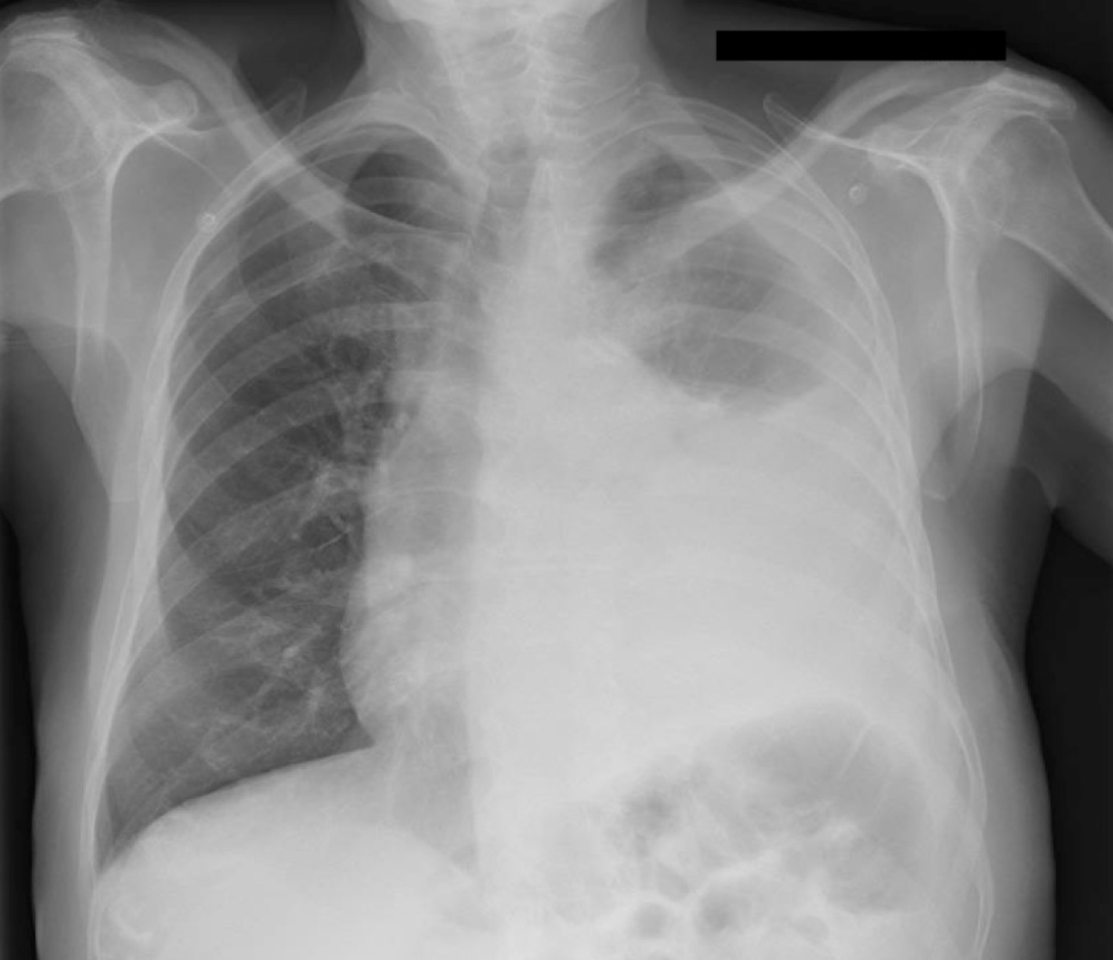
Figure 1 Chest X-ray at admission: Large left pleural effusion with contralateral deviation of the mediastinum.
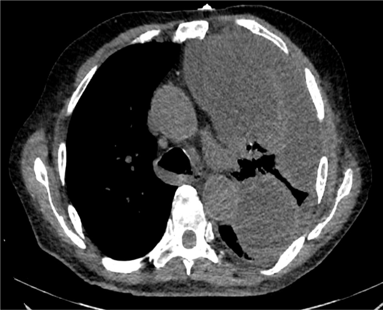
Figure 2 Computed tomography of the chest at admission: Multiloculated left pleural effusion
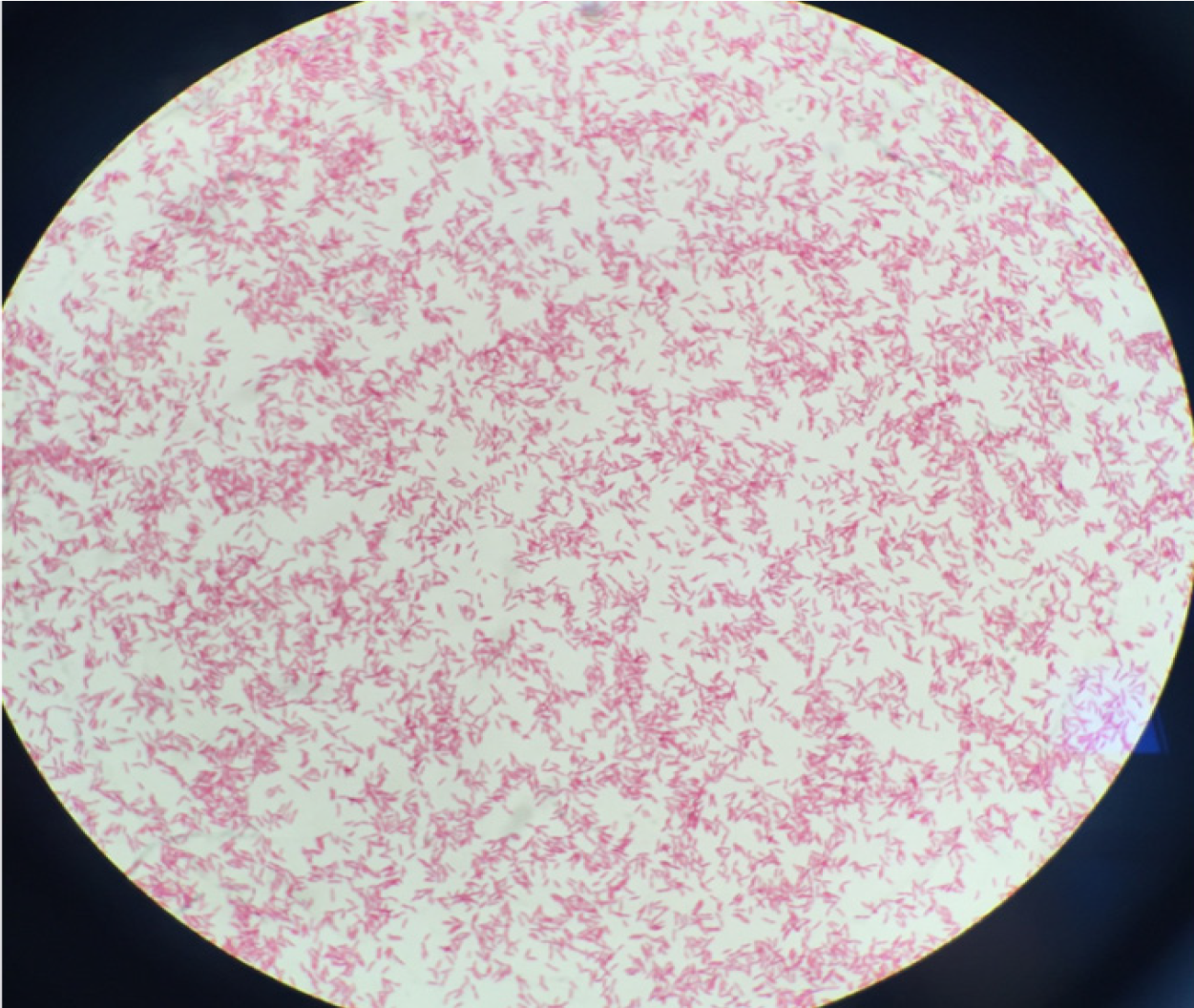
Figure 3 Gram staining of pleural culture confirmed gram-negative bacillus, consistent with Legionella pneumophila.
- Citation: Maillet F, Bonnet N, Billard-Pomares T, El Alaoui Magdoud F, Tandjaoui-Lambiotte Y. Fatal Legionella pneumophila serogroup 1 pleural empyema: A case report. World J Crit Care Med 2019; 8(6): 99-105
- URL: https://www.wjgnet.com/2220-3141/full/v8/i6/99.htm
- DOI: https://dx.doi.org/10.5492/wjccm.v8.i6.99


