©The Author(s) 2017.
World J Crit Care Med. May 4, 2017; 6(2): 135-139
Published online May 4, 2017. doi: 10.5492/wjccm.v6.i2.135
Published online May 4, 2017. doi: 10.5492/wjccm.v6.i2.135
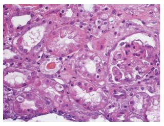
Figure 1 Renal biopsy.
The image shows cortico-medullar renal tissue with endothelial congestion, occasional interposition of mesangial cellularity that in a segmental and focal way produces occlusion of vascular capillary lights; aneurysmal dilation of the glomerular capillaries and some lights with polymorphonuclear and fragmented erythrocytes, as well as histological changes compatible with thrombotic microangiopathic involvement in the initial acute phase are also observed (Hematoxilin-Eosine, 40 ×).
- Citation: Pérez-Cruz FG, Villa-Díaz P, Pintado-Delgado MC, Fernández_Rodríguez ML, Blasco-Martínez A, Pérez-Fernández M. Hemolytic uremic syndrome in adults: A case report. World J Crit Care Med 2017; 6(2): 135-139
- URL: https://www.wjgnet.com/2220-3141/full/v6/i2/135.htm
- DOI: https://dx.doi.org/10.5492/wjccm.v6.i2.135


