©The Author(s) 2015.
World J Crit Care Med. Aug 4, 2015; 4(3): 213-229
Published online Aug 4, 2015. doi: 10.5492/wjccm.v4.i3.213
Published online Aug 4, 2015. doi: 10.5492/wjccm.v4.i3.213
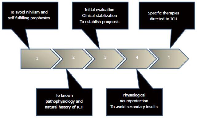
Figure 1 Steps to consider in the approach to the critically ill patient with spontaneous intracerebral hemorrhage.
ICH: Intracerebral hemorrhage.
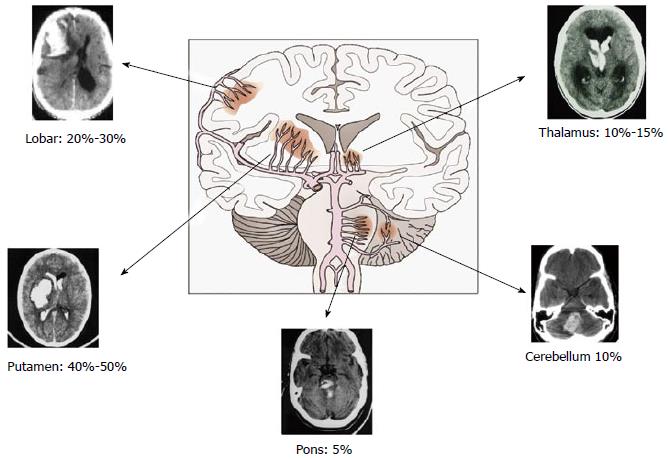
Figure 2 Typical sites of bleeding in spontaneous intracerebral hemorrhage.
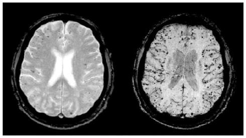
Figure 3 Micro bleeds in magnetic resonance image.
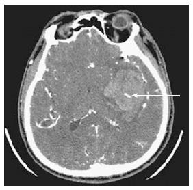
Figure 4 Spot sign.
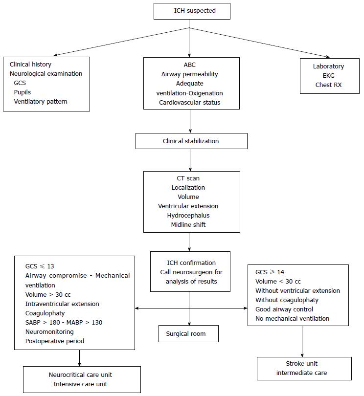
Figure 5 Initial approach of the patients with suspected intracerebral hemorrhage.
GCS: Glasgow Coma Scale; ABC: Airway, breathing, circulation; EKG: Electrocardiogram; CT: Computed tomography; ICH: Intracerebral hemorrhage; SABP: Systolic arterial blood pressure; MABP: Mean arterial blood pressure.
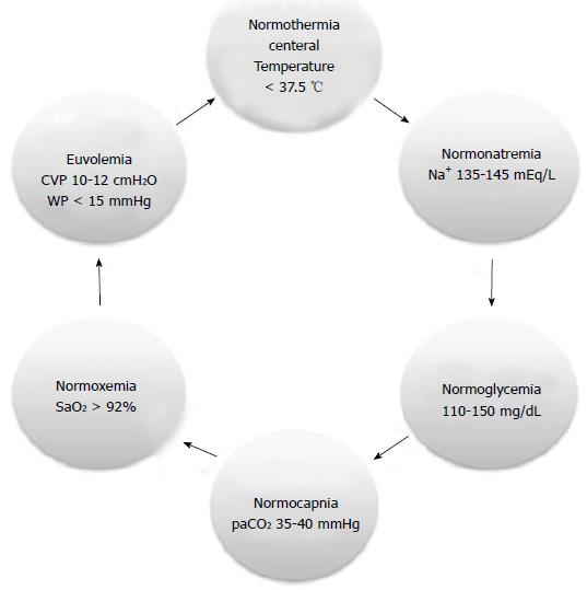
Figure 6 Physiological neuroprotection 6 N rules.
CVP: Central venous pressure; WP: Wedge pressure; SaO2: Oxygen arterial saturation; Na+: Serum sodium levels; paCO2: Carbon dioxide arterial levels.
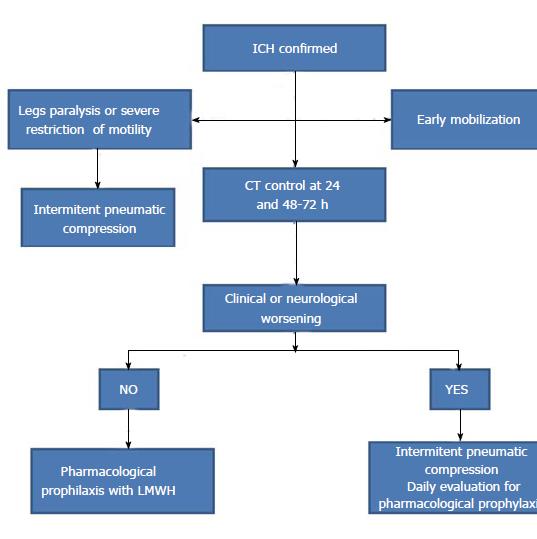
Figure 7 Algorithm suggested for venous thromboembolism prevention in intracerebral hemorrhage.
ICH: Intracerebral hemorrhage; CT: Computed tomography; LMWH: Low molecular weight heparin.
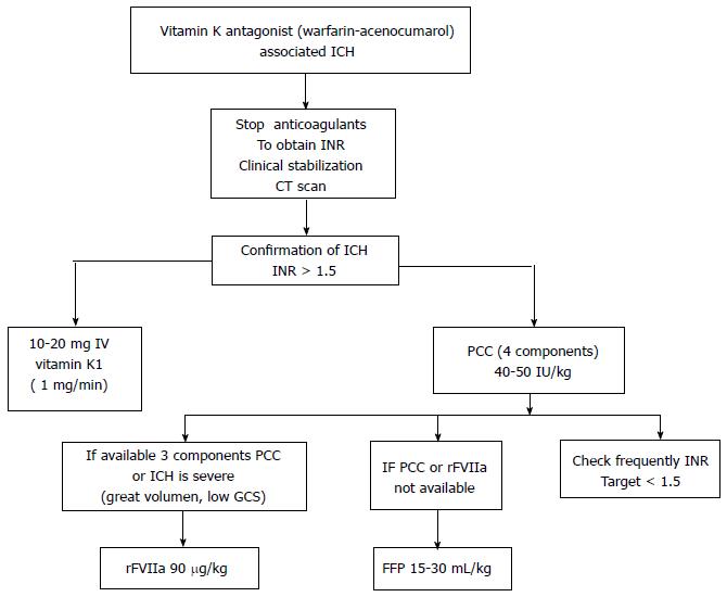
Figure 8 Algorithm to urgent reversal therapy in vitamin K antagonist related intracerebral hemorrhage.
ICH: Intracerebral hemorrhage; INR: International normalization ratio; CT: Computed tomography; IV: Intravenous; PCC: Prothrombin concentrate complex; IU: International units; GCS: Glasgow coma scale; rFVIIa: Activated recombinant seven-factor; FFP: Fresh frozen plasma.
- Citation: Godoy DA, Piñero GR, Koller P, Masotti L, Napoli MD. Steps to consider in the approach and management of critically ill patient with spontaneous intracerebral hemorrhage. World J Crit Care Med 2015; 4(3): 213-229
- URL: https://www.wjgnet.com/2220-3141/full/v4/i3/213.htm
- DOI: https://dx.doi.org/10.5492/wjccm.v4.i3.213


