©The Author(s) 2015.
World J Crit Care Med. Aug 4, 2015; 4(3): 179-191
Published online Aug 4, 2015. doi: 10.5492/wjccm.v4.i3.179
Published online Aug 4, 2015. doi: 10.5492/wjccm.v4.i3.179
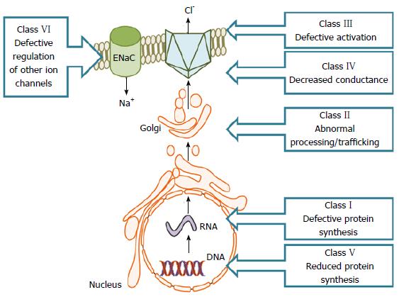
Figure 1 Classification of cystic fibrosis transmembrane conductance regulator mutations.
CFTR mutations are classified into six groups according to their effect on CFTR function. Class I mutations affect biosynthesis, while class II mutations affect protein processing. Milder mutations such as class II to Class V impair CFTR channel function. CFTR: Cystic fibrosis transmembrane conductance regulator.
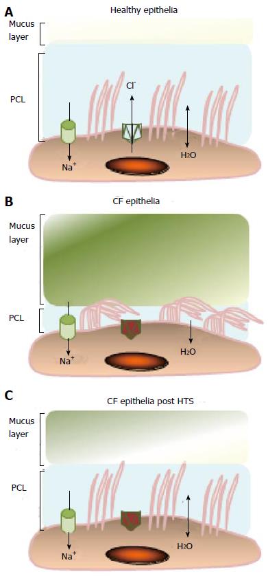
Figure 2 Effects of hypertonic saline on the airway surface liquid in cystic fibrosis.
A: In healthy airway epithelia, CFTR plays a vital role in regulating hydration of the airway surface liquid (ASL) constructed of the periciliary layer (PCL) and the mucus layer; B: Due to defective CFTR in individuals with CF, Cl- secretion is impaired and Na+ absorption through ENaC is increased resulting in dehydration of the ASL and accumulation of thick mucus causing reduced PCL height; C: Treatment with hypertonic saline assists osmosis of water into the ASL and thus rehydrates the mucus and partially restores the PCL allowing for easier clearance of mucus. CFTR: Cystic fibrosis transmembrane conductance regulator.
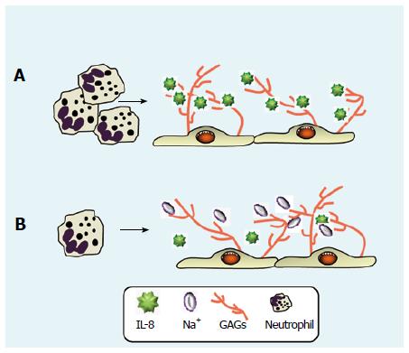
Figure 3 Hypertonic saline reduces levels of interleukin-8 in cystic fibrosis airway samples thereby reducing neutrophil migration.
A: The chemokine IL-8 is a key mediator of inflammation in patients with CF and increases neutrophil migration to the airways. GAGs possess the ability to influence the chemokine profile of the CF lung by binding IL-8 and protecting it from proteolytic degradation; B: HTS functions to disrupt IL-8: GAG complexes, rendering the chemokine susceptible to proteolytic degradation. Clinical application of HTS may serve to decrease the inflammatory burden in the CF lung in vivo. CF: Cystic fibrosis; GAGs: Glycosaminoglycans; HTS: Hypertonic saline; IL-8: Interleukin-8.
Figure 4 Schematic illustration of the NADPH oxidase of resting and formyl peptides activated cells.
The neutrophil NADPH oxidase generates superoxide (O2-) and secondary oxygen-derived toxic products in response to bacteria or a variety of soluble stimuli (fMLP). A: The enzyme is dormant in resting neutrophils. The active site of this enzyme is located in an integral membrane cytochrome, b558, which consists of the two subunits gp91phox and p21phox (subunits); B: Stimulation of the neutrophil by fMLP induces activation and phosphorylation (P) of a number of kinases including Akt; C: P21rac is converted into the active GTP-bound form and the phosphorylation of the cytosolic components (p67phox, p47phox and p40phox) occurs; D: These subunits then translocate to the membrane where they interact with cytochrome b558 to initiate reactive oxygen species production. fMLP: Formyl peptides.
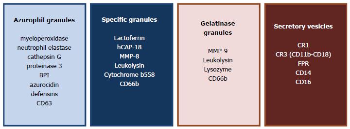
Figure 5 Components of neutrophil granules.
The second mechanism of bacterial killing is mediated by enzymes stored in granules. Essential serine proteases stored in primary granules include NE, cathepsin G and proteinase 3. Other bactericidal proteins of primary granules include defensins, azurocidin and bactericidal permeability-increasing (BPI) protein, which mutually function to destabilise bacterial membranes. Additional antibacterial proteins stored in secondary or specific granules include lactoferrin, the 18-kDa human cathelicidin antimicrobial protein (hCAP18) and lysozyme. Lactoferrin, an iron binding protein displays antimicrobial properties by limiting iron availability and exhibits direct antmicrobial and antifungal properties independent of iron-binding. LL-37, the CX-terminal peptide of hCAP-18, disrupts the integrity of bacterial membranes and can neutralise bacterial endotoxins. The gelatinase or tertiary granules contain mainly gelatinase (MMP-9) whose main function is to degrade type V collagen in the extracellular matrix to aid neutrophil migration. In addition to these three granule types, neutrophils also contain secretory vesicles that contain a reservoir of essential receptors and integrins. All are degranulated to the outside of the cell or into the phagocytic vacuole.
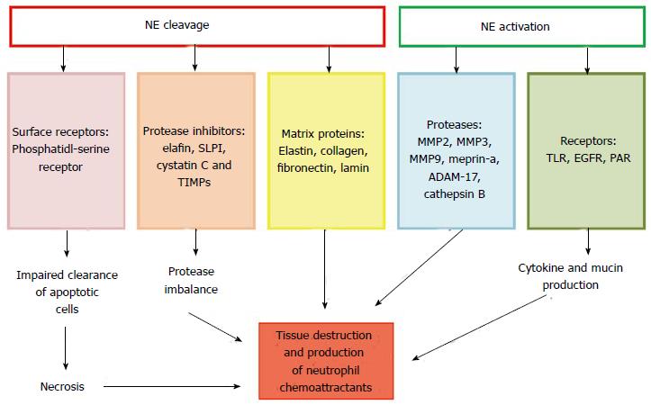
Figure 6 Potential effects of active neutrophil elastase in cystic fibrosis.
Excessive NE activity can lead to proteolysis causing protease-antiprotease imbalance by cleaving protease inhibitors. Cleavage of matrix proteins and surface receptors leads to tissue damage and prolonged immune response, respectively. NE can further activate pro-inflammatory molecules (MMPs and ADAM-17) and receptors. These cumulative effects exacerbate tissue destruction and hyper-inflammation. NE: Neutrophil elastase; CF: Cystic fibrosis; MMPs: Matrix metalloproteinases.
- Citation: Reeves EP, McCarthy C, McElvaney OJ, Vijayan MSN, White MM, Dunlea DM, Pohl K, Lacey N, McElvaney NG. Inhaled hypertonic saline for cystic fibrosis: Reviewing the potential evidence for modulation of neutrophil signalling and function. World J Crit Care Med 2015; 4(3): 179-191
- URL: https://www.wjgnet.com/2220-3141/full/v4/i3/179.htm
- DOI: https://dx.doi.org/10.5492/wjccm.v4.i3.179


