Copyright
©The Author(s) 2024.
World J Crit Care Med. Jun 9, 2024; 13(2): 91212
Published online Jun 9, 2024. doi: 10.5492/wjccm.v13.i2.91212
Published online Jun 9, 2024. doi: 10.5492/wjccm.v13.i2.91212
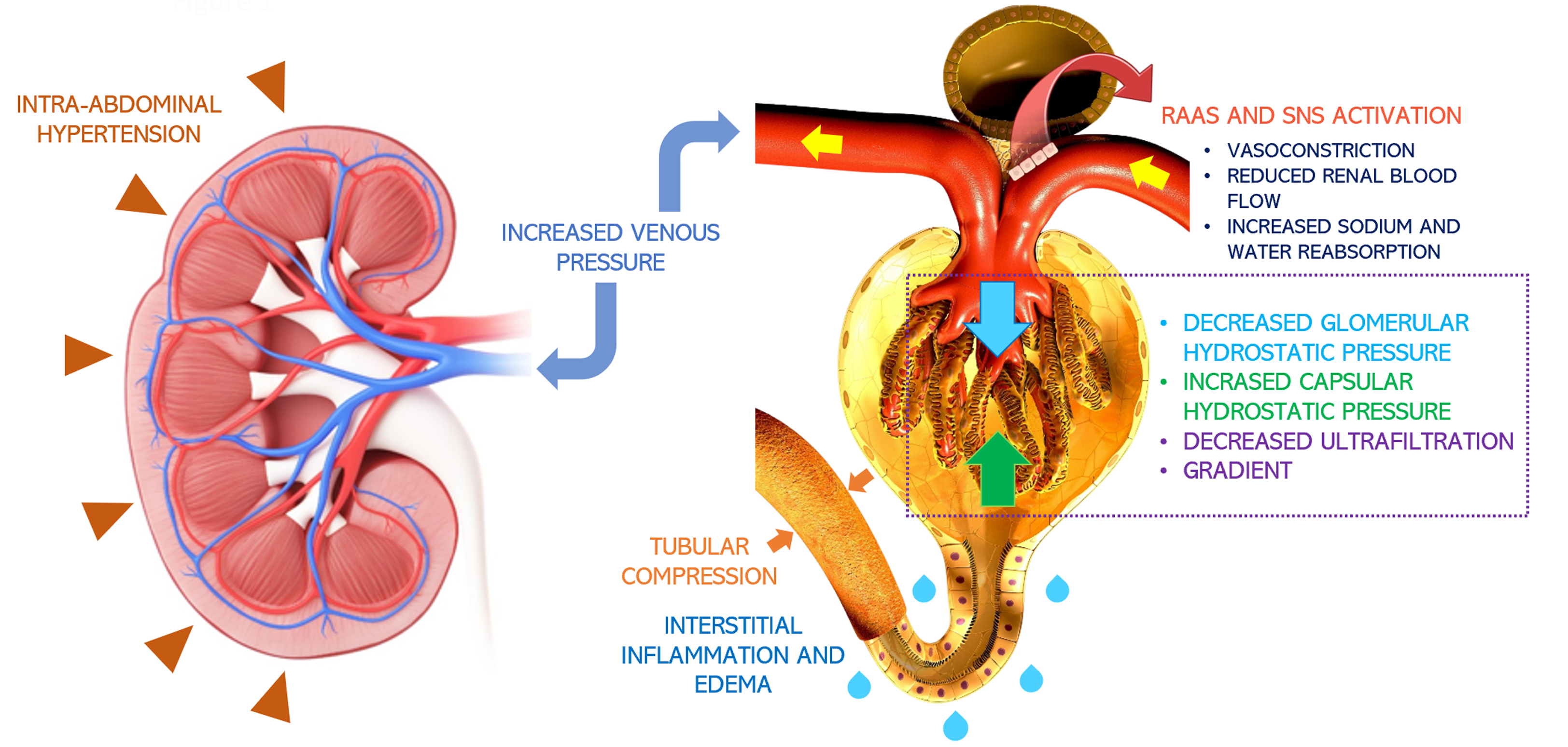
Figure 1 Pathophysiology of congestive nephropathy: Congestion-induced acute renal dysfunction is mediated by retrograde transmission of central venous pressure to the kidneys, leading to development of interstitial edema, inflammation, and activation of the renin-angiotensin-aldosterone system and sympathetic nervous system[66].
This further results in global cessation of glomerular filtration. Intra-abdominal hypertension adds to the problem by simulating a tamponade pathophysiology together with increased interstitial pressures. Citation: Argaiz ER, Romero-Gonzalez G, Rola P, Spiegel R, Haycock KH, Koratala A. Bedside Ultrasound in the Management of Cardiorenal Syndromes: An Updated Review. Cardiorenal Med 2023; 13: 372-384. Copyright© The Author(s) 2023. Published by S. Karger AG, Basel, authors’ prior work published under CC BY-NC License.
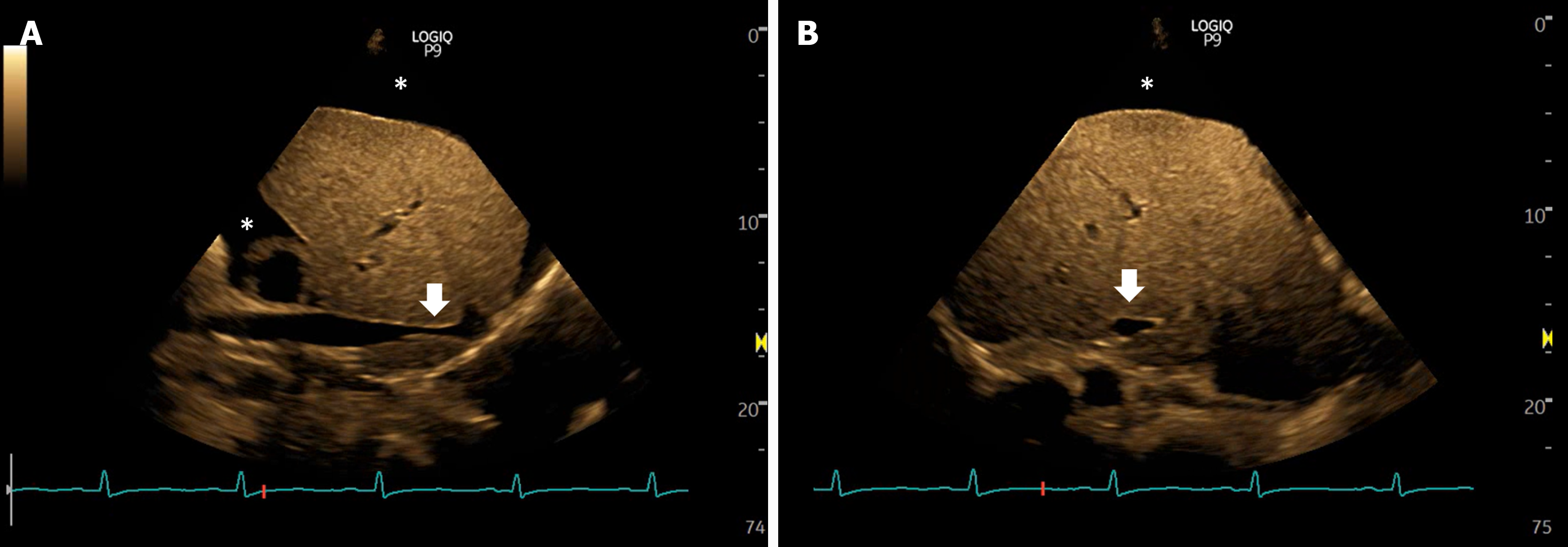
Figure 2 Views of the inferior vena cava in a case of intra-abdominal hypertension showing narrowing or slit-like appearance of the intrahepatic portion (arrows), sometimes described as nipping.
Asterisk indicates ascites. A: Long axis views; B: Transverse views.
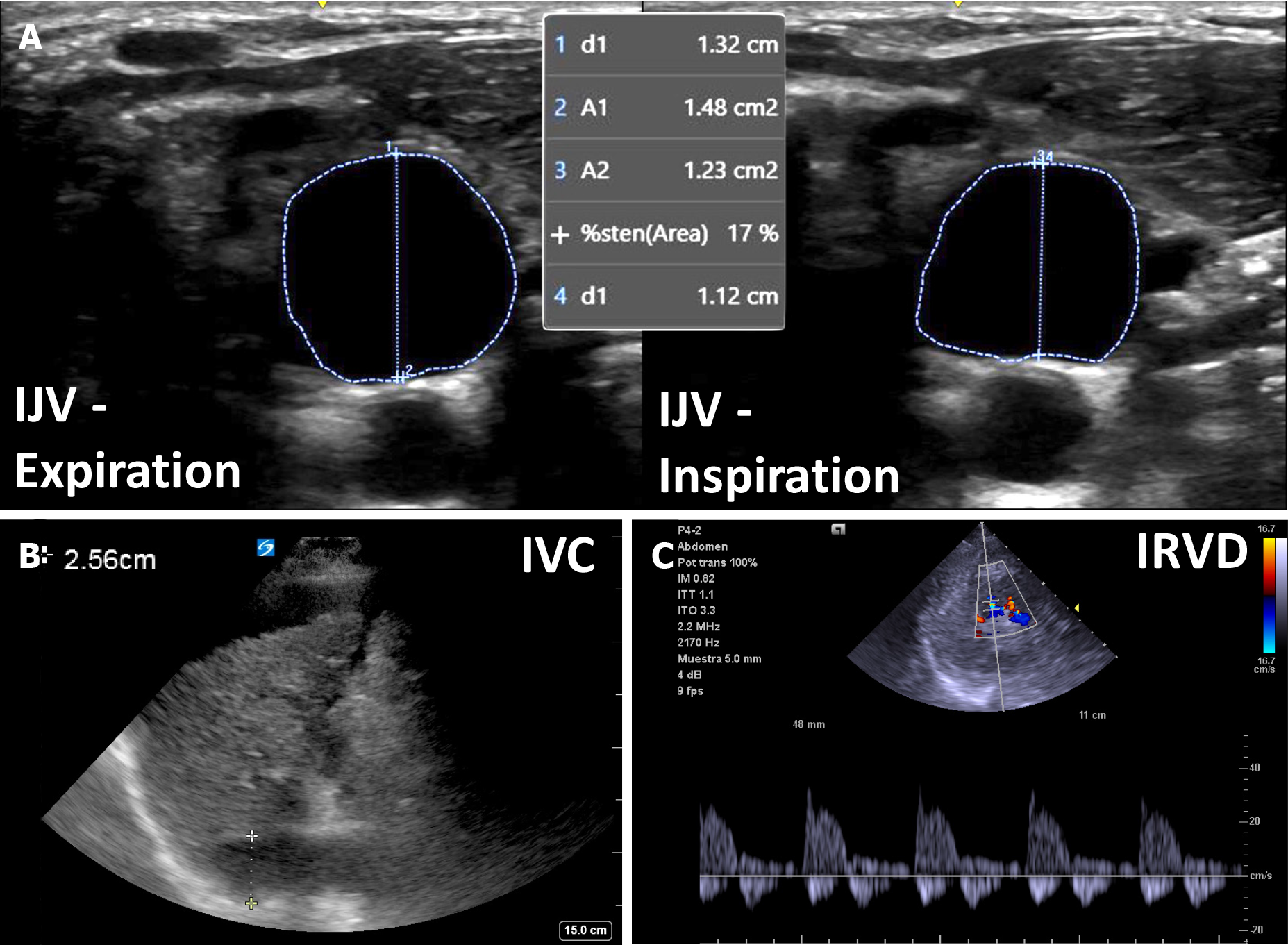
Figure 3 An example of venous congestion in cirrhosis.
A: Plethoric internal jugular vein with less than 25% antero-posterior inspiratory collapse; B: Plethoric inferior vena cava (> 2.0 cm); C: Biphasic intra-renal venous Doppler. IJV: Internal jugular vein; IVC: Inferior vena cava; IRVD: Intra-renal venous Doppler.
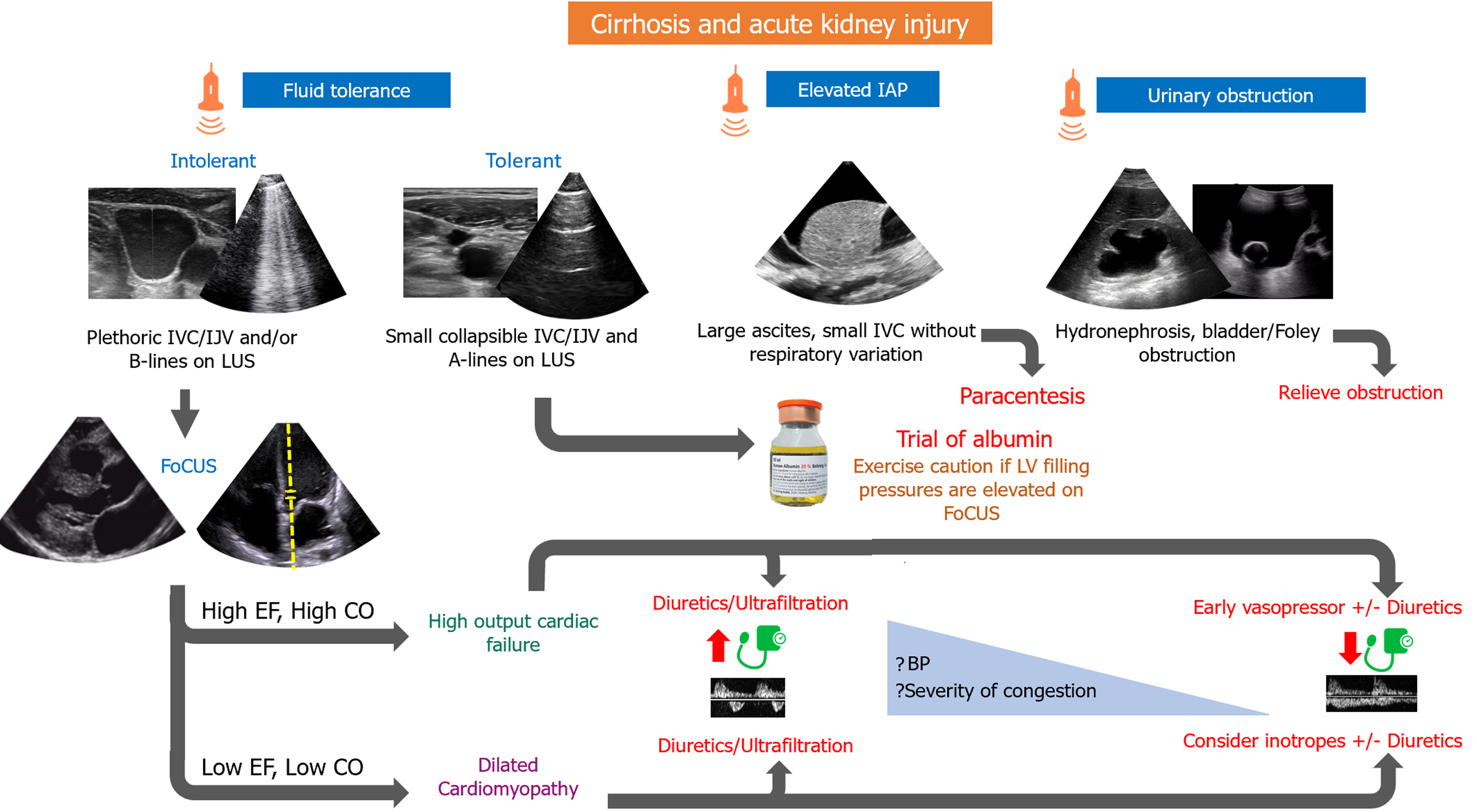
Figure 4 Proposed algorithm for management of cirrhosis and acute kidney injury.
IAP: Intra-abdominal pressure; IJV: Internal jugular vein; IVC: Inferior vena cava; LUS: Lung ultrasound; EF: Ejection fraction; CO: Cardiac output; LV: Left ventricular; BP: Blood pressure; FoCUS: Focused cardiac ultrasound.
- Citation: Aguirre-Villarreal D, Leal-Villarreal MAJ, García-Juárez I, Argaiz ER, Koratala A. Sound waves and solutions: Point-of-care ultrasonography for acute kidney injury in cirrhosis. World J Crit Care Med 2024; 13(2): 91212
- URL: https://www.wjgnet.com/2220-3141/full/v13/i2/91212.htm
- DOI: https://dx.doi.org/10.5492/wjccm.v13.i2.91212


