©2012 Baishideng Publishing Group Co.
World J Crit Care Med. Feb 4, 2012; 1(1): 15-22
Published online Feb 4, 2012. doi: 10.5492/wjccm.v1.i1.15
Published online Feb 4, 2012. doi: 10.5492/wjccm.v1.i1.15
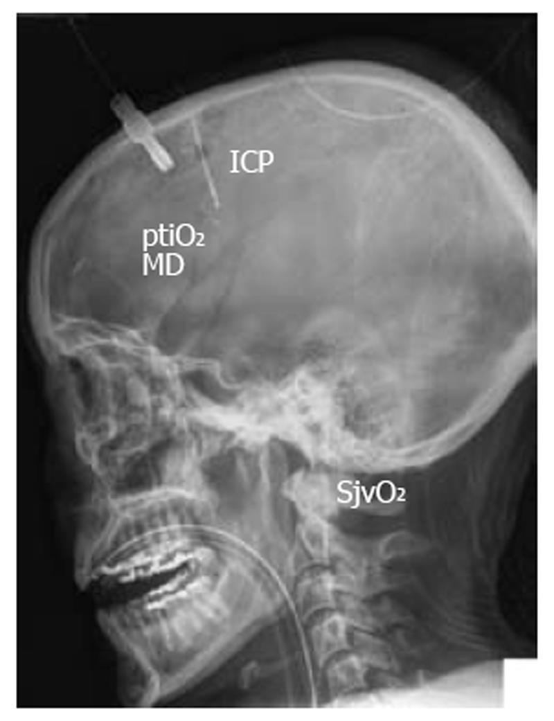
Figure 1 Illustrative lateral X-ray view showing positioning of invasive neuromonitoring to assess intracranial pressure, brain tissue oxygen pressure, brain metabolism using microdialysis, and jugular venous oxygen saturation.
ICP: Intracranial pressure; ptiO2: Tissue oxygen pressure; SjvO2: Jugular venous oxygen saturation.
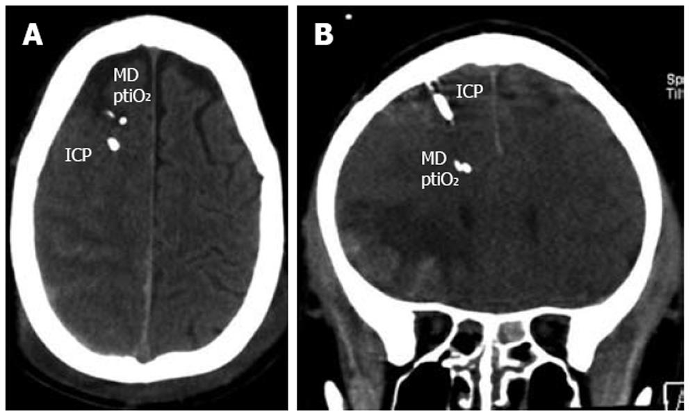
Figure 2 Illustrative examples showing positioning of intracranial pressure, microdialysis catheter, and brain tissue oxygen pressure sensor in the more severely injured hemisphere (A) and within close proximity of a contusion (B).
ICP: Intracranial pressure; ptiO2: Tissue oxygen pressure.
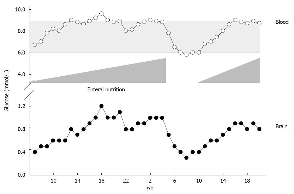
Figure 3 Illustrative case showing the influence of enteral nutrition on arterial blood and brain glucose levels.
Based on low brain glucose, enteral nutrition was gradually increased resulting in elevated blood and brain glucose. Due to surgery, enteral nutrition was stopped. This resulted in a decrease in blood and brain glucose. Restarting of enteral nutrition increased blood and brain glucose again.
- Citation: Stover JF. Contemporary view on neuromonitoring following severe traumatic brain injury. World J Crit Care Med 2012; 1(1): 15-22
- URL: https://www.wjgnet.com/2220-3141/full/v1/i1/15.htm
- DOI: https://dx.doi.org/10.5492/wjccm.v1.i1.15


