©The Author(s) 2015.
World J Surg Proced. Mar 28, 2015; 5(1): 127-136
Published online Mar 28, 2015. doi: 10.5412/wjsp.v5.i1.127
Published online Mar 28, 2015. doi: 10.5412/wjsp.v5.i1.127
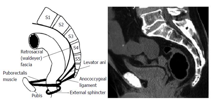
Figure 1 Schematic illustration of sacrococcyx, rectum and important ingredients of retrorectal space.
(Simplified and reproduced from Nicholls et al[42]) and counterpart tomoscan.
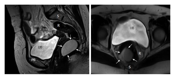
Figure 2 Coronal and axial T2A magnetic resonance imaging planes of a cystic lesion (arrows) in the retrorectal space.
UB: Urinary bladder; PB: Pubic bone; R: Rectum; S: Sacrococcyx.
Figure 3 Macroscopic appearance of dermoid cyst (A), epithelial inclusion cyst (B) and schwannoma (C).
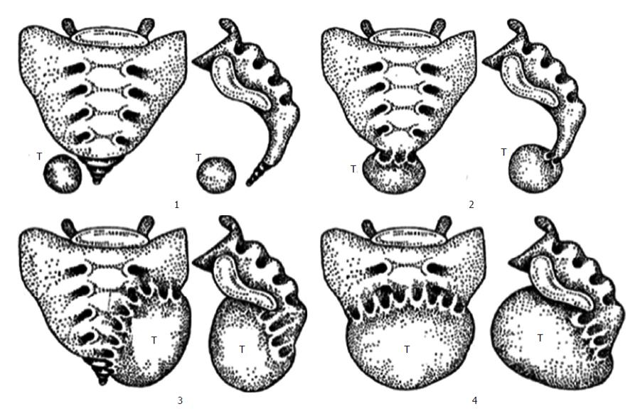
Figure 4 Classification of retrorectal space depending on its relationship with the sacrum (Losanoff et al[20]).
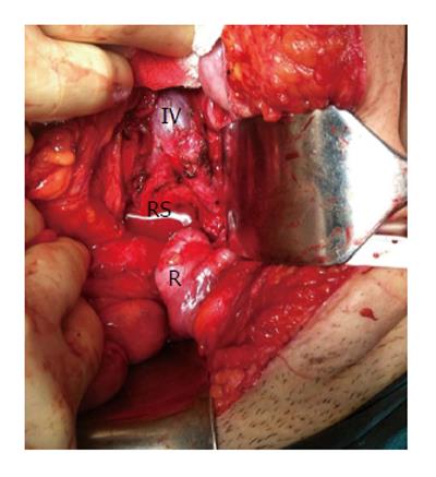
Figure 5 Laparotomy for retrorectal space.
Iliac vessels and RS can easily be reached after lateralization of rectum. IV: Iliac vessels; RS: Retrorectal Space; R: Rectum.
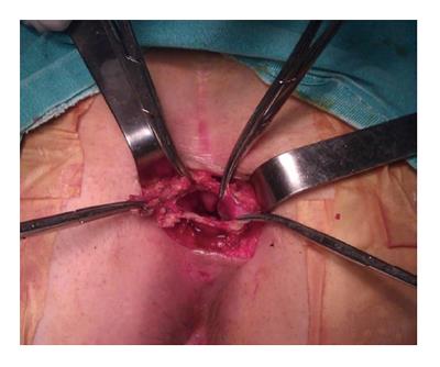
Figure 6 Excision of cystic lesion located low level in retrorectal space by using posterior approach.
- Citation: Uçar AD, Erkan N, Yıldırım M. Surgical treatment of retrorectal (presacral) tumors. World J Surg Proced 2015; 5(1): 127-136
- URL: https://www.wjgnet.com/2219-2832/full/v5/i1/127.htm
- DOI: https://dx.doi.org/10.5412/wjsp.v5.i1.127


