Review Open Access
Copyright ©2014 Baishideng Publishing Group Inc. All rights reserved.
World J Immunol. Jul 27, 2014; 4(2): 42-51
Published online Jul 27, 2014. doi: 10.5411/wji.v4.i2.42
Lysosomal acid lipase is critical for myeloid-derived suppressive cell differentiation, development, and homeostasis
Cong Yan, Center for Immunobiology, Indiana University School of Medicine, Indianapolis, IN 46202, United States
Cong Yan, Hong Du, Department of Pathology and Laboratory Medicine, Indiana University School of Medicine, Indianapolis, IN 46202, United States
Cong Yan, Hong Du, IU Simon Cancer Center, Indiana University School of Medicine, Indianapolis, IN 46202, United States
Author contributions: Yan C and Du H wrote and edited the review.
Supported by National Institutes of Health, No.CA138759, CA152099, to Yan C; HL087001, to Du H, and HL-061803 and HL-067862 to Yan C and Du H
Correspondence to: Dr. Cong Yan, Department of Pathology and Laboratory Medicine, Indiana University School of Medicine, 975 W Walnut Street, IB424G, Indianapolis, IN 46202, United States. coyan@iupui.edu
Telephone: +1-317-2786005 Fax: +1-317-2788198
Received: February 26, 2014
Revised: April 2, 2014
Accepted: June 18, 2014
Published online: July 27, 2014
Processing time: 151 Days and 13.4 Hours

Abstract

Lysosomal acid lipase (LAL) cleaves cholesteryl esters (CE) and triglycerides (TG) to generate cholesterol and free fatty acid in lysosomes of cells. The downstream metabolic products of fatty acids are ligands for activation of peroxisome proliferator-activated receptor gamma (PPARγ). Accumulation of CEs and TGs is resulted from lack of functional LAL in lysosomes of cells, especially in myeloid cells. One characteristic phenotype in LAL knock-out (lal-/-) mice is systemic elevation of myeloid-derived suppressive cells (MDSCs). MDSCs infiltrate into multiple distal organs, alter T cell development, and suppress T cell proliferation and lymphokine production in lal-/- mice, which lead to severe pathogeneses in multiple organs. The gene transcriptional profile analysis in MDSCs from the bone marrow has identified multiple defects responsible for MDSCs malformation and malfunction in lal-/- mice, including G protein signaling, cell cycles, glycolysis metabolism, mitochondrial bioenergetics, mTOR pathway etc. In a separate gene transcriptional profile analysis in the lung of lal-/- mice, matrix metalloproteinase 12 (MMP12) and apoptosis inhibitor 6 (Api6) are highly overexpressed due to lack of ligand synthesis for PPARγ. PPARγ negatively regulates MMP12 and Api6. Blocking the PPAR signaling by overexpression of a dominant negative PPARγ (dnPPARγ) form, or overexpressing MMP12 or Api6 in myeloid or lung epithelial cells in inducible transgenic mouse models results in elevated MDSCs and inflammation-induced tumorigenesis. These studies demonstrate that LAL and its downstream effectors are critical for MDSCs development, differentiation and malfunction.

Key Words: Lysosomal acid lipase; Myeloid-derived suppressor cells; Immunosuppression; Myeloid-derived suppressive cell development; Hematopoiesis

Core tip: Neutral lipid metabolism is essential for myeloid cell proliferation and differentiation. This review summarizes the most recent discoveries that lysosomal acid lipase (LAL), an enzyme hydrolysing cholesteryl esters and triglycerides in lysosomes, plays a critical role in myeloid-derived suppressive cells (MDSCs) development, differentiation, and immune suppressive function. Both LAL knock-out and myeloid specific rescue of LAL knock-out mice are used in the studies. Doxycycline-inducible bitransgenic mouse models of LAL downstream genes are also generated to study MDSCs malformation and malfunction. The molecular pathways/mechanisms to connect LAL and MDSCs are characterized by microarray analyses of gene transcriptional profiles.


HISTORY OF LYSOSOMAL ACID LIPASE

Lysosomal acid lipase (LAL) cleaves cholesteryl esters (CE) and triglycerides (TG) in cell lysosomes. Mutation in the LAL gene results in Wolman disease (WD) of early infantile onset, and cholesteryl ester storage disease (CESD) of late onset. WD was first described by Dr. Wolman[1] in 1956 as severe malnutrition, hepatosplenomegaly, calcified adrenal glands, and death of children within the first few months of life. Affected WD infants display massive accumulations of CE and TG in the lysosomes of hepatocytes and Kupffer cells, as well as in macrophages throughout the viscera, which lead to liver failure, severe hepatosplenomegaly, steatorrhea, pulmonary fibrosis[2,3], and adrenal calcification and insufficiency[4,5]. Lipid engorged macrophages in intestinal villi lead to severe malabsorption and cachexia[2,4]. The average life span of WD is 3.5 mo[6]. CESD was initially described by Fredrickson, Schiff, Langeron, and Infante and their colleagues in 1967[7-10] and named by Partin and Schubert based on phenotype that exhibited hepatomegaly with increased hepatic levels of cholesteryl esters in 1969. CESD can be a more indolent progressive disease, which shows microvesicular steatosis leading to fibrosis and cirrhosis in the liver, increases atherosclerosis and premature demise[11-13]. Wolman disease and CESD result from allelic mutations at the LAL locus on human chromosome 10q23.2-q23.3 and are autosomal recessive traits. The gene spans 45 kb, has 10 exons, and contains no unusual structures, except for a large intron 3. The LIPA mutations found in Wolman disease include deletions and insertions that lead to premature stop codons and the consequent loss of LAL protein and activity[14]. The mutations found in CESD are usually missense mutations, either heteroallelic or homoallelic with another mutant LIPA gene[14].

Recently, some evidence started to emerge, showing altered mononuclear phagocyte differentiation [increased CD14+CD16+ and CD14+CD33+ cells, subsets of human myeloid-derived suppressive cells, or myeloid-derived suppressive cells (MDSCs)] in humans that were heterozygote carriers of LAL mutations[15]. Furthermore, patients with mutations in the LAL gene have been reported to be associated with carcinogenesis[16]. These clinical observations support the extensive characterization in animal models as described below.

LAL PROPERTIES

LAL is a key player in the modulation of cholesterol metabolism in all cells. On the surface membranes of various cells, there are multiple receptors that can deliver LDL-bound cholesteryl esters/triglycerides to lysosomes, but LAL is the only lipase in the lysosomes that hydrolyzes cholesteryl esters and triglycerides. Once cleaved by LAL, the free cholesterol and fatty acids enter the cytosol from lysosome. In LAL deficiency, cholesteryl esters and triglycerides cannot be cleaved; therefore, free cholesterol and fatty acids cannot leave the lysosome[17,18]. Cells sense this as an intracellular (cytosolic) cholesterol deficiency, and the cholesterol biosynthetic pathway is up-regulated to compensate.

Synthesized in the rough endoplasmic reticulum, LAL is a typical soluble lysosomal hydrolase, which is co-translationally glycosylated when it emerges into the endoplasmic reticulum lumen[18,19]. Following the removal of the leader sequence (21 amino acids), LAL is decorated with oligosaccharides that are remodeled during transit through the Golgi apparatus. The N-linked oligosaccharides are remodeled from high mannosyl to complex forms, with a mannose 6-phosphate being added, which serves as the lysosomal sorting targeting signal. The mannose 6-phosphate receptor system is used to deliver the newly synthesized LAL to the lysosome. LAL is not known to require cofactors for optimal hydrolysis, and it functions as a monomer. Unmodified mature protein (378 amino acids) has a predicted molecular weight approximately 42.5 kDa. Different molecular weights have been reported for purified human LAL[20-24]. Occupancy of the LAL N-glycosylation is essential for enzyme stability, i.e., protection from rapid degradation[25].

LAL has significant similarity to other acidic lipases, for example, lingual lipase and gastric lipases that cleave similar substrates in the stomach. However, LAL is distinct from other lipases, including hormone-sensitive lipase, pancreatic lysophospholipid lipase, lecithin cholesterol acyl transferase, lipoprotein lipase, hepatic lipase, and pancreatic lipase[26]. All such lipases share a motif, Gly-X-Ser-X-Gly, that is an essential pentapeptide in the active site[27,28]. This pentapeptide occurs twice in LAL at serine 99 and serine 153, and specific mutation of serine 153 identified this residue as important to catalytic activity[23]. Like other lipases, LAL also has a catalytic triad of Ser153, Asp423 and His353[27].

GENE KNOCK-OUT PHENOTYPES AND MDSCS IN MICE

A Lipa knock-out mouse (lal-/-) has been created to understand the functional roles of LAL in disease pathophysiology, lipid metabolism, and therapeutic approaches[29,30]. The lal-/- phenotype resembles human CESD. It’s histopathologic and biochemical phenotypes are similar to human WD. The lal-/- mice are normal appearing at birth, but develop liver enlargement by 4 wk and have a grossly enlarged abdomen with hepatosplenomegaly, lymph node enlargement, and intestinal villus infiltration by foamy macrophages by 16 wk. Massive accumulation of CE and TG and macrophage storage develops in these and other organs[29,31-34]. Enzyme therapy has been studied in this model using human recombinant LAL (rhLAL) produced in several different eukaryotic systems[24,35,36]. These studies clearly show the potential for correction of the manifestations if enzyme therapy is begun early in the course of the disease[36,37].

Many phenotypes of seemingly unrelated diseases in various organs co-exist in lal-/- mice. Therefore, these diseases must share common cellular and molecular mechanisms that link these pathological processes. Extensive characterization of lal-/- mice shows that elevation of systemic MDSCs is a major manifestation in association with most of the pathogenic conditions (e.g., > 70% in the bone marrow and > 40% in the blood), suggesting that MDSCs play a central role in mediating LAL deficiency-induced pathogenic progression[29,31-34,36,38-41]. MDSCs was originally identified in tumor pathogenesis[42]. Recent studied have linked this cell population to many other chronic inflammatory diseases[43-50]. MDSCs are a mixture of myeloid cells that express CD11b and Gr-1 antigens in mice. In certain disease conditions (cancer), MDSCs are categorized into granulocytic (CD11b+, Ly6G+) and monocytic (CD11b+Ly6C+) MDSC[51]. Interestingly, most gated lal-/- CD11b+ cells show Ly6C+ and Ly6G+ double positive, making them CD11b+Ly6C+ Ly6G+ cells[34]. Normally, healthy immature myeloid lineage cells differentiate into dendritic cells (DCs), macrophages, or granulocytes in response to environmental changes. However, this process is blocked by LAL deficiency, leading to accumulation and expansion of MDSCs with immune suppressive function[51-53]. This is similar to what has been observed in the tumor environment[54]. It is conceivable that through paracrine and autocrine mechanisms, abnormally elevated MDSCs generate and secrete growth factors, chemokines and cytokines to influence cell differentiation, cell proliferation, cell apoptosis and gene expression in residing organ tissues, contributing to the physiological progression of various diseases. Direct cell-cell contact by MDSCs and other cells through the juxtacrine mechanism also contributes to this pathogenic process.

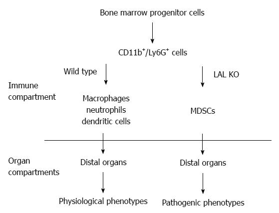
The functional roles of LAL in myeloid cells have been specifically evaluated by creating a myeloid-specific doxycycline-inducible c-fms-rtTA/(tetO)7-CMV-hLAL; lal-/- triple mouse model, in which human LAL is expressed in myeloid cells under the control of the 7.2 kb c-fms promoter/intron2 regulatory sequence in lal-/- mice[32,34,55]. The hLAL expression in myeloid lineage cells in this triple mouse model significantly reduced systemic MDSCs accumulation[34], reversed aberrant gene expression, and ameliorated pathogenic phenotypes[32]. Therefore, the normal biological function of myeloid cells requires normal neutral lipid metabolism (Figure 1).

Figure 1
Figure 1 The functional role of Lysosomal acid lipase in myeloid lineage cells. In the wild type mice, the CD11b+Ly6G+ cells are myeloid lineage precursors for monocytes/macrophages, neutrophils, and dendritic cells, which participate in the normal physiological functions of the distal organs (e.g., lung, liver, etc.), such as clearance of invading pathogens. The lysosomal acid lipase (LAL) activity is essential for normal myeloid lineage cell development, differentiation and function. LAL deficiency leads to neutral lipid accumulation in myeloid cells and blocks CD11b+Ly6G+ cells from further differentiation into mature myeloid lineage cells. The accumulated CD11b+Ly6G+ cells possess various malfunctions that participate in the pathogenic conditions in the residing organs. MDSCs: Myeloid-derived suppressive cells.
MDSCS DIFFERENTIATION AND DEVELOPMENT

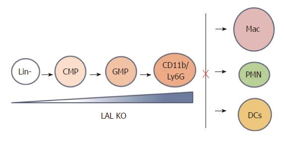
The myeloid linage cells undergo the sequentially differentiated and proliferated from hematopoietic stem cells through an increasingly lineage-restricted intermediate progenitors including common myeloid progenitors (CMPs) and granulocyte-macrophage progenitors (GMPs) in the bone marrow[56,57]. The number and frequency of primitive LSK (Lin-/Sca-1+/c-kit+), CMP, and GMP populations in the bone marrow, systemic myeloid cell distribution are changed in lal-/- mice, leading to an expansion in CD11b+/Gr-1+ MDSCs[41]. Both increased proliferation and decreased apoptosis contribute to the expansion of MDSCs in lal-/- mice. Lal-/- mice also display increased numbers of high proliferative potential colony-forming cells (HPP-CFC), colony-forming unite of granulocyte and macrophage progenitor cells (CFU-GM), colony-forming unite of granulocytes (CFU-G) and colony-forming unite of macrophages (CFU-M) colonies from cultured bone marrow cells. When lal-/- bone marrow cells are transplanted into wild type mice, the donor CD11b+/GR-1+ myeloid cells in the blood, spleen, lung and bone marrow of recipient mice are increased, confirming that the MDSCs increase is primarily due to the intrinsic defect in myeloid lineage progenitor cells. In addition to the intrinsic progenitor problem, the environment in lal-/- mice also contributes to myeloid cell hyperexpansion, since the donor CD11b+/GR-1+ myeloid cell population in lal-/- recipient mice that are transplanted with wild type bone marrow cells is expanded. Therefore, the lal-/- environment does not normally support hematopoiesis. Deregulated bone marrow progenitor cell differentiation is a primary cause for expansion of lal-/- MDSCs, which is attributed to both cell-autonomous and environmental factors. Taken together, LAL expression in myeloid lineage cells is critical to maintain hematopoiesis and myelopoiesis. After MDSCs infiltration into distal organs, at least two mechanisms can explain how the cell-autonomous defect and environmental factors influence each other. Firstly, MDSCs and other regional cells in distal organs influence each other by the paracrine mechanism as both sides secrete cytokines and chemokines. Secondly, MDSCs and other cells can influence each other by direct contact (juxtacrine mechanism). Starting at the GMP stage, hLAL expression in myeloid cells reverses abnormal myeloid development in the bone marrow, and reduces systemic expansion of MDSCs in c-fms-rtTA/(tetO)7-CMV-hLAL; lal-/- triple mice. In addition, differentiation from Lin- progenitor cells to CD11b+GR-1+ cells is abnormally increased in lal-/- mice (Figure 2). This further supports that the cell-autonomous effect of MDSCs expansion in lal-/- mice. Myeloid hLAL expression in c-fms-rtTA/(tetO)7-CMV-hLAL; lal-/- triple mice successfully reverses this abnormality[32]. The environmental effects on MDSCs malformation are further supported by an observation that when the Stat3 pathway is overly activated in lung epithelial cells[58], secretion of Stat3-induced pro-inflammatory cytokines in epithelial cells reversed mature myeloid lineage cells to MDSCs[59].

Figure 2
Figure 2 Lysosomal acid lipase is required for normal myeloid lineage cell development and differentiation. Lysosomal acid lipase (LAL) deficiency leads to increased myeloid-derived suppressive cells differentiation from Lin- progenitor cells in the bone barrow, and decreased differentiation to mature macrophages, neutrophils, and dendritic cells in other compartments. Lin-: Lineage negative progenitor; CMP: Common myeloid progenitor; GMP: Granulocyte-macrophage progenitor; Mac: Macrophage; PMN: Polymorphonuclear cell, or neutrophil; DC: Dendritic cell.
MDSCS IMMUNOSUPPRESSION

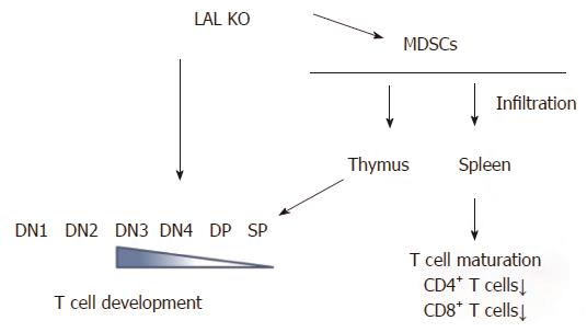
In contrast to myeloid lineage cells, T cells are systemically decreased in lal-/- mice. Lal-/- T cells behave abnormally. In response to stimulation of anti-CD3 plus anti-CD28 antibodies, or phorbol-12-myristate-13-acetate (agonist to activate PKC) and ionomycin (calcium ionophore), there is severely diminished T cell proliferation, decreased CD69 expression, and decreased expression of T cell lymphokines. LAL deficiency does not drive effector T cells into either Th1 or Th2 status[33]. The thymus is the most important organ for T cell development, which is divided into different developmental stages that are marked by CD4-CD8- double negative (DN) 1 to 4 stages, CD4+CD8+ double positive (DP) stage and CD4+ or CD8+ single positive (SP) stage. The earliest stage for thymocyte paucity appears at the DN4 (CD25-CD44-) stage in the lal-/- thymus. After this developmental point, thymocytes are declining at all stages, suggesting that the blockage of T cell development initially occurs at the DN3 to DN4 transition (Figure 3)[33]. Decrease of T cell development and maturation was also observed in lal-/- mice due to the defects in lymphoid progenitors in the bone marrow chimeras study. This notion has been supported by the bone marrow profile analysis, in which common lymphoid progenitor development is blocked in the bone marrow of lal-/- mice[33,41].

Figure 3
Figure 3 Lysosomal acid lipase is required for normal T cell development and differentiation. Lysosomal acid lipase (LAL) deficiency can cause the intrinsic defect in T cell development, starting at the double negative 3 (DN3) stage. In addition, myeloid-derived suppressive cells infiltrate into the thymus and spleen, resulting in blockage of normal T cell development, differentiation, and maturation. DN: CD4 and CD8 double negative; DP: CD4 and CD8 double positive; SP: CD4 or CD8 single positive.

In addition to the above intrinsic defect, extensive analyses have revealed a second mechanism that contributes to systemic reduction of T cell populations. Strikingly, LAL deficiency dramatically increases MDSCs expansion and infiltration in the thymus and the spleen of lal-/- mice, leading to neutral lipid accumulation and abnormal organization of the thymus and spleen[33]. Infiltration of MDSCs in these important T cell organs affects T cell development, differentiation and maturation. Functional analyses have shown that MDSCs from lal-/- mice strongly inhibit proliferation and function of T cells (Figure 3)[34,40,41].

Direct connection between LAL in MDSCs and T cell abnormalities comes from the c-fms-rtTA/(tetO)7-CMV-hLAL;lal-/- triple mouse study. MDSCs expansion and infiltration into the thymus and spleen are reduced in this mouse model. This leads to restoration of T cell proliferation in the spleen and normal T cell development in the thymus[34]. Stat3 and NFκB p65 signaling play a critical role in lal-/- MDSCs immune suppressive function[34]. The above observations are further proved by an MDSCs depletion study, in which anti-Gr-1 antibody treatment recovers T cell numbers in lal-/- mice[34]. lal-/- MDSCs also inhibits T cell lymphokine production, which is resulted from inactivation of the pZAP-70/Syk intracellular signaling, loss of expression of TCR ξ chain and CD69, a failure to respond to TCR stimulation[33]. These defects can also be reversed by myeloid hLAL expression[34]. Lastly, Treg cells inhibit CD4+ T cell lymphokine production and proliferation[60]. LAL deficiency substantially increases CD4+FoxP3+ Treg cells in lal-/-mice[33].

GENE PROFILES IN LAL DEFICIENCY-INDUCED MDSCS

Since LAL controls homeostasis and development of MDSCs, which have profound pathogenic impact on various disease development, it is essential to identify the intrinsic defects that are involved in the MDSCs homeostasis and function for future targeting. In a comprehensive gene transcriptional profile study by Affymetrix GeneChip microarray analysis, multiple pathways have been revealed in lal-/- bone marrow MDSCs. Below are lists of some major (but not limited) changed pathways in lal-/- MDSCs.

Genes of G-protein superfamily

Expression changes of both large and small GTPases have been detected in lal-/- MDSCs, which have diverse functions in cells[61,62]. They include: (1) Rab GTPases, which control vesicle formation, receptor internalization, and trafficking to the nucleus, lysosome and plasma membrane. Rab GTPases regulate cellular proliferation, apoptosis and migration by integrating signaling pathways; (2) Rho GTPases, which organize actin cytoskeleton, cell adhesion and cell motility[63]; and (3) Ras GTPases mediate cell-cycle entry, cell growth, cell survival, cell growth and cellular metabolism by phosphorylating transcription factors through activation of the Raf/Mek/Erk pathway. Activation of Erk and p38 phosphorylation has been observed in lal-/- MDSCs[41].

Histone cluster genes and cell cycle genes

Cell cycle regulating genes are upregulated in lal-/- MDSCs. They include: (1) Histone-variants cluster genes, which favor the epigenetic microenvironment change to promote MDSCs expansion. Histone-variants exchange also contributes to formation of centromeric and telomeric chromatin during cell cycles. Indeed, G1/M phases of lal-/- MDSCs are increased in a cell cycle analysis[64]; (2) Cell cycle related genes[65], including Cdk1, Cdk2, Cdk5, Cdk9, and all Cdk regulatory cyclins (A, B, D, E-type), suggesting constitutive mitogenic signaling and defective responses to anti-mitogenic signals; and (3) Ubiquitination and proteasome enzymes/protein factors, which direct proteins to proteolysis within proteasome for recycling[66].

Metabolism and bioenergetics

Bioenergetic and metabolic genes are abnormally upregulated in lal-/- MDSCs, which control mitochondrial oxidative phosphorylation and energy (ATP production) for cellular activities. These include: (1) lactate dehydrogenase A and B, which produce large quantities of secreted lactate, suggesting that lal-/- MDSCs use an aerobic glycolysis; (2) nitric oxide/reactive oxygen species (ROS) production genes, glutathione peroxidase/glutathione reductase genes, and glucose 6-phosphate dehydrogenase gene, which are involved in production of ROS. The concentration of ROS is significantly increased in lal-/- MDSCs; (3) enzymes and proteins in glycolysis and citric acid cycles; and (4) respiratory chain proteins (NADH dehydrogenases, cytochrome proteins, ATPases and mitochondrial ribosomal proteins).

The mTOR pathway in LAL deficiency induced MDSCs

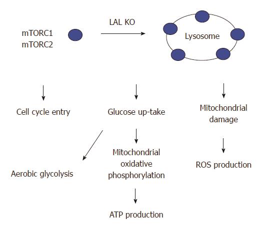
PI3K/thymoma viral proto-oncogene (AKT)/mammalian target of rapamycin (mTOR) is activated in lal-/- MDSCs[64]. mTOR is a lysosomal membrane-bound protein, which controls apoptosis, promotes influx of glucose and amino acids into the cells, stimulates ATP production[67], contributes to cell growth, cell cycle entry, cell survival, and cell motility[68,69]. Lack of the LAL activity changes lipid composition and dynamics on the lysosomal membrane that potentially influence endomembrane trafficking and stimulate the mTOR activity, which in turn coordinates the cellular metabolism[64,69,70]. It has been demonstrated that mTOR plays a critical role in modulating cellular immune functions[71,72], activation of the mTOR pathway contributes to lal-/- MDSCs production and function[40]. mTOR is the catalytic subunit of two distinctive complexes; mTOR complex 1 (mTORC1) and mTOR complex (mTORC2). mTORC1 contains unique regulatory associated proteins of mTOR (RAPTOR) while mTORC2 contains rapamycin-insensitive companion of mTOR (RICTOR)[67,72-75]. Inhibition of mTOR and associated proteins (Raptor, Rictor, and Akt1) corrects lal-/- MDSCs development, increased cell proliferation, decreased cellular apoptosis, and immune suppression in association with decreased ROS production, recovery from impairment of the mitochondrial membrane potential, increased ATP synthesis, and increased cell cycling. Potentially, the mTOR pathway can serve as a target to modulate the emergence of MDSCs in various pathophysiologic states where these cells play an immunosuppressive role (Figure 4).

Figure 4
Figure 4 Lysosomal acid lipase deficiency induces overactivation of the mTOR pathway in myeloid-derived suppressive cells. Lysosomal acid lipase (LAL) is a lysosome-associated enzyme. LAL deficiency increases mTOR complexes anchoring on lysosomes and stimulates the mTOR1 activity to influence the cellular metabolism and proliferation of lal-/- myeloid-derived suppressive cells (MDSCs). These include an increased influx of glucose through aerobic glycolysis, an increased mitochondrial oxidative phosphorylation and ATP production, an impairment of the mitochondrial membrane potential in association with increased reactive oxygen species (ROS) production, and an increased cell cycle entry in lal-/- MDSCs.
The Stat3 and NFκB pathways

Although upregulation of Signal Transducer and Activator of Transcription (Stat) family members and NFκB family members are not detected by microarray analysis, phosphorylation of Stat3 and NFκB has been detected in expanded lal-/- MDSCs[34,41]. Activation of Stat3 directly leads to MDSCs expansion in vivo[58,59].

STUDY OF LAL DOWNSTREAM GENES

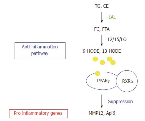
The gene profile study in the lung of lal-/- mice by Affymetrix GeneChip microarray analysis has also been performed. This is because the lung is a lipid rich organ and highly responsive to inflammation. Neutral lipids account for 10% of the composition of pulmonary surfactant that protects alveoli from collapse during respiratory cycles[76]. LAL deficiency results in massive myeloid cell infiltration, hyperplasia and emphysema in the lal-/- lung[32,39]. Comparison between the changed gene lists of bone marrow MDSCs and the whole lung by Affymetrix GeneChip microarray analyses reveals a few overlapping genes. Therefore, LAL performs differential roles in different compartments. LAL exerts its biological effects through its downstream genes. In order to fully understand the LAL functions, it is necessary and essential to characterize its downstream genes. From the whole lung gene list, the two most up-regulated genes matrix metal proteinase 12 (MMP12) and apoptosis inhibitor 6 (Api6) are characterized extensively. The functional role of LAL downstream effector peroxisome proliferator-activated receptor gamma (PPARγ) has also been studied in depth. Figure 5 shows the relationship between LAL and its downstream effectors.

Figure 5
Figure 5 Lysosomal acid lipase and its downstream effector genes. Lysosomal acid lipase (LAL) cleaves cholesteryl esters (CE) and triglycerides (TG) to produce free cholesterol (FC) and fatty acids (FFA) in lysosomes of cells. The lipid derivatives (9-HODE, 13-HODE) of FFA serve as ligands for PPARγ in coupling with retinoid X receptor α (RXRα, which suppresses gene expression of a variety of pro-inflammatory cytokines. The LAL/PPARγ axis serves as an anti-inflammatory pathway. LAL deficiency blocks this metabolic pathway to provoke up-regulation of pro-inflammatory cytokines (e.g., Api6, MMP12). TGF: Transforming growth factor beta; IL: Interleukin; MCP: Monocyte chemotactic protein; TNF: Tumor necrosis factor; NF: Nuclear factor.
PPARγ

Involvement of the receptor network in the metabolic programming of myeloid lineage cells is essential to the innate immune system[77,78]. PPARγ is of high interest for several reasons. Firstly, the metabolites of LAL hydrolysis, 9-hydroxyoctadecanoic acids (9-HODE) or 13-HODE from linoleic acid, serve as ligands for PPARγ. Upon binding to the ligands, PPAR interacts with the retinoid X receptor (RXR) to form the PPARγ/RXR dimer on target genes. Secondly, PPARγ plays an important role in anti-inflammation of various tissues[77,79,80]. It has been shown that PPARγ agonists suppress gene expression of inflammatory cytokines[79]. In the lal-/- lung, these pro-inflammatory cytokines are up-regulated (Figure 5)[39]. Therefore, LAL deficiency causes inactivation of PPARγ by depleting ligand production. Using the lung as a model system, reintroduction of LAL downstream metabolic derivative 9-HODE (a natural occurring ligand for PPARγ) and a synthetic ligand compound ciglitazone for PPARγ improves the inflammatory status and pathogenesis in the lal-/- lung. Therefore, the ligands/PPARγ axis controls inflammation-triggered elevated gene expression and pathogenesis in the lal-/- mice[31].

To directly evaluate functional role of LAL downstream effector PPARγ in myeloid cells, dominant negative PPARγ (dnPPARγ) is overexpressed in a myeloid-specific c-fms-rtTA/(TetO)7-CMV-dnPPARγ bitransgenic mouse model[81]. In this bitransgenic system, total numbers and frequencies of LK, LSK, CMP and GMP progenitor cells in the bone marrow are abnormally elevated. DnPPARγ overexpression leads to up-regulation of IL-1β, IL-6 and TNFα in the blood plasma. MDSCs from this bitransgenic mouse model inhibit the proliferation and lymphokine production of wild type CD4+ T cells in vitro. Both CD4+ and CD8+ T cell populations are decreased in doxycycline-induced dnPPARγ expressed mice. Bone marrow transplantation reveals that a myeloid autonomous defect is responsible for MDSC expansion, immunosuppression and tumorigenesis in this myeloid-specifically expressed dnPPARγ bitransgenic mice. Multiple forms of carcinoma and sarcoma in various organs (the lung, liver, spleen and lymph nodes) are observed in this mouse model. Therefore, the LAL/hormonal ligands/PPARγ axis is critical to control inflammation and the induction of various tumors. Disruption of this pathway in myeloid cells, either by blocking ligand synthesis (as in lal -/- mice), or inhibition of PPARγ (as in c-fms-rtTA/(TetO)7-CMV-dnPPARγ bitransgenic mice) can initiate up-regulation of inflammatory molecules which cause hematopoietic progenitors skewing towards myeloid lineage expansion to form MDSCs.

Matrix metalloproteinases12

Zinc-dependent MMPs act as modulators for inflammation and innate immunity by activating, deactivating or modifying the activities of signaling cytokines, chemokines and receptors through proteolytic and nonproteolytic functions[82-84]. Among MMPs, MMP12 is a 22-kDa secretory proteinase that is predominantly expressed in macrophages as previously reported[85]. MMP12 degrades extracellular matrix components, such as type IV collagen, fibronectin, laminin, gelatin, vitronectin, entactin, heparin, and chondroitin sulphates, to facilitate tissue remodeling[86]. The expression of MMP12 in macrophages is induced in the lung of cigarette smokers[87]. Inactivation of the MMP12 gene in knock-out mice demonstrates a critical role of MMP12 in smoking-induced chronic obstructive pulmonary disease (COPD)[88], a disease highly related to lung cancer. From clinical studies, MMP12 correlates with early cancer-related deaths in non-small cell lung cancer (NSCLC), especially with those associated with tobacco cigarette smoke exposure[89,90]. In the lal-/- lung, MMP-12 is the highest upregulated gene[31]. In the lal-/- lung, both macrophages and lung epithelial alveolar type II (AT II) cells are responsible for MMP-12 increase[31,91,92]. Both myeloid-specific and lung epithelia-specific MMP12 bitransgenic mouse models have been created to study the functional roles of this LAL/PPARγ downstream molecule.

In the myeloid-specific c-fms-rtTA/(TetO)7-CMV-MMP12 bitransgenic mouse model, induction of MMP12 abnormally elevates numbers and frequencies of CMP and GMP populations in the bone marrow, similar to that observed in lal-/- mice. Addition of activated MMP12 is able to stimulate wild type Lin- progenitor cells to differentiate into the MDSC population, suggesting that MMP12 directly exerts its effect on hematopoietic progenitor cells. The MDSCs are systemically increased in multiple organs of MMP12 bitransgenic mice. MDSCs from MMP12-overexpred bitransgenic mice suppress T cell proliferation and function. MMP12 directly stimulates differentiation of CD11b+Gr-1+ cells from Lin- progenitor cells. In the lung, the concentration of IL-6 is increased, which aberrantly activates oncogenic Stat3 and increases expression of Stat3 downstream genes in epithelial tumor progenitor cells. As a result, spontaneous emphysema and lung adenocarcinoma are sequentially developed in MMP12-overexpressive bitransgenic mice, suggesting a critical role of MMP12 in the transition from emphysema to lung cancer.

In epithelial-specific CCSP-rtTA/(TetO)7-CMV-MMP12 bitransgenic mice, MMP12 overexpression induces regional MDSCs infiltration and increases epithelial growth. Again, spontaneous emphysema and bronchioalveolar adenocarcinoma are developed sequentially. Importantly, MMP12 upregulation is highly associated with COPD and lung cancer in human patients. Together, these studies support that LAL/PPARγ downstream MMP12 plays a critical role in emphysema to lung cancer transition that is facilitated by inflammation. Clinically, it has been reported that there is a pathophysiological connection between emphysema/COPD and lung cancers[93,94].

Apoptosis inhibitor 6

Apoptosis inhibitor 6 (Api6) belongs to the macrophage scavenger receptor cysteine-rich domain superfamily (SRCR-SF)[95,96]. Api6 expression is the second highest induced gene in the lal-/- lung. Api6 is regulated by LAL metabolic derivatives (e.g., 9-HODE) and PPARγ[31]. In a myeloid-specific c-fms-rtTA/(TetO)7-CMV-Api6 bitransgenic mouse model, many phenotypes are similar to those observed in lal-/- mice. Overexpression of Api6 abnormally elevates MDSCs in the bone marrow, blood and lung with increased cell proliferation and decreased apoptotic activities. Api6 overexpression activates Stat3, Erk1/2 and p38 in myeloid lineage cells. Persistent inflammation in myeloid-specific Api6 bitransgenic mice causes lung adenocarcinoma[97].

Pathogenic overexpression of Api6 is also observed in lal-/- AT II cells. In an epithelial-specific CCSP-rtTA/(TetO)7-CMV-Api6 bitransgenic mice, Api6 overexpression in AT II cells increases pro-inflammatory cytokine/chemokine levels in bronchoalveolar lavage fluid and serum, activates oncogenic signaling and inhibits apoptosis, promotes expansion of MDSCs in lung and blood but not in the bone marrow or spleen. Lung MDSCs from this bitransgenic mouse model suppress T cell proliferation and function, which results in occurrence of emphysema and adenocarcinoma.

CONCLUSION

MDSCs play vital roles in various inflammation-induced chronic diseases. Elimination or reduction of MDSCs populations can slow down disease formation and progression. It is important to identify the molecular pathways in order to effectively block MDSCs homeostasis and function. Extensive studies outlined in this review have shown that the role of LAL in controlling neutral lipid metabolism is a key player in MDSCs development, homeostasis and function, therefore, providing a new avenue to develop therapeutic or immunologic approaches for clinical application. Through studies of the LAL function, defective gene expression patterns have been mapped in lal-/- MDSCs. These provide novel targets for controlling MDSCs and associated diseases by designing small molecule inhibitors. Clinically, small molecule inhibitors for c-kit have been tested to target MDSCs[98]. Using the gene profile list from LAL deficiency-induced MDSCs, more small molecule inhibitors can and will be identified to inhibit MDSCs pathogenic functions in various disease conditions.

ACKNOWLEDGMENTS

The authors thank Miss Katlin Walls for proof-reading the manuscript.

Footnotes

P- Reviewer: Gopinath SCB, Schuurman HJ, Saeki K S- Editor: Ji FF L- Editor: A E- Editor: Wang CH

References
1.  Abramov A, Schorr S, Wolman M. Generalized xanthomatosis with calcified adrenals. AMA J Dis Child. 1956;91:282-286.  [PubMed]  [DOI]  [Cited in This Article: ]
2.  Boldrini R, Devito R, Biselli R, Filocamo M, Bosman C. Wolman disease and cholesteryl ester storage disease diagnosed by histological and ultrastructural examination of intestinal and liver biopsy. Pathol Res Pract. 2004;200:231-240.  [PubMed]  [DOI]  [Cited in This Article: ]  [Cited by in Crossref: 46]  [Cited by in F6Publishing: 38]  [Article Influence: 1.9]  [Reference Citation Analysis (0)]
3.  Krivit W, Peters C, Dusenbery K, Ben-Yoseph Y, Ramsay NK, Wagner JE, Anderson R. Wolman disease successfully treated by bone marrow transplantation. Bone Marrow Transplant. 2000;26:567-570.  [PubMed]  [DOI]  [Cited in This Article: ]  [Cited by in Crossref: 72]  [Cited by in F6Publishing: 79]  [Article Influence: 3.3]  [Reference Citation Analysis (0)]
4.  Assmann G, Seedorf U. Acid lipase deficiency: Wolman disease and cholesteryl ester storage disease. The Metabolic and Molecular Bases of Inherited Disease. 6 ed. New York: McGraw-Hill 1995; 2563-2587.  [PubMed]  [DOI]  [Cited in This Article: ]
5.  Stein J, Garty BZ, Dror Y, Fenig E, Zeigler M, Yaniv I. Successful treatment of Wolman disease by unrelated umbilical cord blood transplantation. Eur J Pediatr. 2007;166:663-666.  [PubMed]  [DOI]  [Cited in This Article: ]  [Cited by in Crossref: 59]  [Cited by in F6Publishing: 63]  [Article Influence: 3.7]  [Reference Citation Analysis (0)]
6.  Reynolds T. Cholesteryl ester storage disease: a rare and possibly treatable cause of premature vascular disease and cirrhosis. J Clin Pathol. 2013;66:918-923.  [PubMed]  [DOI]  [Cited in This Article: ]  [Cited by in Crossref: 35]  [Cited by in F6Publishing: 32]  [Article Influence: 2.9]  [Reference Citation Analysis (0)]
7.  Fredrickson DS, Sloan HR, Ferrans VJ, Demosky SJ. Cholesteryl ester storage disease: a most unusual manifestation of deficiency of two lysosomal enzyme activities. Trans Assoc Am Physicians. 1972;85:109-119.  [PubMed]  [DOI]  [Cited in This Article: ]
8.  Schiff L, Schubert WK, McAdams AJ, Spiegel EL, O’Donnell JF. Hepatic cholesterol ester storage disease, a familial disorder. I. Clinical aspects. Am J Med. 1968;44:538-546.  [PubMed]  [DOI]  [Cited in This Article: ]  [Cited by in Crossref: 46]  [Cited by in F6Publishing: 49]  [Article Influence: 0.9]  [Reference Citation Analysis (0)]
9.  Lageron A, Caroli J, Stralin H, Barbier P. [Cholesterolic polycoria in adults. I. Clinical and histochemical study]. Presse Med. 1967;75:2785.  [PubMed]  [DOI]  [Cited in This Article: ]
10.  Infante R, Polonovski J, Caroli J. [Cholesterolic polycoria in adults. II. Biochemical study]. Presse Med. 1967;75:2829-2832.  [PubMed]  [DOI]  [Cited in This Article: ]
11.  Beaudet AL, Ferry GD, Nichols BL, Rosenberg HS. Cholesterol ester storage disease: clinical, biochemical, and pathological studies. J Pediatr. 1977;90:910-914.  [PubMed]  [DOI]  [Cited in This Article: ]  [Cited by in Crossref: 79]  [Cited by in F6Publishing: 81]  [Article Influence: 1.7]  [Reference Citation Analysis (0)]
12.  Bernstein DL, Hülkova H, Bialer MG, Desnick RJ. Cholesteryl ester storage disease: review of the findings in 135 reported patients with an underdiagnosed disease. J Hepatol. 2013;58:1230-1243.  [PubMed]  [DOI]  [Cited in This Article: ]  [Cited by in Crossref: 257]  [Cited by in F6Publishing: 225]  [Article Influence: 20.5]  [Reference Citation Analysis (0)]
13.  Fouchier SW, Defesche JC. Lysosomal acid lipase A and the hypercholesterolaemic phenotype. Curr Opin Lipidol. 2013;24:332-338.  [PubMed]  [DOI]  [Cited in This Article: ]  [Cited by in Crossref: 62]  [Cited by in F6Publishing: 51]  [Article Influence: 4.6]  [Reference Citation Analysis (0)]
14.  Grabowski GA, Du H. Lysosomal Acid Lipase Deficiencies: The Wolman Disease/Cholesteryl ester storage disease Spectrum. The Online Metabolic and Molecular Bases of inherited Disease (OMMBID). 9th ed. New York: McGraw-Hill 2012; .  [PubMed]  [DOI]  [Cited in This Article: ]
15.  Rothe G, Stöhr J, Fehringer P, Gasche C, Schmitz G. Altered mononuclear phagocyte differentiation associated with genetic defects of the lysosomal acid lipase. Atherosclerosis. 1997;130:215-221.  [PubMed]  [DOI]  [Cited in This Article: ]  [Cited by in Crossref: 14]  [Cited by in F6Publishing: 15]  [Article Influence: 0.6]  [Reference Citation Analysis (0)]
16.  Elleder M, Chlumská A, Hyánek J, Poupĕtová H, Ledvinová J, Maas S, Lohse P. Subclinical course of cholesteryl ester storage disease in an adult with hypercholesterolemia, accelerated atherosclerosis, and liver cancer. J Hepatol. 2000;32:528-534.  [PubMed]  [DOI]  [Cited in This Article: ]  [Cited by in Crossref: 58]  [Cited by in F6Publishing: 61]  [Article Influence: 2.5]  [Reference Citation Analysis (0)]
17.  Brown MS, Sobhani MK, Brunschede GY, Goldstein JL. Restoration of a regulatory response to low density lipoprotein in acid lipase-deficient human fibroblasts. J Biol Chem. 1976;251:3277-3286.  [PubMed]  [DOI]  [Cited in This Article: ]
18.  Sando GN, Ma GP, Lindsley KA, Wei YP. Intercellular transport of lysosomal acid lipase mediates lipoprotein cholesteryl ester metabolism in a human vascular endothelial cell-fibroblast coculture system. Cell Regul. 1990;1:661-674.  [PubMed]  [DOI]  [Cited in This Article: ]
19.  Sando GN, Henke VL. Recognition and receptor-mediated endocytosis of the lysosomal acid lipase secreted by cultured human fibroblasts. J Lipid Res. 1982;23:114-123.  [PubMed]  [DOI]  [Cited in This Article: ]
20.  Sando GN, Rosenbaum LM. Human lysosomal acid lipase/cholesteryl ester hydrolase. Purification and properties of the form secreted by fibroblasts in microcarrier culture. J Biol Chem. 1985;260:15186-15193.  [PubMed]  [DOI]  [Cited in This Article: ]
21.  Ameis D, Merkel M, Eckerskorn C, Greten H. Purification, characterization and molecular cloning of human hepatic lysosomal acid lipase. Eur J Biochem. 1994;219:905-914.  [PubMed]  [DOI]  [Cited in This Article: ]  [Cited by in Crossref: 71]  [Cited by in F6Publishing: 78]  [Article Influence: 2.6]  [Reference Citation Analysis (0)]
22.  Du H, Sheriff S, Bezerra J, Leonova T, Grabowski GA. Molecular and enzymatic analyses of lysosomal acid lipase in cholesteryl ester storage disease. Mol Genet Metab. 1998;64:126-134.  [PubMed]  [DOI]  [Cited in This Article: ]  [Cited by in Crossref: 38]  [Cited by in F6Publishing: 39]  [Article Influence: 1.5]  [Reference Citation Analysis (0)]
23.  Sheriff S, Du H, Grabowski GA. Characterization of lysosomal acid lipase by site-directed mutagenesis and heterologous expression. J Biol Chem. 1995;270:27766-27772.  [PubMed]  [DOI]  [Cited in This Article: ]  [Cited by in Crossref: 101]  [Cited by in F6Publishing: 104]  [Article Influence: 3.6]  [Reference Citation Analysis (0)]
24.  Du H, Cameron TL, Garger SJ, Pogue GP, Hamm LA, White E, Hanley KM, Grabowski GA. Wolman disease/cholesteryl ester storage disease: efficacy of plant-produced human lysosomal acid lipase in mice. J Lipid Res. 2008;49:1646-1657.  [PubMed]  [DOI]  [Cited in This Article: ]  [Cited by in Crossref: 50]  [Cited by in F6Publishing: 53]  [Article Influence: 3.3]  [Reference Citation Analysis (0)]
25.  Zschenker O, Bähr C, Hess UF, Ameis D. Systematic mutagenesis of potential glycosylation sites of lysosomal acid lipase. J Biochem. 2005;137:387-394.  [PubMed]  [DOI]  [Cited in This Article: ]  [Cited by in Crossref: 14]  [Cited by in F6Publishing: 14]  [Article Influence: 0.7]  [Reference Citation Analysis (0)]
26.  Komaromy MC, Schotz MC. Cloning of rat hepatic lipase cDNA: evidence for a lipase gene family. Proc Natl Acad Sci USA. 1987;84:1526-1530.  [PubMed]  [DOI]  [Cited in This Article: ]  [Cited by in Crossref: 111]  [Cited by in F6Publishing: 122]  [Article Influence: 3.3]  [Reference Citation Analysis (0)]
27.  Lohse P, Lohse P, Chahrokh-Zadeh S, Seidel D. Human lysosomal acid lipase/cholesteryl ester hydrolase and human gastric lipase: site-directed mutagenesis of Cys227 and Cys236 results in substrate-dependent reduction of enzymatic activity. J Lipid Res. 1997;38:1896-1905.  [PubMed]  [DOI]  [Cited in This Article: ]
28.  Roussel A, Canaan S, Egloff MP, Rivière M, Dupuis L, Verger R, Cambillau C. Crystal structure of human gastric lipase and model of lysosomal acid lipase, two lipolytic enzymes of medical interest. J Biol Chem. 1999;274:16995-17002.  [PubMed]  [DOI]  [Cited in This Article: ]  [Cited by in Crossref: 130]  [Cited by in F6Publishing: 124]  [Article Influence: 5.0]  [Reference Citation Analysis (0)]
29.  Du H, Duanmu M, Witte D, Grabowski GA. Targeted disruption of the mouse lysosomal acid lipase gene: long-term survival with massive cholesteryl ester and triglyceride storage. Hum Mol Genet. 1998;7:1347-1354.  [PubMed]  [DOI]  [Cited in This Article: ]  [Cited by in Crossref: 115]  [Cited by in F6Publishing: 112]  [Article Influence: 4.3]  [Reference Citation Analysis (0)]
30.  Yan C, Lian X, Dai Y, Wang X, Qu P, White A, Qin Y, Du H. Gene delivery by the hSP-B promoter to lung alveolar type II epithelial cells in LAL-knockout mice through bone marrow mesenchymal stem cells. Gene Ther. 2007;14:1461-1470.  [PubMed]  [DOI]  [Cited in This Article: ]  [Cited by in Crossref: 14]  [Cited by in F6Publishing: 17]  [Article Influence: 1.0]  [Reference Citation Analysis (0)]
31.  Lian X, Yan C, Qin Y, Knox L, Li T, Du H. Neutral lipids and peroxisome proliferator-activated receptor-{gamma} control pulmonary gene expression and inflammation-triggered pathogenesis in lysosomal acid lipase knockout mice. Am J Pathol. 2005;167:813-821.  [PubMed]  [DOI]  [Cited in This Article: ]  [Cited by in Crossref: 51]  [Cited by in F6Publishing: 48]  [Article Influence: 2.5]  [Reference Citation Analysis (0)]
32.  Yan C, Lian X, Li Y, Dai Y, White A, Qin Y, Li H, Hume DA, Du H. Macrophage-specific expression of human lysosomal acid lipase corrects inflammation and pathogenic phenotypes in lal-/- mice. Am J Pathol. 2006;169:916-926.  [PubMed]  [DOI]  [Cited in This Article: ]  [Cited by in Crossref: 54]  [Cited by in F6Publishing: 59]  [Article Influence: 3.3]  [Reference Citation Analysis (0)]
33.  Qu P, Du H, Wilkes DS, Yan C. Critical roles of lysosomal acid lipase in T cell development and function. Am J Pathol. 2009;174:944-956.  [PubMed]  [DOI]  [Cited in This Article: ]  [Cited by in Crossref: 42]  [Cited by in F6Publishing: 49]  [Article Influence: 3.3]  [Reference Citation Analysis (0)]
34.  Qu P, Yan C, Blum JS, Kapur R, Du H. Myeloid-specific expression of human lysosomal acid lipase corrects malformation and malfunction of myeloid-derived suppressor cells in lal-/- mice. J Immunol. 2011;187:3854-3866.  [PubMed]  [DOI]  [Cited in This Article: ]  [Cited by in Crossref: 29]  [Cited by in F6Publishing: 36]  [Article Influence: 2.8]  [Reference Citation Analysis (0)]
35.  Du H, Levine M, Ganesa C, Witte DP, Cole ES, Grabowski GA. The role of mannosylated enzyme and the mannose receptor in enzyme replacement therapy. Am J Hum Genet. 2005;77:1061-1074.  [PubMed]  [DOI]  [Cited in This Article: ]
36.  Du H, Schiavi S, Levine M, Mishra J, Heur M, Grabowski GA. Enzyme therapy for lysosomal acid lipase deficiency in the mouse. Hum Mol Genet. 2001;10:1639-1648.  [PubMed]  [DOI]  [Cited in This Article: ]
37.  Balwani M, Breen C, Enns GM, Deegan PB, Honzík T, Jones S, Kane JP, Malinova V, Sharma R, Stock EO. Clinical effect and safety profile of recombinant human lysosomal acid lipase in patients with cholesteryl ester storage disease. Hepatology. 2013;58:950-957.  [PubMed]  [DOI]  [Cited in This Article: ]  [Cited by in Crossref: 82]  [Cited by in F6Publishing: 89]  [Article Influence: 8.1]  [Reference Citation Analysis (0)]
38.  Du H, Heur M, Duanmu M, Grabowski GA, Hui DY, Witte DP, Mishra J. Lysosomal acid lipase-deficient mice: depletion of white and brown fat, severe hepatosplenomegaly, and shortened life span. J Lipid Res. 2001;42:489-500.  [PubMed]  [DOI]  [Cited in This Article: ]
39.  Lian X, Yan C, Yang L, Xu Y, Du H. Lysosomal acid lipase deficiency causes respiratory inflammation and destruction in the lung. Am J Physiol Lung Cell Mol Physiol. 2004;286:L801-L807.  [PubMed]  [DOI]  [Cited in This Article: ]
40.  Ding X, Du H, Yoder MC, Yan C. Critical role of the mTOR pathway in development and function of myeloid-derived suppressor cells in lal-/- mice. Am J Pathol. 2014;184:397-408.  [PubMed]  [DOI]  [Cited in This Article: ]  [Cited by in Crossref: 25]  [Cited by in F6Publishing: 29]  [Article Influence: 2.6]  [Reference Citation Analysis (0)]
41.  Qu P, Shelley WC, Yoder MC, Wu L, Du H, Yan C. Critical roles of lysosomal acid lipase in myelopoiesis. Am J Pathol. 2010;176:2394-2404.  [PubMed]  [DOI]  [Cited in This Article: ]  [Cited by in Crossref: 33]  [Cited by in F6Publishing: 35]  [Article Influence: 2.5]  [Reference Citation Analysis (0)]
42.  Talmadge JE, Gabrilovich DI. History of myeloid-derived suppressor cells. Nat Rev Cancer. 2013;13:739-752.  [PubMed]  [DOI]  [Cited in This Article: ]  [Cited by in Crossref: 828]  [Cited by in F6Publishing: 918]  [Article Influence: 83.5]  [Reference Citation Analysis (0)]
43.  Okwan-Duodu D, Umpierrez GE, Brawley OW, Diaz R. Obesity-driven inflammation and cancer risk: role of myeloid derived suppressor cells and alternately activated macrophages. Am J Cancer Res. 2013;3:21-33.  [PubMed]  [DOI]  [Cited in This Article: ]
44.  Cuenca AG, Delano MJ, Kelly-Scumpia KM, Moreno C, Scumpia PO, Laface DM, Heyworth PG, Efron PA, Moldawer LL. A paradoxical role for myeloid-derived suppressor cells in sepsis and trauma. Mol Med. 2011;17:281-292.  [PubMed]  [DOI]  [Cited in This Article: ]  [Cited by in Crossref: 221]  [Cited by in F6Publishing: 261]  [Article Influence: 18.6]  [Reference Citation Analysis (0)]
45.  Goh C, Narayanan S, Hahn YS. Myeloid-derived suppressor cells: the dark knight or the joker in viral infections? Immunol Rev. 2013;255:210-221.  [PubMed]  [DOI]  [Cited in This Article: ]  [Cited by in Crossref: 91]  [Cited by in F6Publishing: 96]  [Article Influence: 9.6]  [Reference Citation Analysis (0)]
46.  Natarajan S, Thomson AW. Tolerogenic dendritic cells and myeloid-derived suppressor cells: potential for regulation and therapy of liver auto- and alloimmunity. Immunobiology. 2010;215:698-703.  [PubMed]  [DOI]  [Cited in This Article: ]  [Cited by in Crossref: 32]  [Cited by in F6Publishing: 34]  [Article Influence: 2.4]  [Reference Citation Analysis (0)]
47.  Dilek N, Vuillefroy de Silly R, Blancho G, Vanhove B. Myeloid-derived suppressor cells: mechanisms of action and recent advances in their role in transplant tolerance. Front Immunol. 2012;3:208.  [PubMed]  [DOI]  [Cited in This Article: ]  [Cited by in Crossref: 64]  [Cited by in F6Publishing: 68]  [Article Influence: 5.7]  [Reference Citation Analysis (0)]
48.  Yin B, Ma G, Yen CY, Zhou Z, Wang GX, Divino CM, Casares S, Chen SH, Yang WC, Pan PY. Myeloid-derived suppressor cells prevent type 1 diabetes in murine models. J Immunol. 2010;185:5828-5834.  [PubMed]  [DOI]  [Cited in This Article: ]  [Cited by in Crossref: 116]  [Cited by in F6Publishing: 136]  [Article Influence: 9.7]  [Reference Citation Analysis (0)]
49.  Morales JK, Saleem SJ, Martin RK, Saunders BL, Barnstein BO, Faber TW, Pullen NA, Kolawole EM, Brooks KB, Norton SK. Myeloid-derived suppressor cells enhance IgE-mediated mast cell responses. J Leukoc Biol. 2014;95:643-650.  [PubMed]  [DOI]  [Cited in This Article: ]  [Cited by in Crossref: 18]  [Cited by in F6Publishing: 20]  [Article Influence: 1.8]  [Reference Citation Analysis (0)]
50.  Ostanin DV, Bhattacharya D. Myeloid-derived suppressor cells in the inflammatory bowel diseases. Inflamm Bowel Dis. 2013;19:2468-2477.  [PubMed]  [DOI]  [Cited in This Article: ]  [Cited by in Crossref: 37]  [Cited by in F6Publishing: 38]  [Article Influence: 3.5]  [Reference Citation Analysis (0)]
51.  Gabrilovich DI, Nagaraj S. Myeloid-derived suppressor cells as regulators of the immune system. Nat Rev Immunol. 2009;9:162-174.  [PubMed]  [DOI]  [Cited in This Article: ]  [Cited by in Crossref: 5121]  [Cited by in F6Publishing: 5134]  [Article Influence: 342.3]  [Reference Citation Analysis (0)]
52.  Ostrand-Rosenberg S, Sinha P. Myeloid-derived suppressor cells: linking inflammation and cancer. J Immunol. 2009;182:4499-4506.  [PubMed]  [DOI]  [Cited in This Article: ]  [Cited by in Crossref: 1269]  [Cited by in F6Publishing: 1322]  [Article Influence: 88.1]  [Reference Citation Analysis (0)]
53.  Sica A, Bronte V. Altered macrophage differentiation and immune dysfunction in tumor development. J Clin Invest. 2007;117:1155-1166.  [PubMed]  [DOI]  [Cited in This Article: ]
54.  Lindau D, Gielen P, Kroesen M, Wesseling P, Adema GJ. The immunosuppressive tumour network: myeloid-derived suppressor cells, regulatory T cells and natural killer T cells. Immunology. 2013;138:105-115.  [PubMed]  [DOI]  [Cited in This Article: ]  [Cited by in Crossref: 482]  [Cited by in F6Publishing: 593]  [Article Influence: 53.9]  [Reference Citation Analysis (0)]
55.  Sasmono RT, Oceandy D, Pollard JW, Tong W, Pavli P, Wainwright BJ, Ostrowski MC, Himes SR, Hume DA. A macrophage colony-stimulating factor receptor-green fluorescent protein transgene is expressed throughout the mononuclear phagocyte system of the mouse. Blood. 2003;101:1155-1163.  [PubMed]  [DOI]  [Cited in This Article: ]  [Cited by in Crossref: 498]  [Cited by in F6Publishing: 515]  [Article Influence: 24.5]  [Reference Citation Analysis (0)]
56.  Weissman IL, Shizuru JA. The origins of the identification and isolation of hematopoietic stem cells, and their capability to induce donor-specific transplantation tolerance and treat autoimmune diseases. Blood. 2008;112:3543-3553.  [PubMed]  [DOI]  [Cited in This Article: ]  [Cited by in Crossref: 305]  [Cited by in F6Publishing: 282]  [Article Influence: 17.6]  [Reference Citation Analysis (0)]
57.  Blank U, Karlsson G, Karlsson S. Signaling pathways governing stem-cell fate. Blood. 2008;111:492-503.  [PubMed]  [DOI]  [Cited in This Article: ]  [Cited by in Crossref: 258]  [Cited by in F6Publishing: 273]  [Article Influence: 16.1]  [Reference Citation Analysis (0)]
58.  Li Y, Du H, Qin Y, Roberts J, Cummings OW, Yan C. Activation of the signal transducers and activators of the transcription 3 pathway in alveolar epithelial cells induces inflammation and adenocarcinomas in mouse lung. Cancer Res. 2007;67:8494-8503.  [PubMed]  [DOI]  [Cited in This Article: ]  [Cited by in Crossref: 109]  [Cited by in F6Publishing: 115]  [Article Influence: 6.8]  [Reference Citation Analysis (0)]
59.  Wu L, Du H, Li Y, Qu P, Yan C. Signal transducer and activator of transcription 3 (Stat3C) promotes myeloid-derived suppressor cell expansion and immune suppression during lung tumorigenesis. Am J Pathol. 2011;179:2131-2141.  [PubMed]  [DOI]  [Cited in This Article: ]  [Cited by in Crossref: 68]  [Cited by in F6Publishing: 66]  [Article Influence: 5.1]  [Reference Citation Analysis (0)]
60.  Wang HY, Wang RF. Regulatory T cells and cancer. Curr Opin Immunol. 2007;19:217-223.  [PubMed]  [DOI]  [Cited in This Article: ]  [Cited by in Crossref: 252]  [Cited by in F6Publishing: 245]  [Article Influence: 14.4]  [Reference Citation Analysis (0)]
61.  Neves SR, Ram PT, Iyengar R. G protein pathways. Science. 2002;296:1636-1639.  [PubMed]  [DOI]  [Cited in This Article: ]  [Cited by in Crossref: 947]  [Cited by in F6Publishing: 894]  [Article Influence: 40.6]  [Reference Citation Analysis (0)]
62.  Gavi S, Shumay E, Wang HY, Malbon CC. G-protein-coupled receptors and tyrosine kinases: crossroads in cell signaling and regulation. Trends Endocrinol Metab. 2006;17:48-54.  [PubMed]  [DOI]  [Cited in This Article: ]  [Cited by in Crossref: 75]  [Cited by in F6Publishing: 80]  [Article Influence: 4.4]  [Reference Citation Analysis (0)]
63.  Konstantinopoulos PA, Karamouzis MV, Papavassiliou AG. Post-translational modifications and regulation of the RAS superfamily of GTPases as anticancer targets. Nat Rev Drug Discov. 2007;6:541-555.  [PubMed]  [DOI]  [Cited in This Article: ]  [Cited by in Crossref: 336]  [Cited by in F6Publishing: 355]  [Article Influence: 20.9]  [Reference Citation Analysis (0)]
64.  Yan C, Ding X, Dasgupta N, Wu L, Du H. Gene profile of myeloid-derived suppressive cells from the bone marrow of lysosomal acid lipase knock-out mice. PLoS One. 2012;7:e30701.  [PubMed]  [DOI]  [Cited in This Article: ]  [Cited by in Crossref: 16]  [Cited by in F6Publishing: 18]  [Article Influence: 1.5]  [Reference Citation Analysis (0)]
65.  Malumbres M, Barbacid M. Cell cycle, CDKs and cancer: a changing paradigm. Nat Rev Cancer. 2009;9:153-166.  [PubMed]  [DOI]  [Cited in This Article: ]  [Cited by in Crossref: 2454]  [Cited by in F6Publishing: 2679]  [Article Influence: 178.6]  [Reference Citation Analysis (0)]
66.  Ovaa H. Active-site directed probes to report enzymatic action in the ubiquitin proteasome system. Nat Rev Cancer. 2007;7:613-620.  [PubMed]  [DOI]  [Cited in This Article: ]  [Cited by in Crossref: 34]  [Cited by in F6Publishing: 37]  [Article Influence: 2.2]  [Reference Citation Analysis (0)]
67.  Guertin DA, Sabatini DM. Defining the role of mTOR in cancer. Cancer Cell. 2007;12:9-22.  [PubMed]  [DOI]  [Cited in This Article: ]
68.  Bousquet M, Recher C, Queleen C, Demur C, Payrastre B, Brousset P. Assessment of somatic mutations in phosphatidylinositol 3-kinase gene in human lymphoma and acute leukaemia. Br J Haematol. 2005;131:411-413.  [PubMed]  [DOI]  [Cited in This Article: ]
69.  Zoncu R, Efeyan A, Sabatini DM. mTOR: from growth signal integration to cancer, diabetes and ageing. Nat Rev Mol Cell Biol. 2011;12:21-35.  [PubMed]  [DOI]  [Cited in This Article: ]  [Cited by in Crossref: 2855]  [Cited by in F6Publishing: 3094]  [Article Influence: 221.0]  [Reference Citation Analysis (0)]
70.  Korolchuk VI, Saiki S, Lichtenberg M, Siddiqi FH, Roberts EA, Imarisio S, Jahreiss L, Sarkar S, Futter M, Menzies FM. Lysosomal positioning coordinates cellular nutrient responses. Nat Cell Biol. 2011;13:453-460.  [PubMed]  [DOI]  [Cited in This Article: ]  [Cited by in Crossref: 703]  [Cited by in F6Publishing: 649]  [Article Influence: 49.9]  [Reference Citation Analysis (0)]
71.  Thomson AW, Turnquist HR, Raimondi G. Immunoregulatory functions of mTOR inhibition. Nat Rev Immunol. 2009;9:324-337.  [PubMed]  [DOI]  [Cited in This Article: ]  [Cited by in Crossref: 617]  [Cited by in F6Publishing: 661]  [Article Influence: 44.1]  [Reference Citation Analysis (0)]
72.  Weichhart T, Säemann MD. The multiple facets of mTOR in immunity. Trends Immunol. 2009;30:218-226.  [PubMed]  [DOI]  [Cited in This Article: ]  [Cited by in Crossref: 201]  [Cited by in F6Publishing: 191]  [Article Influence: 12.7]  [Reference Citation Analysis (0)]
73.  Heitman J, Movva NR, Hall MN. Targets for cell cycle arrest by the immunosuppressant rapamycin in yeast. Science. 1991;253:905-909.  [PubMed]  [DOI]  [Cited in This Article: ]
74.  Sabatini DM, Erdjument-Bromage H, Lui M, Tempst P, Snyder SH. RAFT1: a mammalian protein that binds to FKBP12 in a rapamycin-dependent fashion and is homologous to yeast TORs. Cell. 1994;78:35-43.  [PubMed]  [DOI]  [Cited in This Article: ]
75.  Brown EJ, Albers MW, Shin TB, Ichikawa K, Keith CT, Lane WS, Schreiber SL. A mammalian protein targeted by G1-arresting rapamycin-receptor complex. Nature. 1994;369:756-758.  [PubMed]  [DOI]  [Cited in This Article: ]
76.  Yan C, Du H. Alveolus formation: what have we learned from genetic studies? J Appl Physiol. 2004;97:1543-1548.  [PubMed]  [DOI]  [Cited in This Article: ]
77.  Kiss M, Czimmerer Z, Nagy L. The role of lipid-activated nuclear receptors in shaping macrophage and dendritic cell function: From physiology to pathology. J Allergy Clin Immunol. 2013;132:264-286.  [PubMed]  [DOI]  [Cited in This Article: ]  [Cited by in Crossref: 99]  [Cited by in F6Publishing: 113]  [Article Influence: 10.3]  [Reference Citation Analysis (0)]
78.  Nagy L, Szanto A, Szatmari I, Széles L. Nuclear hormone receptors enable macrophages and dendritic cells to sense their lipid environment and shape their immune response. Physiol Rev. 2012;92:739-789.  [PubMed]  [DOI]  [Cited in This Article: ]  [Cited by in Crossref: 153]  [Cited by in F6Publishing: 164]  [Article Influence: 13.7]  [Reference Citation Analysis (0)]
79.  Jiang C, Ting AT, Seed B. PPAR-gamma agonists inhibit production of monocyte inflammatory cytokines. Nature. 1998;391:82-86.  [PubMed]  [DOI]  [Cited in This Article: ]
80.  Ricote M, Li AC, Willson TM, Kelly CJ, Glass CK. The peroxisome proliferator-activated receptor-gamma is a negative regulator of macrophage activation. Nature. 1998;391:79-82.  [PubMed]  [DOI]  [Cited in This Article: ]
81.  Wu L, Yan C, Czader M, Foreman O, Blum JS, Kapur R, Du H. Inhibition of PPARγ in myeloid-lineage cells induces systemic inflammation, immunosuppression, and tumorigenesis. Blood. 2012;119:115-126.  [PubMed]  [DOI]  [Cited in This Article: ]  [Cited by in Crossref: 71]  [Cited by in F6Publishing: 77]  [Article Influence: 5.9]  [Reference Citation Analysis (0)]
82.  Page-McCaw A, Ewald AJ, Werb Z. Matrix metalloproteinases and the regulation of tissue remodelling. Nat Rev Mol Cell Biol. 2007;8:221-233.  [PubMed]  [DOI]  [Cited in This Article: ]
83.  Kessenbrock K, Plaks V, Werb Z. Matrix metalloproteinases: regulators of the tumor microenvironment. Cell. 2010;141:52-67.  [PubMed]  [DOI]  [Cited in This Article: ]  [Cited by in Crossref: 3192]  [Cited by in F6Publishing: 3623]  [Article Influence: 258.8]  [Reference Citation Analysis (0)]
84.  Marco M, Fortin C, Fulop T. Membrane-type matrix metalloproteinases: key mediators of leukocyte function. J Leukoc Biol. 2013;94:237-246.  [PubMed]  [DOI]  [Cited in This Article: ]  [Cited by in Crossref: 32]  [Cited by in F6Publishing: 34]  [Article Influence: 3.1]  [Reference Citation Analysis (0)]
85.  Werb Z, Gordon S. Elastase secretion by stimulated macrophages. Characterization and regulation. J Exp Med. 1975;142:361-377.  [PubMed]  [DOI]  [Cited in This Article: ]
86.  Gronski TJ, Martin RL, Kobayashi DK, Walsh BC, Holman MC, Huber M, Van Wart HE, Shapiro SD. Hydrolysis of a broad spectrum of extracellular matrix proteins by human macrophage elastase. J Biol Chem. 1997;272:12189-12194.  [PubMed]  [DOI]  [Cited in This Article: ]
87.  Shapiro SD, Kobayashi DK, Ley TJ. Cloning and characterization of a unique elastolytic metalloproteinase produced by human alveolar macrophages. J Biol Chem. 1993;268:23824-23829.  [PubMed]  [DOI]  [Cited in This Article: ]
88.  Hautamaki RD, Kobayashi DK, Senior RM, Shapiro SD. Requirement for macrophage elastase for cigarette smoke-induced emphysema in mice. Science. 1997;277:2002-2004.  [PubMed]  [DOI]  [Cited in This Article: ]
89.  Hofmann HS, Hansen G, Richter G, Taege C, Simm A, Silber RE, Burdach S. Matrix metalloproteinase-12 expression correlates with local recurrence and metastatic disease in non-small cell lung cancer patients. Clin Cancer Res. 2005;11:1086-1092.  [PubMed]  [DOI]  [Cited in This Article: ]
90.  Qu P, Du H, Wang X, Yan C. Matrix metalloproteinase 12 overexpression in lung epithelial cells plays a key role in emphysema to lung bronchioalveolar adenocarcinoma transition. Cancer Res. 2009;69:7252-7261.  [PubMed]  [DOI]  [Cited in This Article: ]  [Cited by in Crossref: 67]  [Cited by in F6Publishing: 72]  [Article Influence: 4.8]  [Reference Citation Analysis (0)]
91.  MacIvor DM, Shapiro SD, Pham CT, Belaaouaj A, Abraham SN, Ley TJ. Normal neutrophil function in cathepsin G-deficient mice. Blood. 1999;94:4282-4293.  [PubMed]  [DOI]  [Cited in This Article: ]
92.  Shapiro SD, Senior RM. Matrix metalloproteinases. Matrix degradation and more. Am J Respir Cell Mol Biol. 1999;20:1100-1102.  [PubMed]  [DOI]  [Cited in This Article: ]
93.  Lee G, Walser TC, Dubinett SM. Chronic inflammation, chronic obstructive pulmonary disease, and lung cancer. Curr Opin Pulm Med. 2009;15:303-307.  [PubMed]  [DOI]  [Cited in This Article: ]  [Cited by in Crossref: 121]  [Cited by in F6Publishing: 128]  [Article Influence: 8.5]  [Reference Citation Analysis (0)]
94.  Sohal SS, Ward C, Danial W, Wood-Baker R, Walters EH. Recent advances in understanding inflammation and remodeling in the airways in chronic obstructive pulmonary disease. Expert Rev Respir Med. 2013;7:275-288.  [PubMed]  [DOI]  [Cited in This Article: ]  [Cited by in Crossref: 65]  [Cited by in F6Publishing: 70]  [Article Influence: 7.0]  [Reference Citation Analysis (0)]
95.  Gebe JA, Llewellyn M, Hoggatt H, Aruffo A. Molecular cloning, genomic organization and cell-binding characteristics of mouse Spalpha. Immunology. 2000;99:78-86.  [PubMed]  [DOI]  [Cited in This Article: ]
96.  Miyazaki T, Hirokami Y, Matsuhashi N, Takatsuka H, Naito M. Increased susceptibility of thymocytes to apoptosis in mice lacking AIM, a novel murine macrophage-derived soluble factor belonging to the scavenger receptor cysteine-rich domain superfamily. J Exp Med. 1999;189:413-422.  [PubMed]  [DOI]  [Cited in This Article: ]
97.  Qu P, Du H, Li Y, Yan C. Myeloid-specific expression of Api6/AIM/Sp alpha induces systemic inflammation and adenocarcinoma in the lung. J Immunol. 2009;182:1648-1659.  [PubMed]  [DOI]  [Cited in This Article: ]
98.  Kao J, Ko EC, Eisenstein S, Sikora AG, Fu S, Chen SH. Targeting immune suppressing myeloid-derived suppressor cells in oncology. Crit Rev Oncol Hematol. 2011;77:12-19.  [PubMed]  [DOI]  [Cited in This Article: ]  [Cited by in Crossref: 106]  [Cited by in F6Publishing: 100]  [Article Influence: 7.1]  [Reference Citation Analysis (0)]