©2013 Baishideng Publishing Group Co.
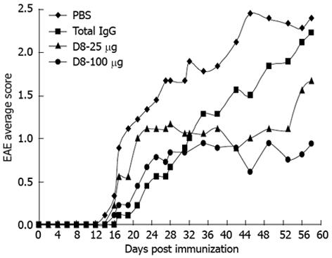
Figure 1 Anti-eo-2 neutralizing mAb treatment effect on progressive experimental autoimmune encephalomyelitis clinical course.
Progressive experimental autoimmune encephalomyelitis (EAE) was induced in female C57BL/6 mice by immunization with two following subcutaneous injections of MOG35-55 peptide, emulsified in CFA, with an interval of 1 wk. EAE-induced mice were injected daily intraperitoneally with 25 μg or 100 μg D8, mouse IgG, or with vehicle control only (PBS) starting from day 0 post immunization, and monitored for EAE clinical score. A significant improvement in EAE clinical score compared to total IgG treated mice was observed only with the higher concentration of D8 on day 56, in which treatment with D8 100 μg led to a decline of 61.5% in average clinical score (n = 10 in each group, P = 0.04, one way ANOVA).
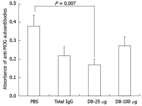
Figure 2 The effect of anti-eo-2 neutralizing mAb treatment on the level of anti-MOG35-55 autoantibodies in experimental autoimmune encephalomyelitis sera.
Experimental autoimmune encephalomyelitis (EAE) mice daily treated with PBS, total mouse IgG, 25 μg or 100 μg D8, were sacrificed on day 58 and their sera were assessed for the presence of anti-myelin oligodendrocyte glycoprotein (anti-MOG) autoantibodies using ELISA. No significant effect in the level of anti-MOG IgG antibodies was accepted in both D8 treated groups compared to the IgG treated group (values presented are A450 nm, n = 9 in each group).
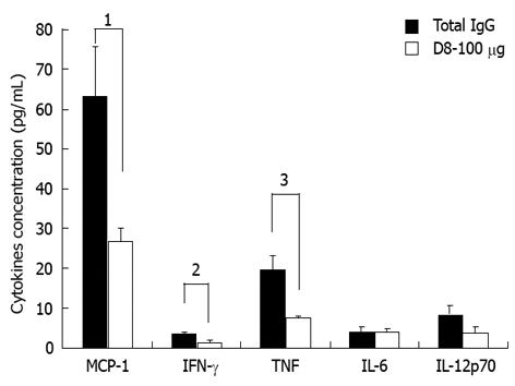
Figure 3 Anti-eo-2 neutralizing mAb treatment effect on pro-inflammatory cytokines profile.
Experimental autoimmune encephalomyelitis (EAE) mice sera from total IgG and 100 μg D8 groups were assessed for the presence of interleukin (IL)-6, interferon (IFN)-γ, tumor necrosis factor (TNF)-α, IL-12p70 and macrophage chemoattractant protein (MCP)-1 using the BD™ Cytometric Bead Array Mouse Inflammation Kit. Treatment of EAE-induced mice with D8 100 μg led to a significant decrease of 57.3% in serum levels of MCP-1, 61.2% in serum levels of IFN-γ and 60.8% in levels of TNF-α, compared to IgG treatment (n = 6 in each group, 1P = 0.03, 2P = 0.02, 3P = 0.005, two-tailed Student’s t-test).
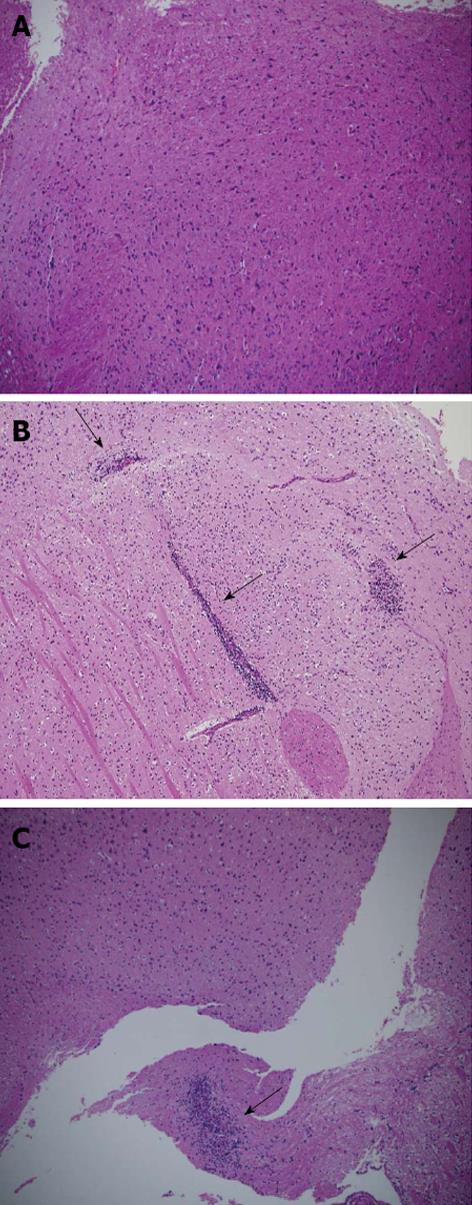
Figure 4 The extent of cellular infiltration in anti-eo-2 mAb treated experimental autoimmune encephalomyelitis mice is low compared with PBS treated experimental autoimmune encephalomyelitis mice and their healthy littermates.
A: Hematoxylin and eosin staining of representative brain sections from healthy C57BL/6 mice;B: PBS treated experimental autoimmune encephalomyelitis (EAE) mice; C: EAE mice treated with 100 μg D8. Lower extent of cellular infiltration in D8 100 μg treated group is observed compared with the IgG treated group. Arrows indicate inflammatory infiltration. Magnification × 200.
- Citation: Mausner-Fainberg K, Karni A, George J, Entin-Meer M, Afek A. Eotaxin-2 blockade ameliorates experimental autoimmune encephalomyelitis. World J Immunol 2013; 3(1): 7-14
- URL: https://www.wjgnet.com/2219-2824/full/v3/i1/7.htm
- DOI: https://dx.doi.org/10.5411/wji.v3.i1.7


