©The Author(s) 2016.
World J Clin Pediatr. Aug 8, 2016; 5(3): 262-272
Published online Aug 8, 2016. doi: 10.5409/wjcp.v5.i3.262
Published online Aug 8, 2016. doi: 10.5409/wjcp.v5.i3.262
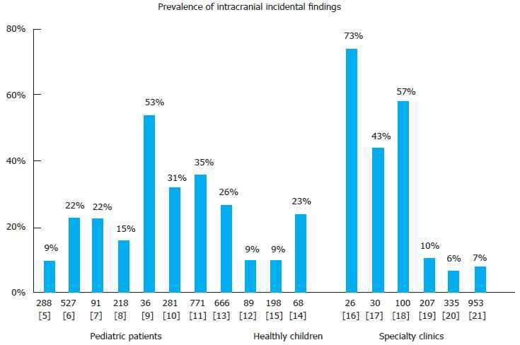
Figure 1 A comparative prevalence of incidental findings on pediatric magnetic resonance imaging of the studies.
The numbers above bracket represents number of children in the study; Number in the brackets represents the reference for the study. The subjects in the studies’ reference[5-13] except[12] on the left were from General Neurology Clinic. The studies’[16-21] on the right were from Specialty Clinics other than neurology. Three studies in middle; ref.[12,15] were from research in healthy children and the study[14] was community-based in healthy population.
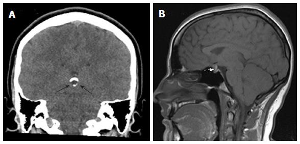
Figure 2 Three out of four incidental findings in a single 16-year-old girl who presented with right facial nerve palsy.
The left maxillary sinusitis is not shown. A: Coronal non-contrasted computerized tomography of the brain shows a partially calcified cystic pineal lesion (black arrows); B: A true midline sagittal magnetic resonance image contrast enhancing pituitary mass measuring 13 mm × 10 mm × 10 mm (white arrow) and cerebellar ectopia, 7 mm (black arrow). There was no enhancement of facial nerve in vicinity of the right internal carotid canal.
- Citation: Gupta SN, Gupta VS, White AC. Spectrum of intracranial incidental findings on pediatric brain magnetic resonance imaging: What clinician should know? World J Clin Pediatr 2016; 5(3): 262-272
- URL: https://www.wjgnet.com/2219-2808/full/v5/i3/262.htm
- DOI: https://dx.doi.org/10.5409/wjcp.v5.i3.262


