©The Author(s) 2016.
World J Clin Pediatr. Feb 8, 2016; 5(1): 1-15
Published online Feb 8, 2016. doi: 10.5409/wjcp.v5.i1.1
Published online Feb 8, 2016. doi: 10.5409/wjcp.v5.i1.1
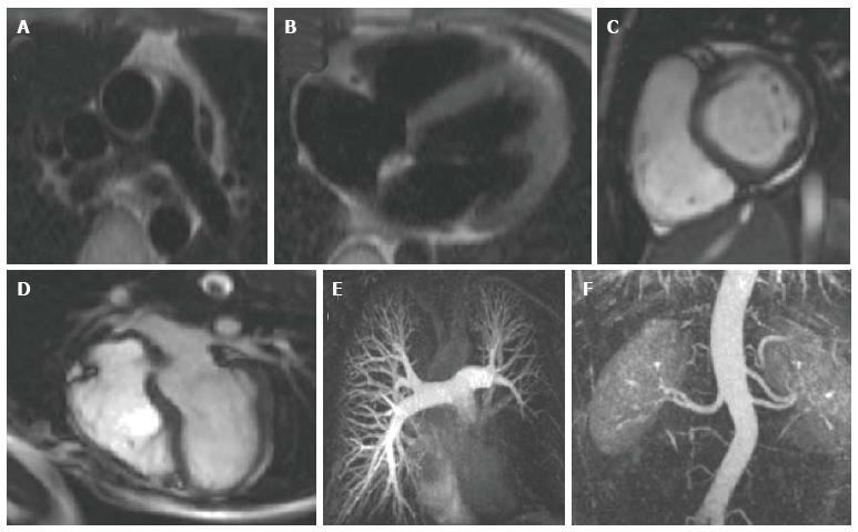
Figure 1 Examples of images produced by individual cardiovascular magnetic resonance pulse sequences.
A and B are black blood spin echo images showing the ascending and descending aorta at the main pulmonary artery level A and the 4 cardiac chambers B; C and D are bright blood SSFP cine images showing short axis and 4 chamber views respectively, while E and F are examples of 3D contrast-enhanced MRA; E is a contrast-enhanced MRA of the pulmonary tree in a patient with a large sarcoma. There is no opacification of the arterial supply of the left lower lobe, indicating complete occlusion of the lower branch of the left pulmonary artery; F is a contrast-enhanced MRA of the descending aorta for assessment of renal anatomy, demonstrating an accessory renal artery to left kidney (a common normal variant). SSFP: Steady state free precession; MRA: Magnetic resonance angiography.
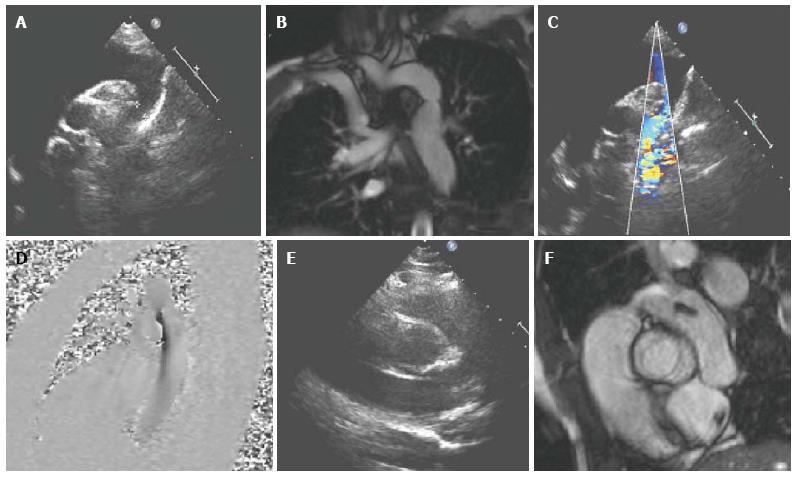
Figure 2 A 17-year-old girl with coarctation of the descending aorta, imaged by transthoracic echocardiography (A,C,E) and cardiovascular magnetic resonance (B,D,F).
Imaging of the aortic arch using TTE can be challenging, depending on the suprasternal image quality A and especially in older children with poorer acoustic windows. CMR allows accurate measurement of the dimensions of the area of stenosis B and assessment of the remainder of the thoracic aorta and head and neck vessels. Flow velocity mapping D, similar to Doppler echocardiography C is used to measure peak velocity through the stenosed area and assess for a diastolic tail. In this case, the bicuspid aortic valve was not well visualised on TTE E but was clearly seen on CMR F. CMR: Cardiovascular magnetic resonance; TTE: Transthoracic echocardiography.
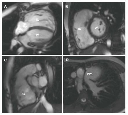
Figure 3 A 10-year-old girl with tetralogy of Fallot repaired at 1 year of age.
She had a 1 year history of increasing exertional breathlessness and reduced exercise capacity. Her CMR showed near-free pulmonary regurgitation with a dilated right ventricle and a reduced right ventricular ejection fraction. In view of these findings, she was offered a pulmonary valve replacement. A shows a 4 chamber SSFP cine image; B a short axis through the mid ventricle; C a right ventricular outflow tract view and D a transverse section through the thorax at main pulmonary artery level showing the dilated main pulmonary artery. RV: Right ventricle; LV: Left ventricle; MPA: Main pulmonary artery; SSFP: Steady state free precession; CMR: Cardiovascular magnetic resonance.
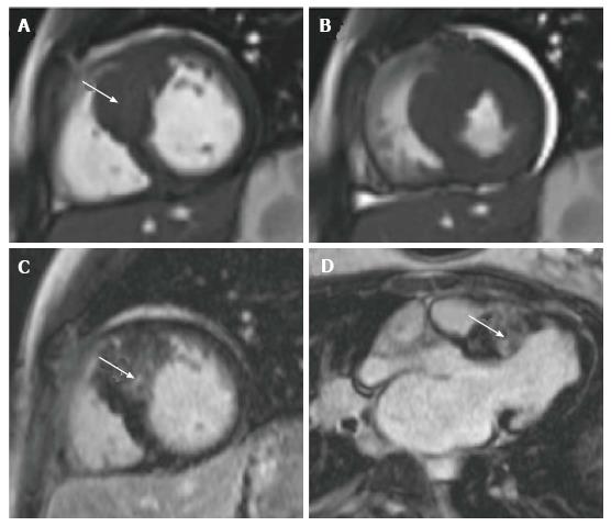
Figure 4 A new diagnosis of hypertrophic cardiomyopathy in a 12-year-old girl who presented as an out of hospital arrest with documented ventricular fibrillation.
A shows a short axis view of the left ventricle at end systole at basal level. There is asymmetrical septal hypertrophy (arrowed) with a maximal wall thickness of 26 mm; B shows the same ventricular position in end systole; C and D are late gadolinium enhancement images demonstrating extensive patchy fibrosis of the hypertrophied septum (arrowed).
- Citation: Mitchell FM, Prasad SK, Greil GF, Drivas P, Vassiliou VS, Raphael CE. Cardiovascular magnetic resonance: Diagnostic utility and specific considerations in the pediatric population. World J Clin Pediatr 2016; 5(1): 1-15
- URL: https://www.wjgnet.com/2219-2808/full/v5/i1/1.htm
- DOI: https://dx.doi.org/10.5409/wjcp.v5.i1.1


