Copyright
©2013 Baishideng Publishing Group Co.
World J Clin Pediatr. Nov 8, 2013; 2(4): 70-76
Published online Nov 8, 2013. doi: 10.5409/wjcp.v2.i4.70
Published online Nov 8, 2013. doi: 10.5409/wjcp.v2.i4.70
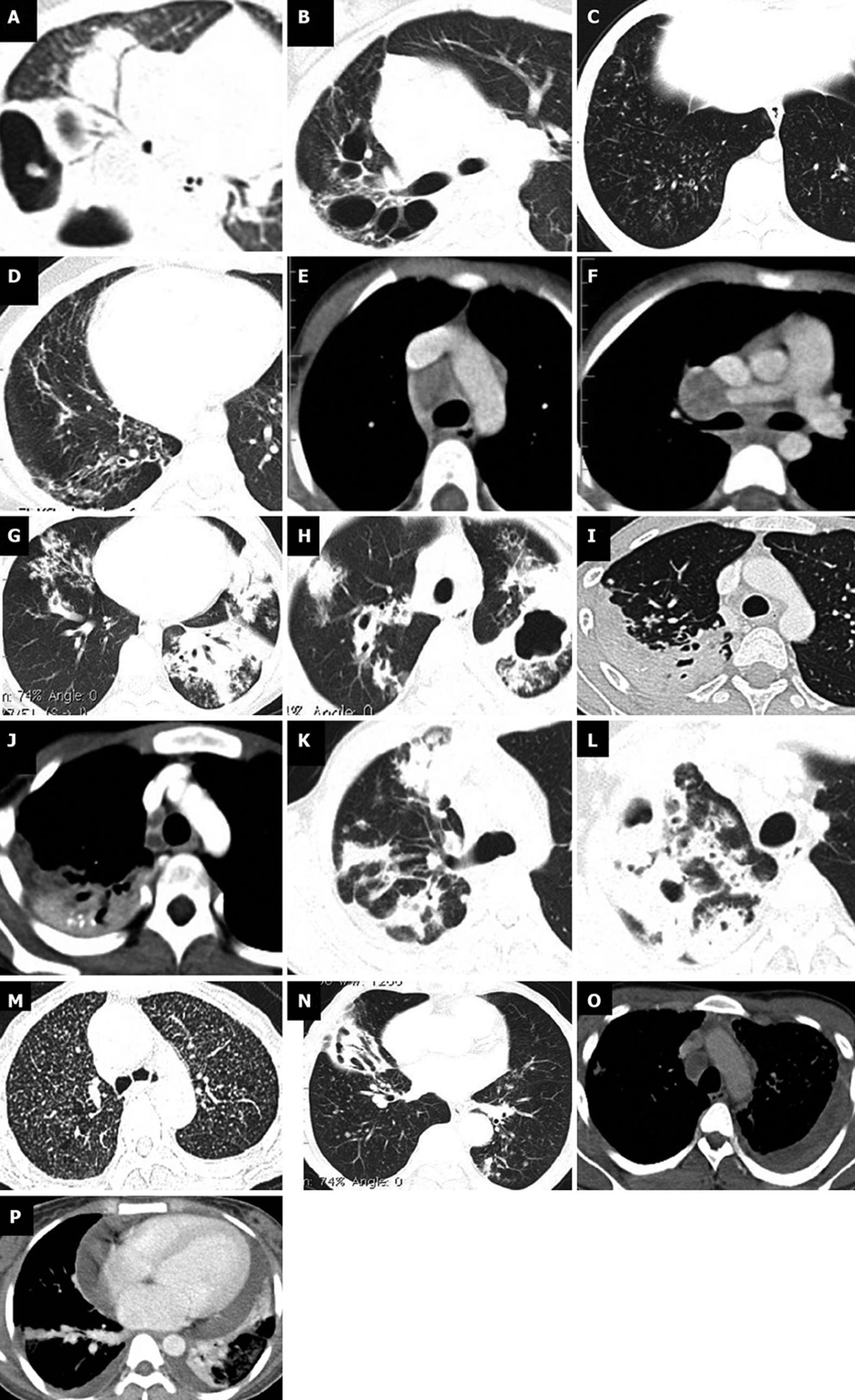
Figure 1 Computed tomography findings in children (less than 10 years), adolescents (10-18 years), and adults (above 18 years) with chest tuberculosis.
A-F: Children; A: Parenchymal consolidation in right lung with adjacent thick walled cavities; B: Multiple cavities with adjacent fibrosis in right upper lobe; C: Tiny low density centrilobular nodules seen diffusely in both lungs; D: Fibrobronchiectatic changes in right middle and lower lobes; E-F: Enlarged low attenuating necrotic nodes in right paratracheal, and right hilar location repectively; G-J: Adolescents; G: Parenchymal consolidation with centrilobular nodules in left lung, nodules in right middle lobe; H: Thick walled cavities with surrounding consolidation and air space nodules in both upper lobes; I-J: Pleural based fibrotic changes with calcification in right upper lobe with necrotic right paratracheal node; K-P: Adults; K: Multifocal consolidation in right upper lobe; L: Thick walled cavities in right upper lobe; M: Miliary TB- multiple tiny nodules distributed randomly in both lungs; N: Fibrobronchietasis in right middle lobe with nodules in left lower lobe; O: Necrotic right paratracheal node with empyema in left side; P: Pericardial and bilateral pleural effusion.
-
Citation: Veedu PT, Bhalla AS, Vishnubhatla S, Kabra SK, Arora A, Singh D, Gupta AK. Pediatric
vs adult pulmonary tuberculosis: A retrospective computed tomography study. World J Clin Pediatr 2013; 2(4): 70-76 - URL: https://www.wjgnet.com/2219-2808/full/v2/i4/70.htm
- DOI: https://dx.doi.org/10.5409/wjcp.v2.i4.70


