
















Copyright: ©Author(s) 2026.
World J Clin Pediatr. Dec 9, 2026; 15(4): 122166
Published online Dec 9, 2026. doi: 10.5409/wjcp.122166
Published online Dec 9, 2026. doi: 10.5409/wjcp.122166
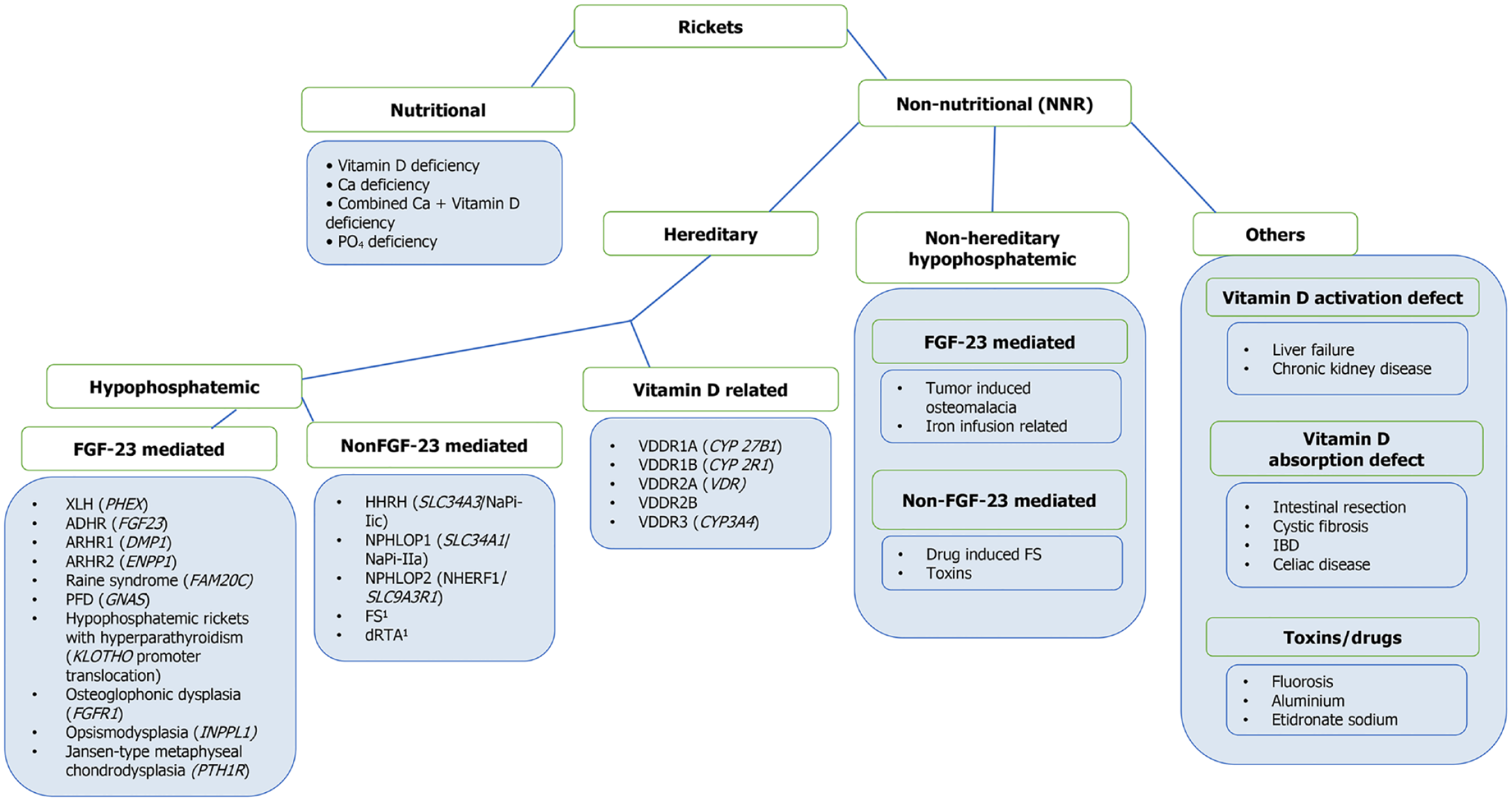
Figure 1 Etiological classification of rickets.
1Multiple known causative genes. FGF-23: Fibroblast growth factor 23; XLH: X-linked hypophosphatemic ricket; PHEX: Phosphate regulating endopeptidase homolog X-linked; ADHR: Autosomal dominant hypophosphatemic rickets; ARHR1: Autosomal recessive hypophosphatemic rickets type 1; DMP1: Dentin matrix protein 1; ARHR2: Autosomal recessive hypophosphatemic rickets type 2; ENPP1: Ectonucleotide pyrophosphatase/phosphodiesterase 1; FAM20C: Family with sequence similarity 20-member C; PFD: Polyostotic fibrous dysplasia; GNAS: Guanine nucleotide-binding protein alpha-stimulating; FGFR1: Fibroblast growth factor receptor 1; INPPL1: Inositol polyphosphate phosphatase-like 1; PTH1R: Parathyroid hormone 1 receptor; Ca: Calcium; PO4: Phosphorus; HHRH: Hypophosphatemic rickets with hypercalciuria; SLC34A3: Solute carrier family 34 member 3; NPHLOP1: Nephrolithiasis and osteoporosis type 1; SLC34A1: Solute carrier family 34 member 1; NPHLOP2: Nephrolithiasis and osteoporosis type 2; NHERF1: Na+/H+ exchanger regulatory factor 1; SLC9A3R1: Solute carrier family 9 member A3 regulator 1; FS: Fanconi syndrome; dRTA: Distal renal tubular acidosis; VDDR1A: Vitamin D dependent rickets type 1A; CYP27B1: Cytochrome P450 family 27 subfamily B member 1; VDDR1B: Vitamin D dependent rickets type 1B; CYP2R1: Cytochrome P450 family 2 subfamily R member 1; VDDR2A: Vitamin D dependent rickets type 2A; VDDR2B: Vitamin D dependent rickets type 2B; VDR: Vitamin D receptor; VDDR3: Vitamin D dependent rickets type 3; NNR: Non-nutritional ricket; IBD: Inflammatory bowel disease.
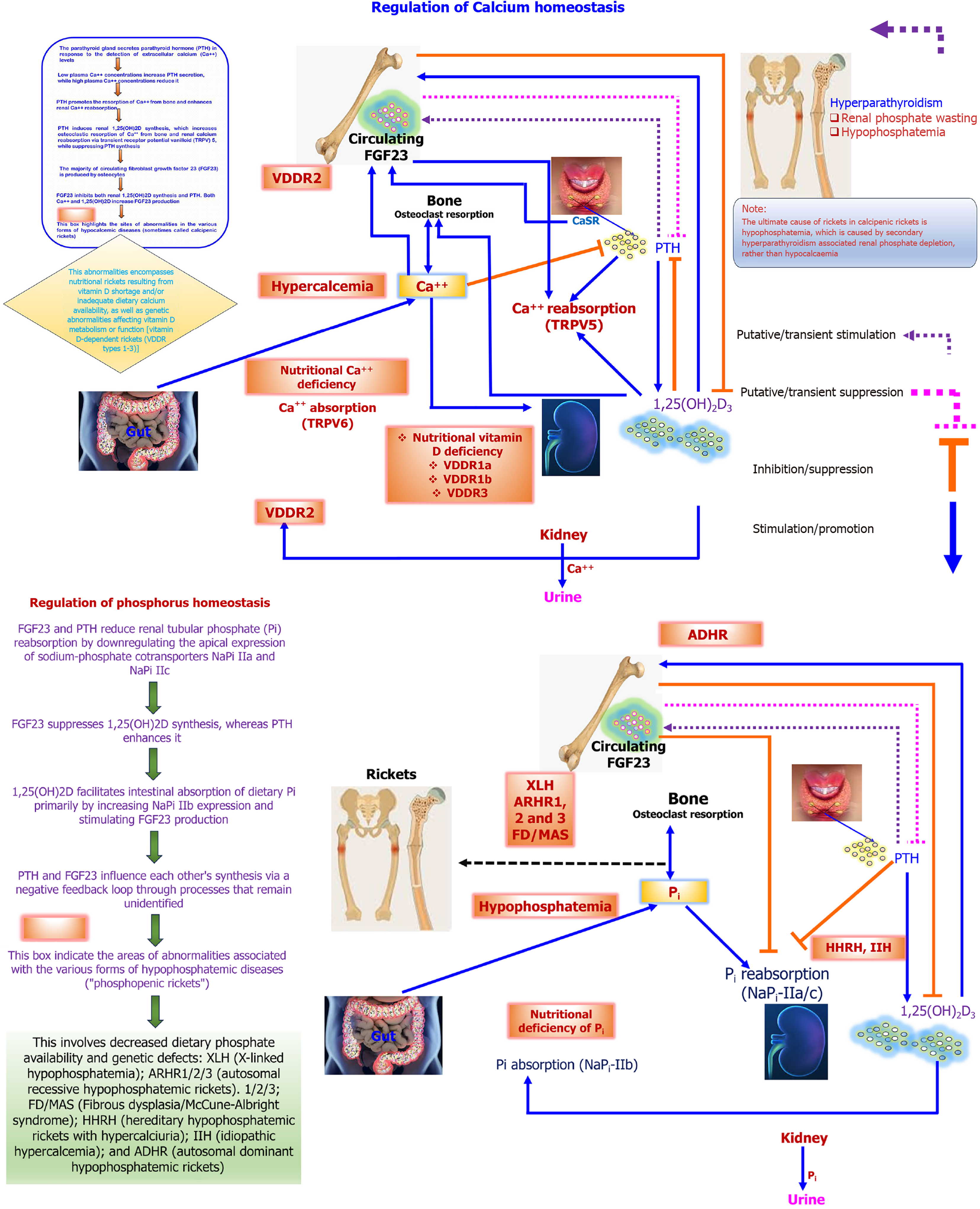
Figure 2 Regulation of calcium and phosphate homeostasis and disorders associated with rickets.
Schematic representation of calcium (Ca) and phosphate homeostasis regulated by parathyroid hormone (PTH), 1,25-dihydroxyvitamin D [1,25(OH)2D], and fibroblast growth factor 23 (FGF-23). In Ca regulation (upper panel), PTH increases bone resorption, renal Ca2+ reabsorption via transient receptor potential vanilloid 5, and 1,25(OH)2D synthesis, while 1,25(OH)2D enhances intestinal absorption via transient receptor potential vanilloid 6. In phosphate regulation (lower panel), FGF-23 and PTH reduce renal phosphate reabsorption by downregulating sodium-phosphate cotransporters NaPi-IIa (solute carrier family 34 member 1) and NaPi-IIc (solute carrier family 34 member 3), while 1,25(OH)2D increases intestinal phosphate absorption. Disorders shown include vitamin D-dependent rickets (vitamin D dependent rickets type 1A: Cytochrome P450 family 27 subfamily B member 1; vitamin D dependent rickets type 1B: Cytochrome P450 family 2 subfamily R member 1; vitamin D dependent rickets type 2: Vitamin D receptor), X-linked hypophosphatemia (X-linked hypophosphatemic rickets; phosphate regulating endopeptidase homolog X-linked), autosomal dominant hypophosphatemic rickets (autosomal dominant hypophosphatemic rickets; FGF-23), autosomal recessive hypophosphatemic rickets (autosomal recessive hypophosphatemic rickets; dentin matrix protein 1, ectonucleotide pyrophosphatase/phosphodiesterase 1), fibrous dysplasia/McCune-Albright syndrome (fibrous dysplasia/McCune-Albright syndrome), and hereditary hypophosphatemic rickets with hypercalciuria (hereditary hypophosphatemic rickets with hypercalciuria; solute carrier family 34 member 3). PTH: Parathyroid hormone; Ca: Calcium; 1,25(OH)2D: 1,25-dihydroxyvitamin D; TRPV5: Transient receptor potential vanilloid 5; CaSR: Calcium-sensing receptor; FGF-23: Fibroblast growth factor 23; VDDR: Vitamin D dependent rickets; VDDR2: Vitamin D dependent rickets type 2; TRPV6: Transient receptor potential vanilloid 6; 1,25(OH)2D3:1,25-dihydroxyvitamin D3; VDDR1A: Vitamin D dependent rickets type 1A; VDDR1B: Vitamin D dependent rickets type 1B; VDDR3: Vitamin D dependent rickets type 3; Pi: Phosphate; XLH: X-linked hypophosphatemic ricket; ARHR: Autosomal recessive hypophosphatemic ricket; FD: Fibrous dysplasia; MAS: McCune-Albright syndrome; IIH: Idiopathic hypercalciuria; ADHR: Autosomal dominant hypophosphatemic ricket.
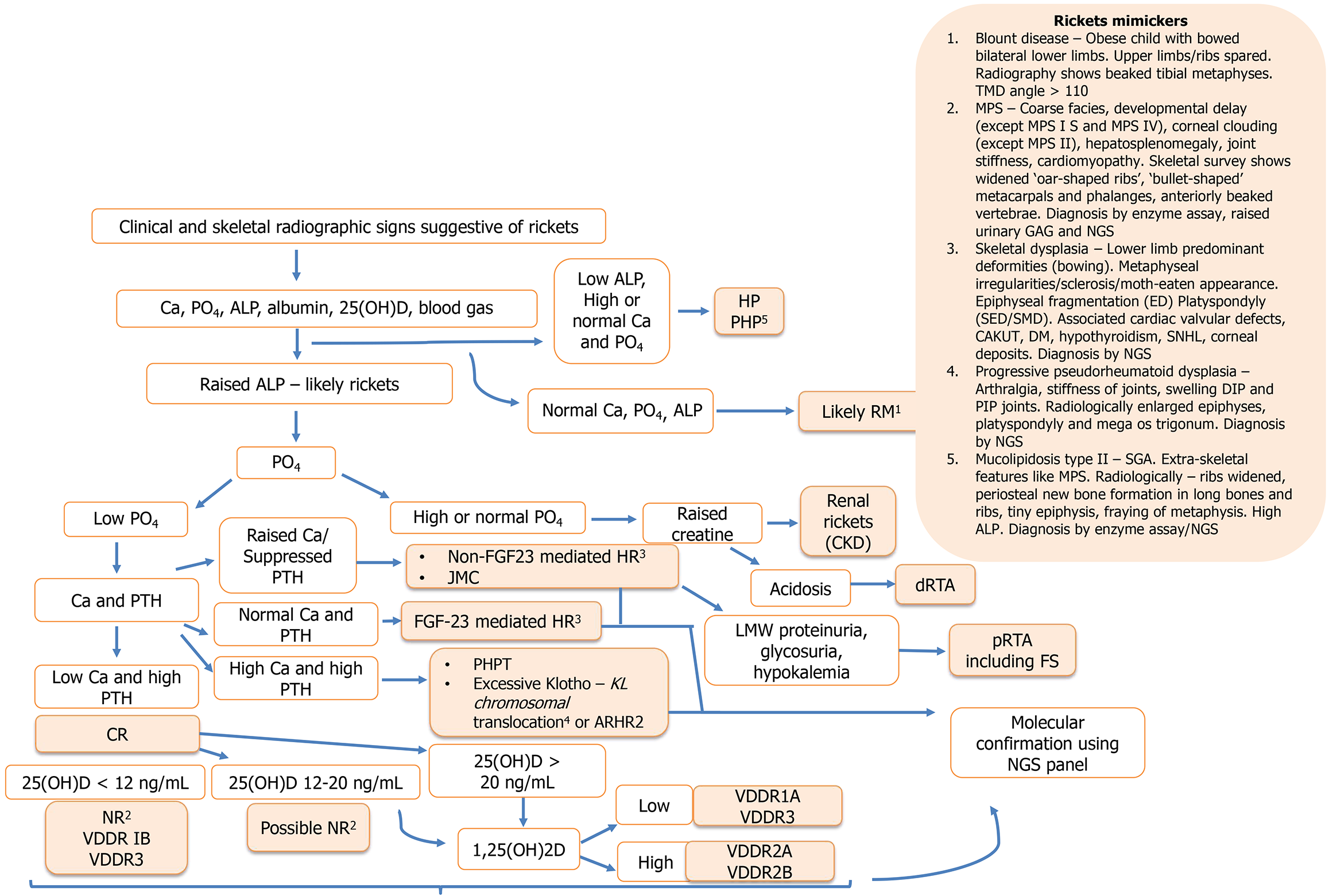
Figure 3 Algorithm for etiological diagnosis of rickets using biochemical tests.
1Exception: Raised alkaline phosphatase in mucolipidosis. 2In the clinical context of established nutritional deficiency or risk factors for vitamin D deficiency. 3Differentiation between fibroblast growth factor 23 (FGF-23) mediated hypophosphatemic ricket and non FGF-23 mediated hypophosphatemic ricket may be made using FGF-23 assays or urinary Ca creatinine ratio or 1,25-dihydroxyvitamin D. 4Requires polymerase chain reaction for diagnosis. 5Rule out hypothyroidism and growth hormone deficiency. Ca: Calcium; PO4: Phosphorus; ALP: Alkaline phosphatase; 25(OH)D: 25-hydroxyvitamin D; PTH: Parathyroid hormone; CR: Calciopenic ricket; NR: Nutritional ricket; VDDR1B: Vitamin D dependent rickets type 1B; VDDR3: Vitamin D dependent rickets type 3; FGF-23: Fibroblast growth factor 23; HR: Hypophosphatemic rickets; JMC: Jansen metaphyseal chondrodysplasia; KL: Klotho; ARHR2: Autosomal recessive hypophosphatemic rickets 2; 1,25(OH)2D: 1,25-dihydroxyvitamin D; HP: Hypophosphatasia; PHP: Pseudo hypophosphatasia; RM: Rickets mimicker; CKD: Chronic kidney disease; dRTA: Distal renal tubular acidosis; pRTA: Proximal renal tubular acidosis; FS: Fanconi syndrome; LMW: Low-molecular-weight; NGS: Next-generation sequencing; VDDR1A: Vitamin D dependent rickets type 1A; VDDR3: Vitamin D dependent rickets type 3; VDDR2A: Vitamin D dependent rickets type 2A; VDDR2B: Vitamin D dependent rickets type 2B; TMD: Tibia metaphyseal-diaphyseal; MPS: Mucopolysaccharidosis; GAG: Glycosaminoglycans; ED: Epiphyseal dysplasia; SED: Spondyloepiphyseal dysplasia; SMD: Spondylometaphyseal dysplasia; CAKUT: Congenital anomalies of the kidney and urinary tract; DM: Diabetes mellitus; SNHL: Sensorineural hearing loss; DIP: Distal interphalangeal joint; PIP: Proximal interphalangeal joint; SGA: Small for gestational age.
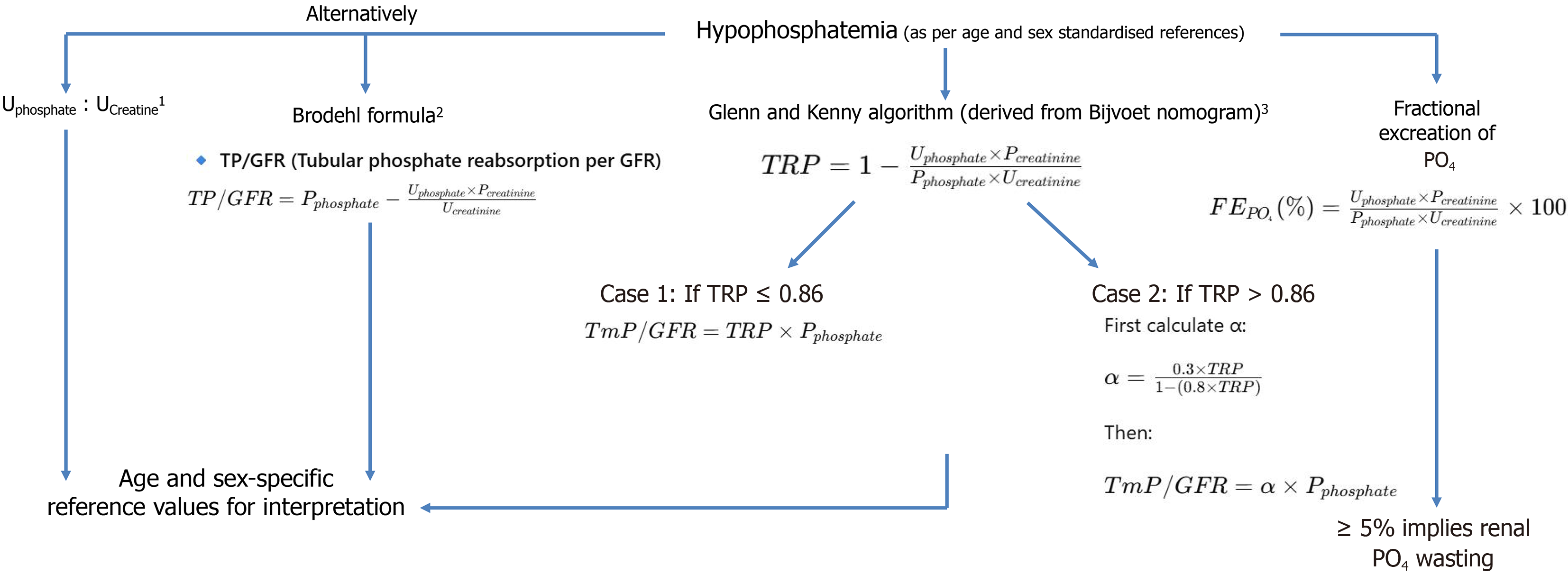
Figure 4 Indices to ascertain renal phosphorus wasting in hypophosphatemia.
1Falsely low values in severe hypophosphatemia. 2May be used across all ranges of TRP and falsely low values in severe hypophosphatemia. 3Limitations: (1) Ideally requires oral phosphorus Loading; (2) Nomogram established for adults - children have physiologically higher serum phosphorus levels; (3) Overestimates tubular maximum reabsorption of phosphate/glomerular filtration rate if TRP > 0.8; and (4) Falsely low values in severe hypophosphatemia. UPhosphate: Urinary phosphate; UCreatinine: Urinary creatinine; PPhosphate: Plasma phosphate, PCreatinine: Plasma creatinine; TP: Tubular phosphate; GFR: Glomerular filtration rate; TRP: Tubular reabsorption of phosphate; PO4: Phosphorus; TmP: Tubular maximum reabsorption of phosphate.
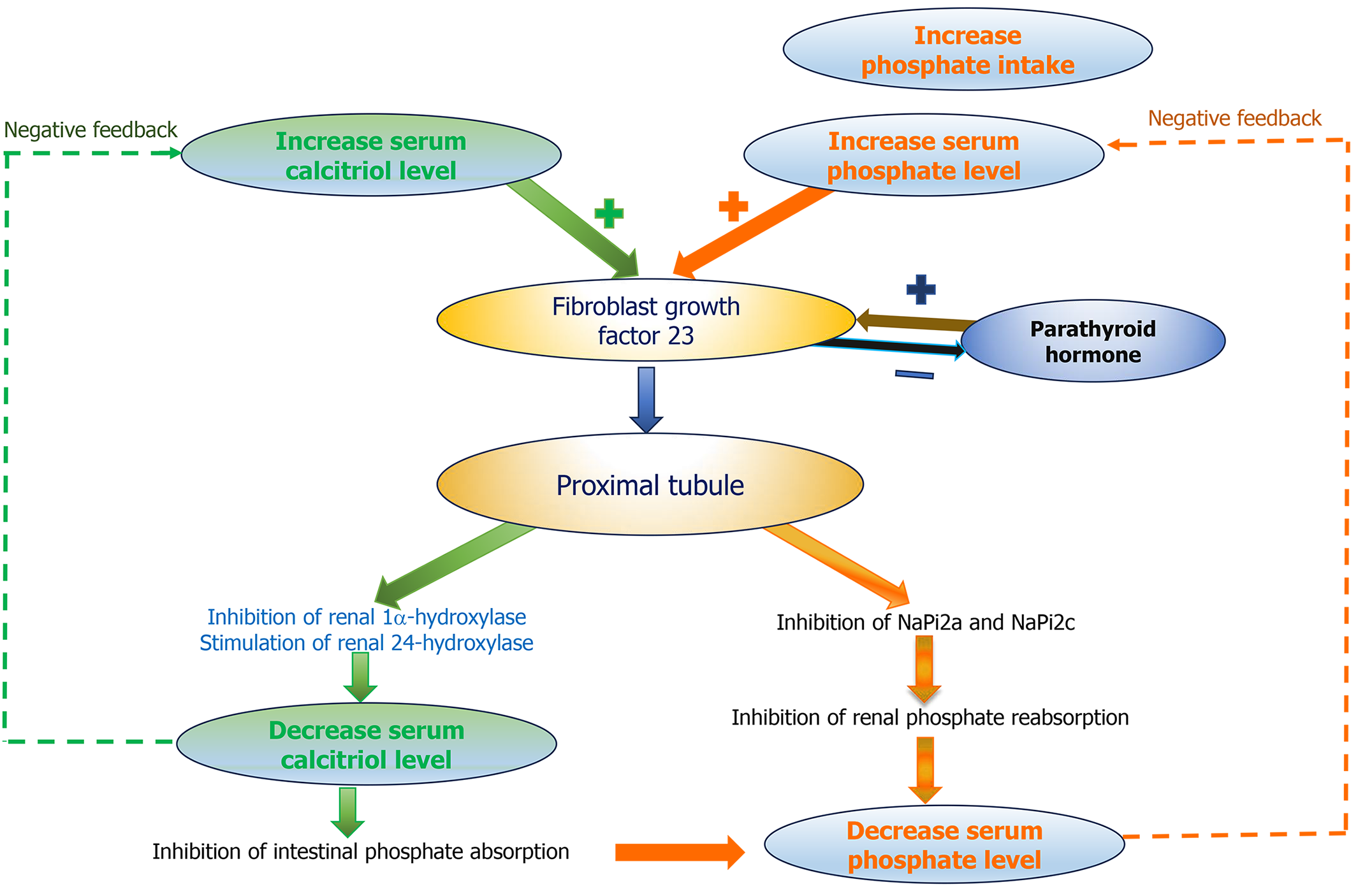
Figure 5 Actions of fibroblast growth factor 23 in phosphate homeostasis.
Schematic showing regulation of phosphate and calcitriol (1,25-dihydroxyvitamin D) by fibroblast growth factor 23 (FGF-23). Increased serum phosphate and calcitriol stimulate FGF-23 secretion. FGF-23 acts on the renal proximal tubule to inhibit sodium-phosphate cotransporters NaPi-IIa (solute carrier family 34 member 1) and NaPi-IIc (solute carrier family 34 member 3), reducing phosphate reabsorption and lowering serum phosphate. It also suppresses renal 1α-hydroxylase (cytochrome P450 family 27 subfamily B member 1) and stimulates 24-hydroxylase (cytochrome P450 family 24 subfamily A member 1), decreasing calcitriol levels and intestinal phosphate absorption. Parathyroid hormone shows bidirectional interaction with FGF-23 in a negative feedback loop to maintain phosphate and calcitriol homeostasis.
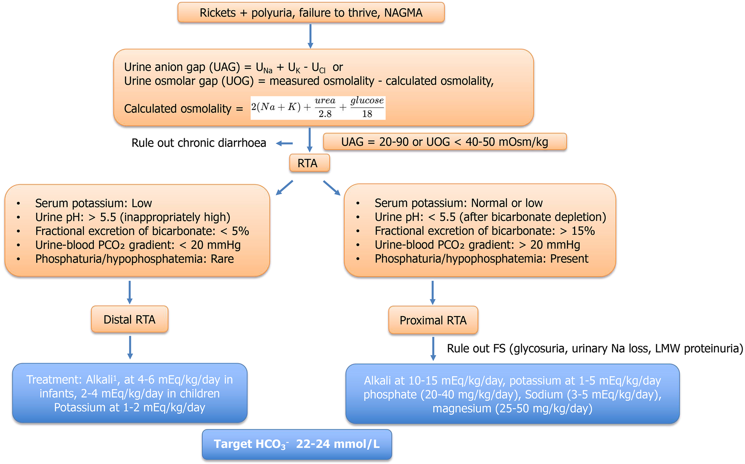
Figure 6 Approach to rickets due to renal tubular acidosis.
1Preparations: Syrups - potrate (bicarbonate and potassium 2 mEq/mL), nodosis (0.8 mEq/mL). Tablets: Sodamint (3.6 mEq/300 mg, 6 mEq/500 mg, 7.8/650 mg), acidose 500 mg. NAGMA: Normal anion gap metabolic acidosis; UAG: Urine anion gap; UOG: Urine osmolar gap; PCO2: Partial pressure of carbon dioxide; RTA: Renal tubular acidosis; FS: Fanconi syndrome; LMW: Low molecular weight.
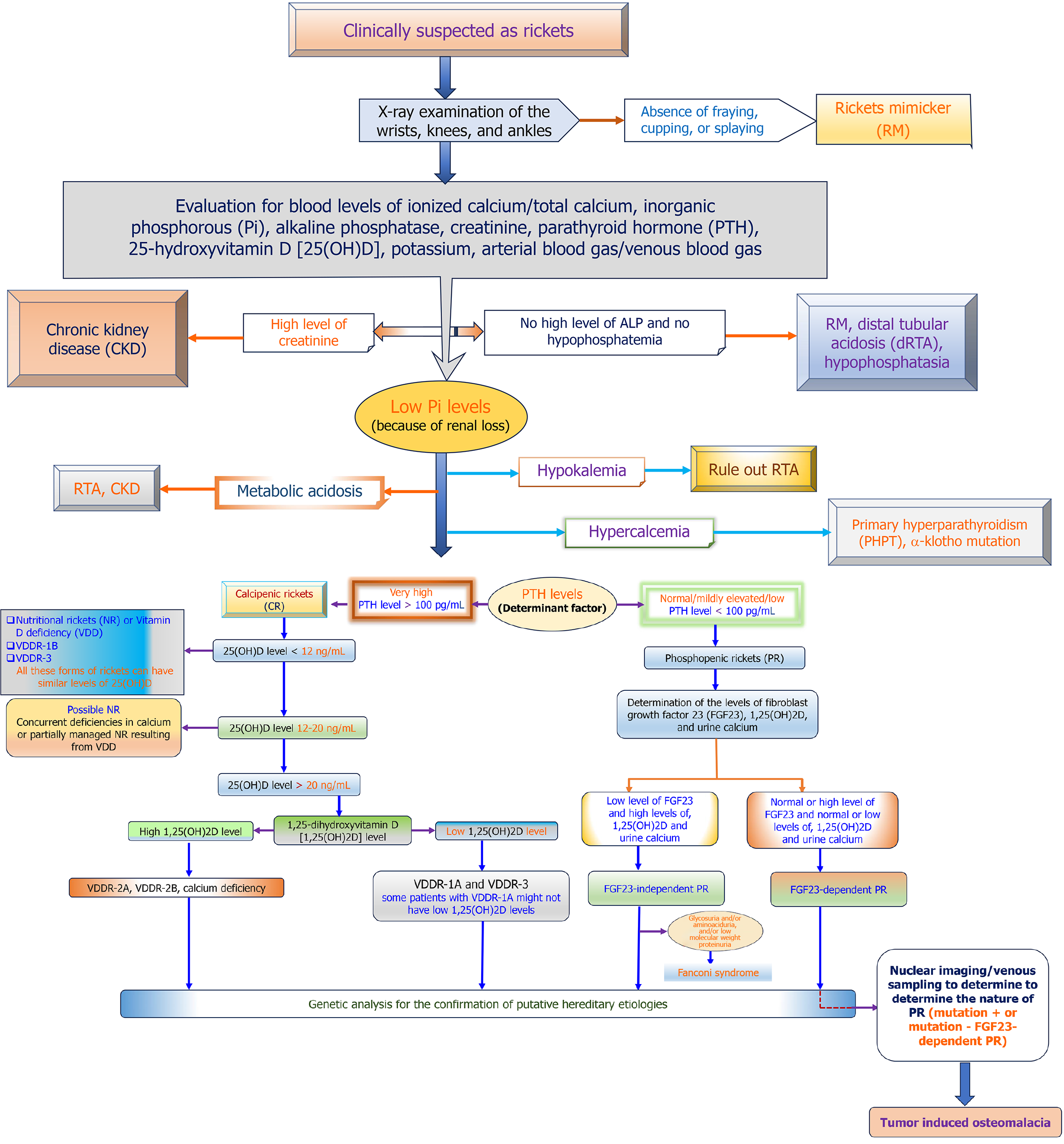
Figure 7 Diagnostic algorithm including hereditary and non-hereditary etiologies of rickets[17].
Pi: Phosphate; PTH: Parathyroid hormone; 25(OH)D: 25-hydroxyvitamin D; CKD: Chronic kidney disease; RTA: Renal tubular acidosis; RM: Rickets mimicker; PHPT: Primary hyperparathyroidism; NR: Nutritional rickets; VDD: Vitamin D deficiency; VDDR: Vitamin D dependent rickets; VDDR1B: Vitamin D dependent rickets type 1B; VDDR3: Vitamin D dependent rickets type 3; 1,25(OH)2D: 1,25-dihydroxyvitamin D; CR: Calcipenic rickets; PTH: Parathyroid hormone; PR: Phosphopenic rickets; FGF-23: Fibroblast growth factor 23.
- Citation: Pathak PP, Ray S. Non-nutritional rickets: Approach, precision medicine, and outcomes. World J Clin Pediatr 2026; 15(4): 122166
- URL: https://www.wjgnet.com/2219-2808/full/v15/i4/122166.htm
- DOI: https://dx.doi.org/10.5409/wjcp.122166