
















Copyright: ©Author(s) 2026.
World J Clin Pediatr. Dec 9, 2026; 15(4): 117885
Published online Dec 9, 2026. doi: 10.5409/wjcp.117885
Published online Dec 9, 2026. doi: 10.5409/wjcp.117885
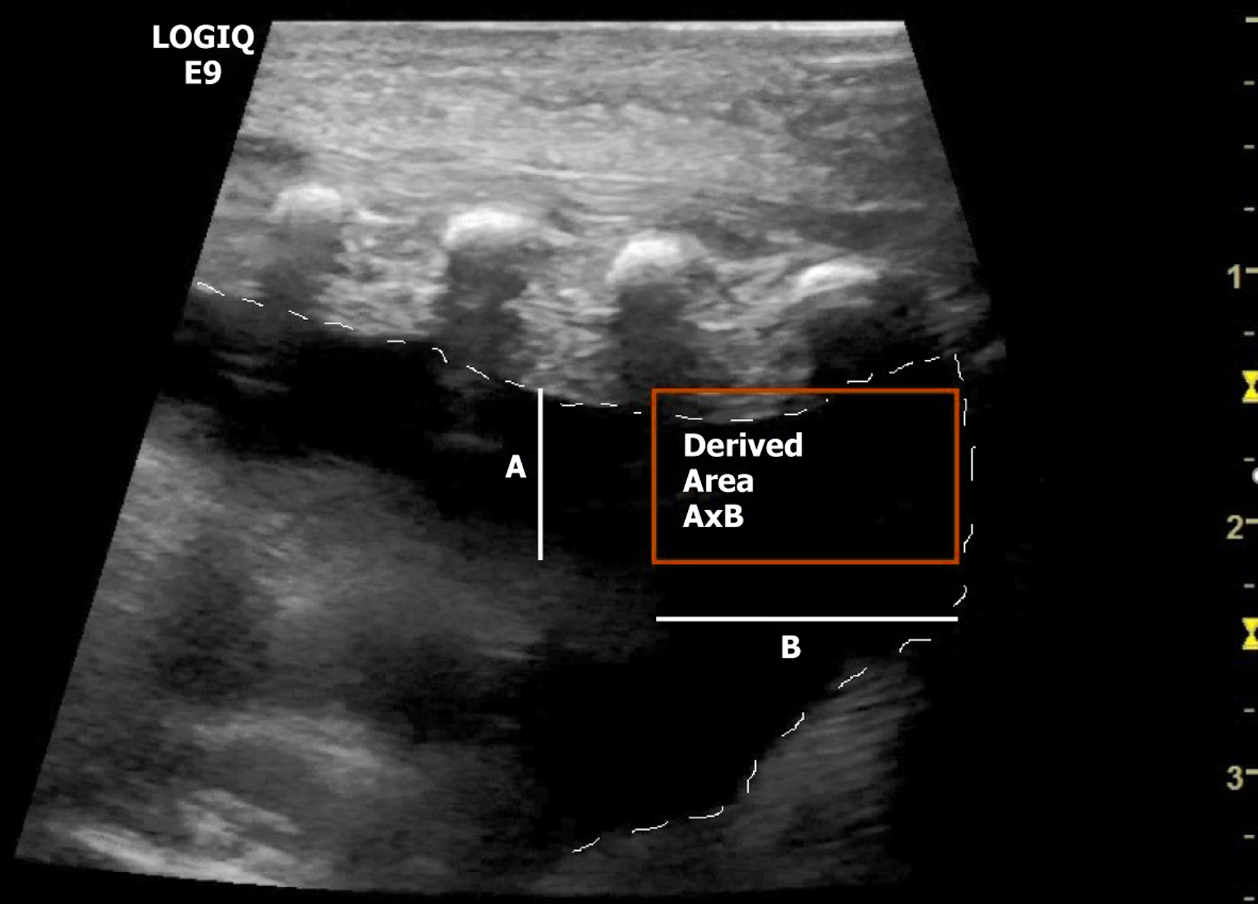
Figure 1 Illustration of the ultrasound measurements.
The lung-chest wall distance (line A) and the lung-diaphragm distance (line B) are measured in centimeters to determine the derived area (orange rectangle) as a geometric approximation.
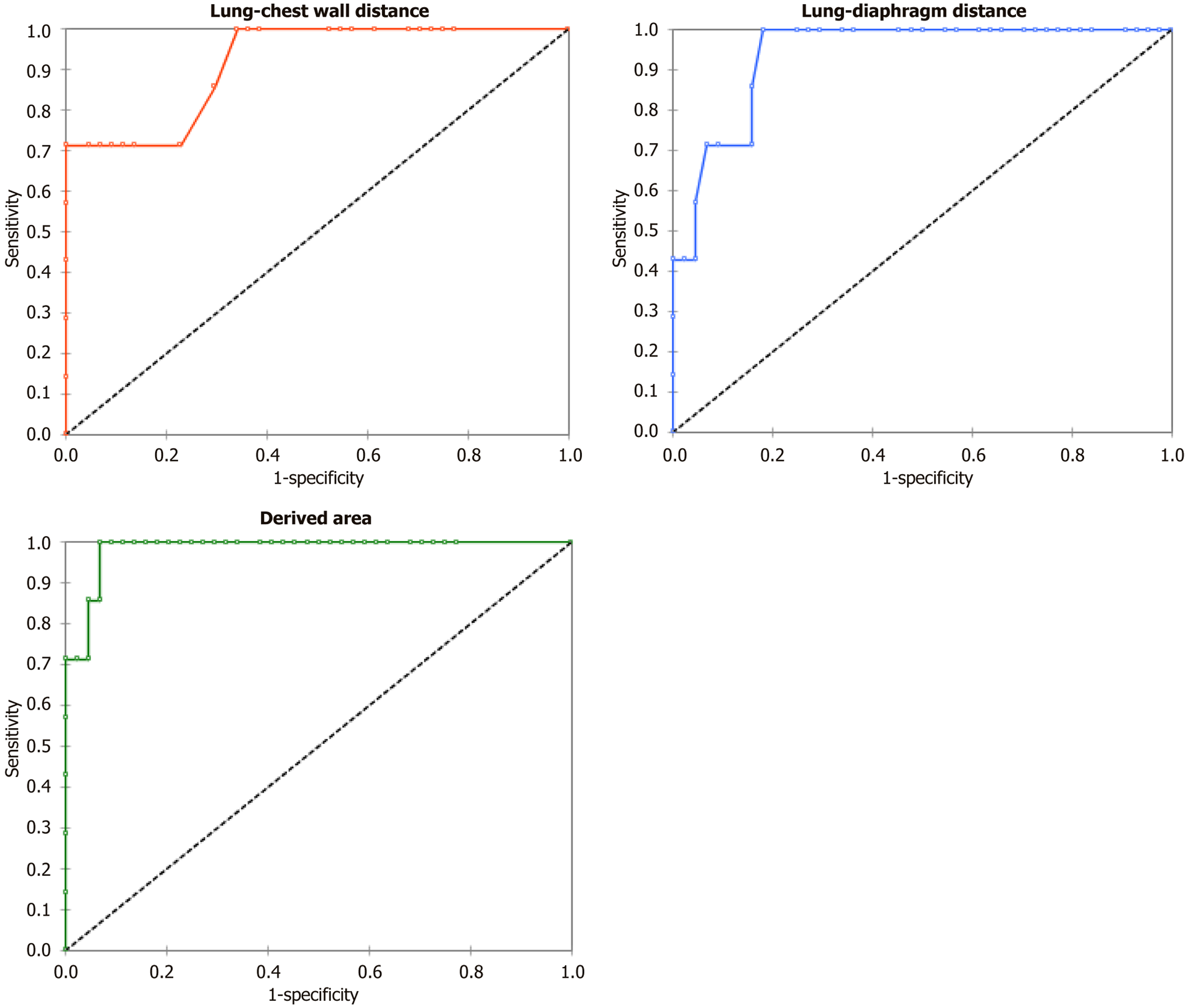
Figure 2 Receiver operating characteristic curves for lung-chest wall distance, lung-diaphragm distance, and derived area.
Area under the curve (AUC) of 0.984 (95%CI: 0.954-1.000) for the derived area, 0.938 (95%CI: 0.869-1.000) for the lung-diaphragm distance, and 0.917 (95%CI: 0.810-1.000) for the lung-chest wall distance.
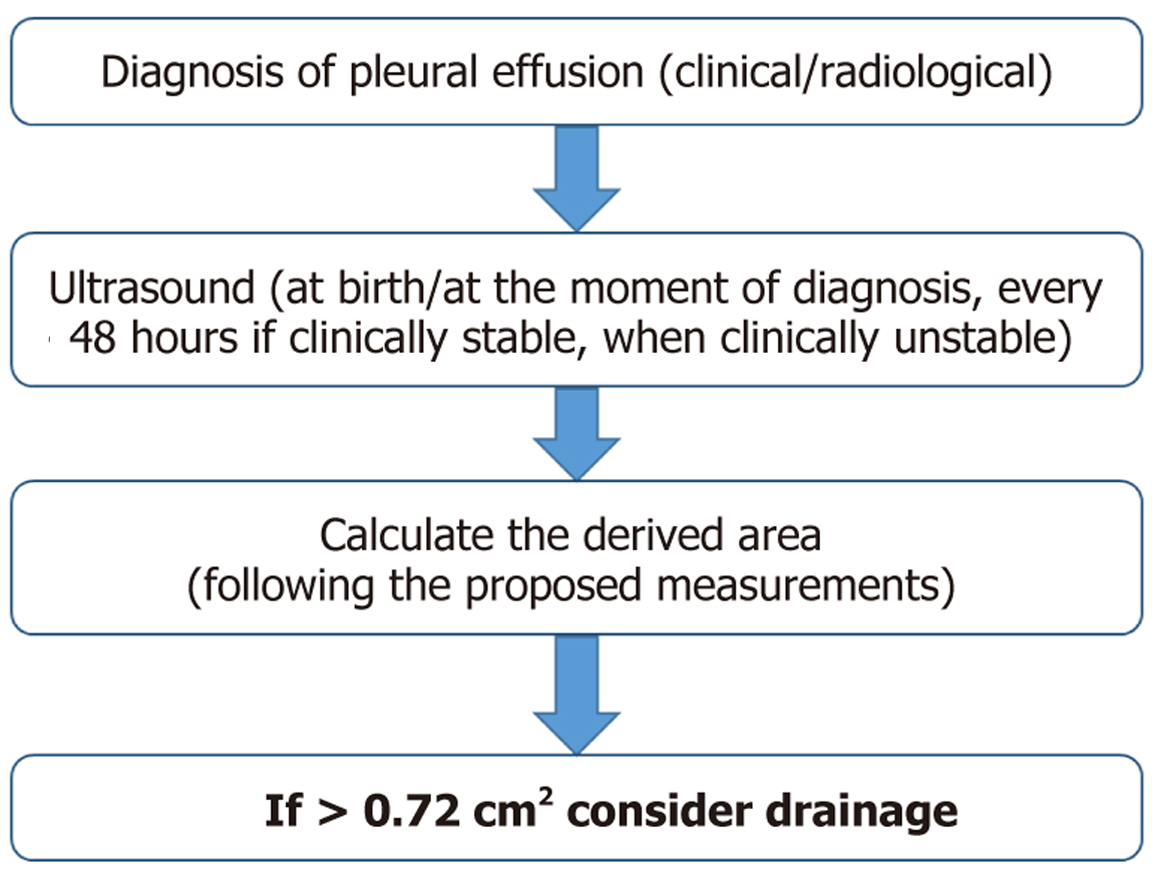
Figure 3
Hypothetical algorithm for future prospective testing.
- Citation: Perri A, Sbordone A, Fattore S, De Matteis A, Rotunno G, Marrocco GA, Nobile S, Giordano L, Tana M, Vento G. Neonatal chylothorax and ultrasound: Diagnosis, measurements, and correlations with management. World J Clin Pediatr 2026; 15(4): 117885
- URL: https://www.wjgnet.com/2219-2808/full/v15/i4/117885.htm
- DOI: https://dx.doi.org/10.5409/wjcp.117885