
















Copyright: ©Author(s) 2026.
World J Clin Pediatr. Sep 9, 2026; 15(3): 121760
Published online Sep 9, 2026. doi: 10.5409/wjcp.121760
Published online Sep 9, 2026. doi: 10.5409/wjcp.121760
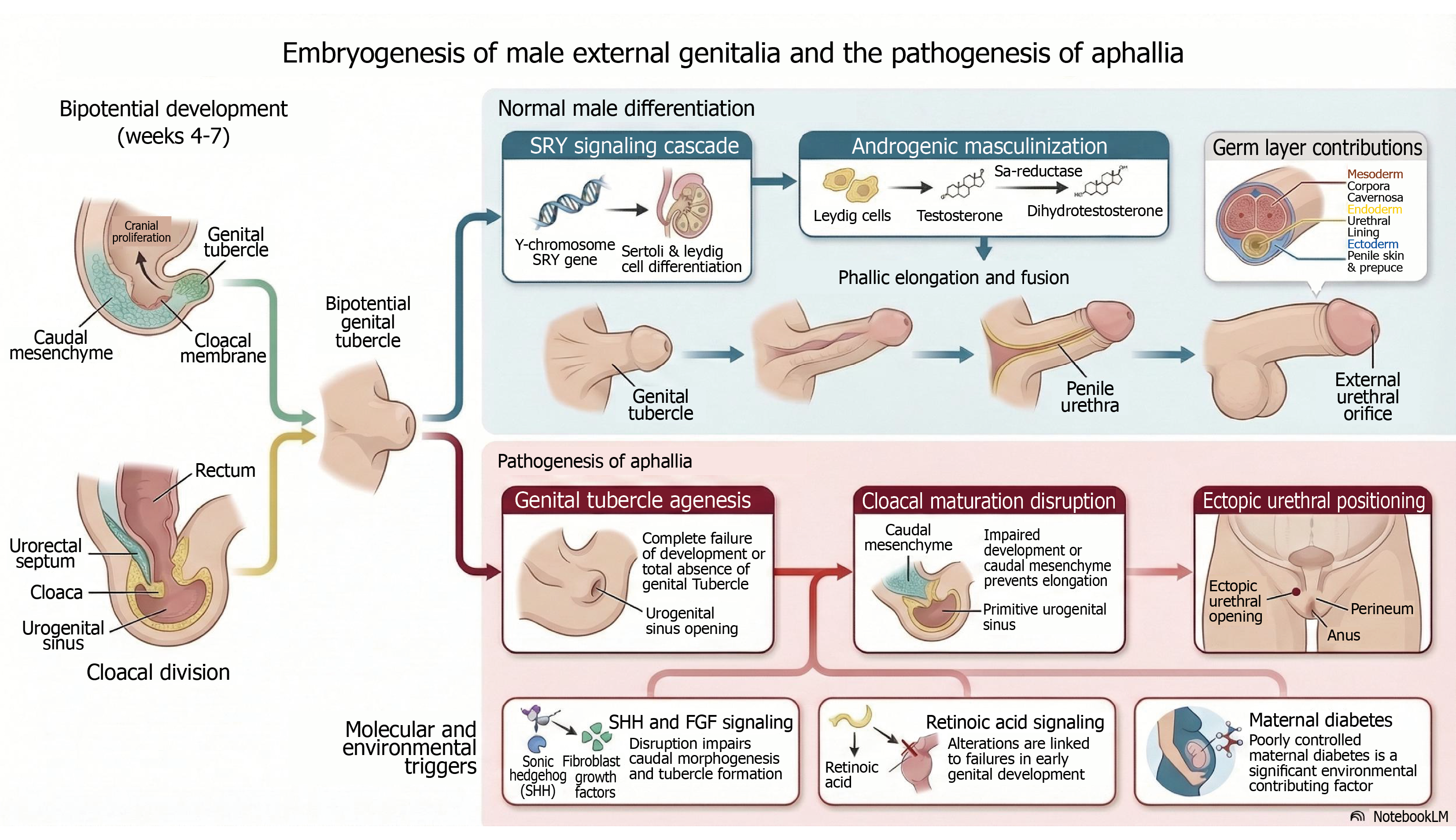
Figure 1 Embryogenesis of male external genitalia and pathogenesis of aphallia, illustrating normal genital tubercle development, androgen-driven differentiation and mechanisms leading to genital tubercle agenesis and ectopic urethral positioning.
The figure layout was assisted by an artificial intelligence-based tool (NotebookLM); however, all scientific content, conceptualization, labeling and annotations were independently developed by the authors. SRY: Sex determining region Y; DHT: Dihydrotestosterone; SHH: Sonic hedgehog; FGF: Fibroblast growth factors.
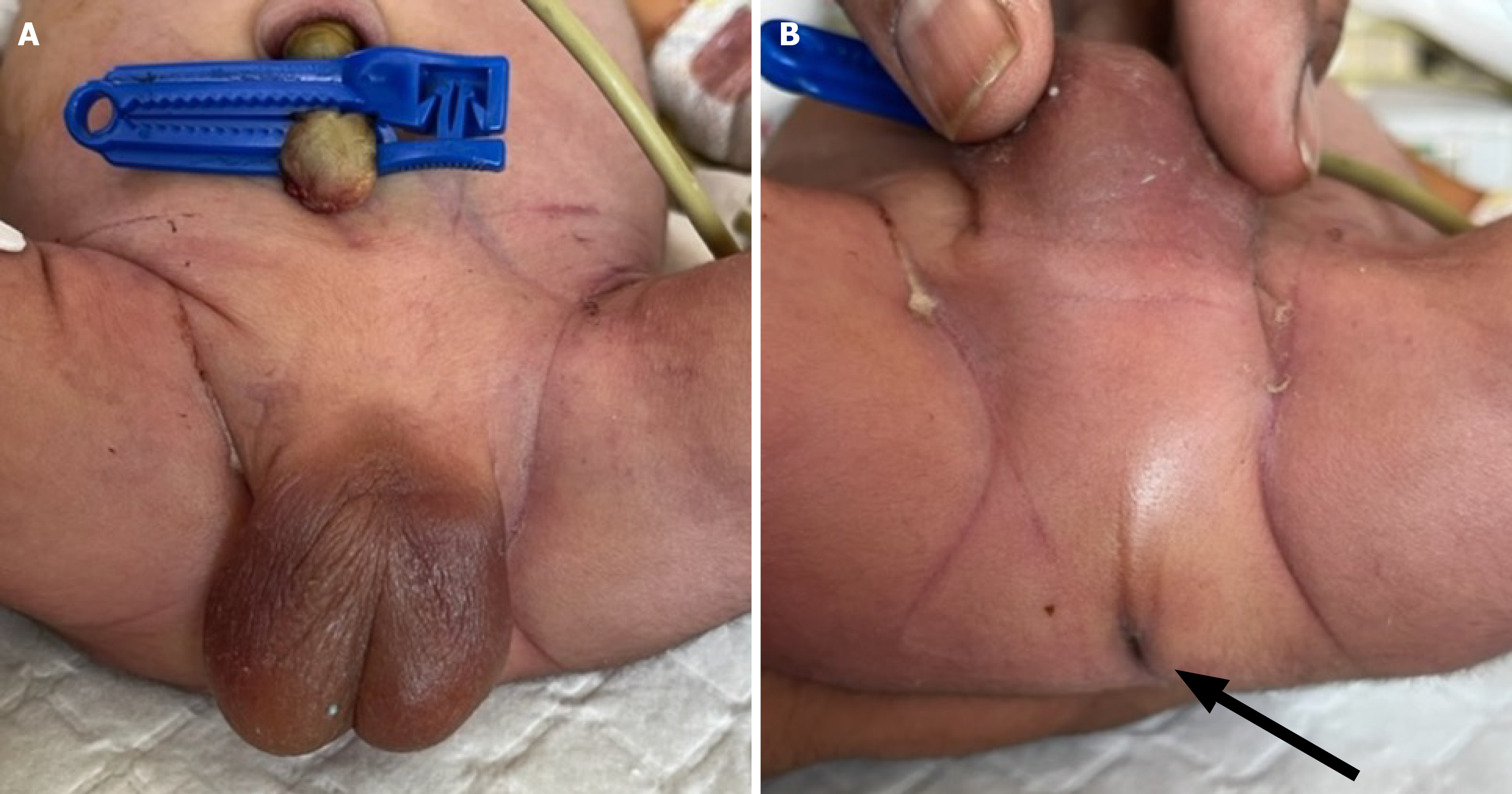
Figure 2 An illustrative case of aphallia.
A: Both testes in well‐developed scrotum, absent phallus and without any urinary opening or fistula; B: Narrow anal opening (black arrow marked) with no urinary fistula visualized at anal verge.
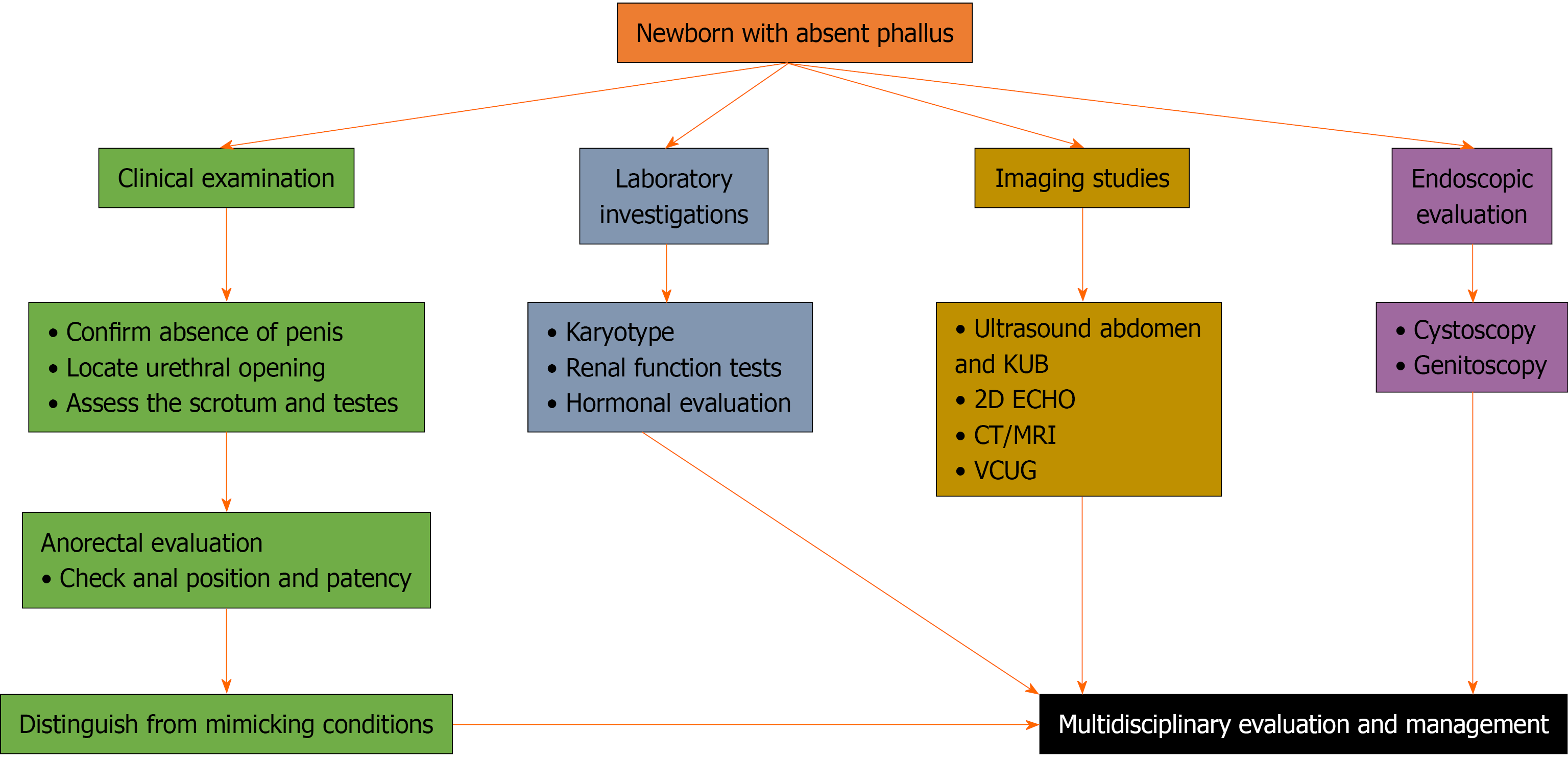
Figure 3 Diagnostic evaluation algorithm for a newborn with aphallia.
Outlining stepwise clinical assessment, systemic investigations, imaging studies and endoscopic evaluation, culminating in multidisciplinary management. KUB: Kidney, ureter and bladder; 2D ECHO: 2-dimensional echocardiogram; CT: Computed tomography; MRI: Magnetic resonance imaging; VCUG: Voiding cystourethrography.
- Citation: Nahid E, Ahmad MF, Shafique MA. Congenital aphallia: From embryology to management - a focused minireview. World J Clin Pediatr 2026; 15(3): 121760
- URL: https://www.wjgnet.com/2219-2808/full/v15/i3/121760.htm
- DOI: https://dx.doi.org/10.5409/wjcp.121760