©The Author(s) 2025.
World J Clin Pediatr. Dec 9, 2025; 14(4): 110750
Published online Dec 9, 2025. doi: 10.5409/wjcp.v14.i4.110750
Published online Dec 9, 2025. doi: 10.5409/wjcp.v14.i4.110750
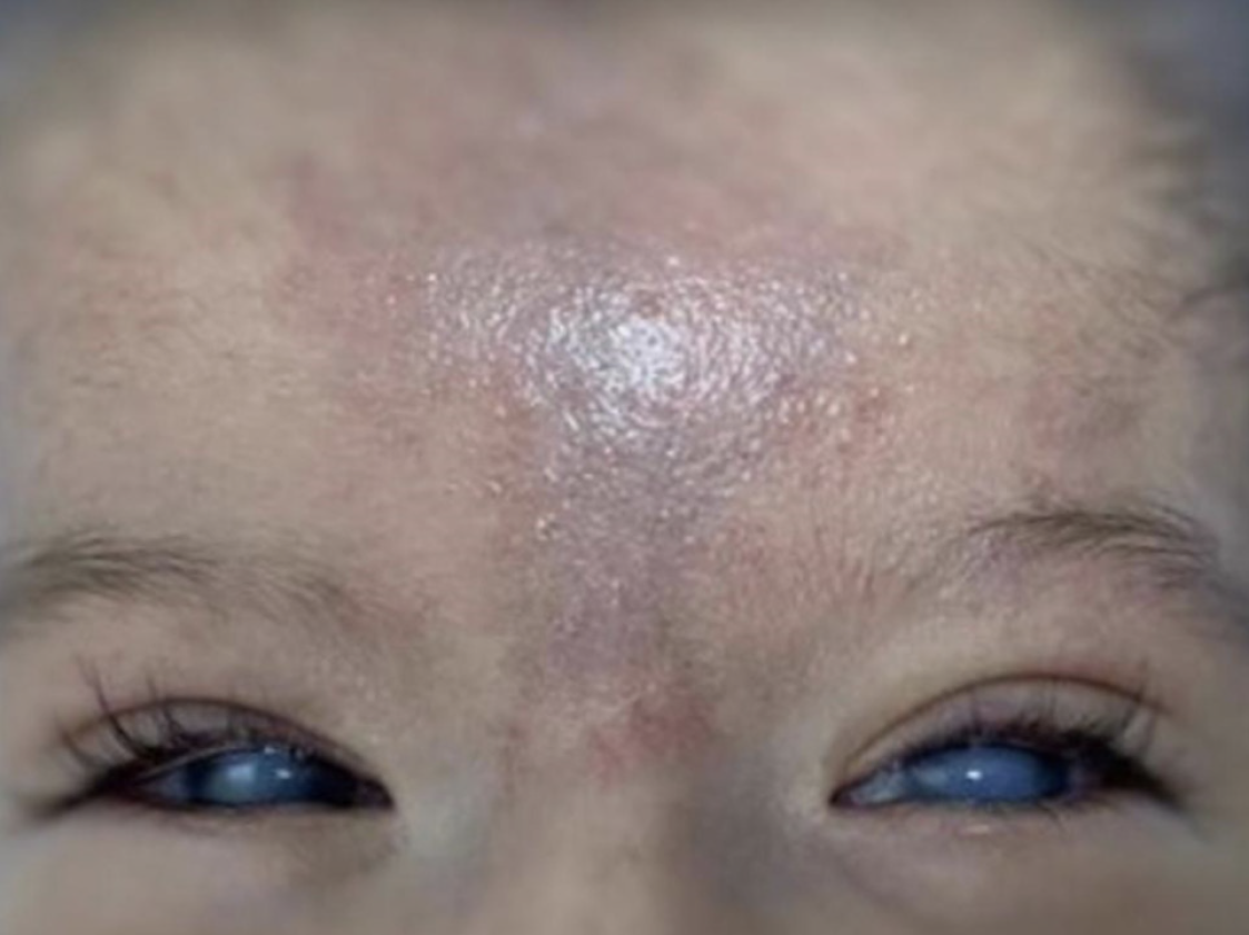
Figure 1 A visual representation of sclerocornea, corneal haze, blue sclera, and hemangioma.
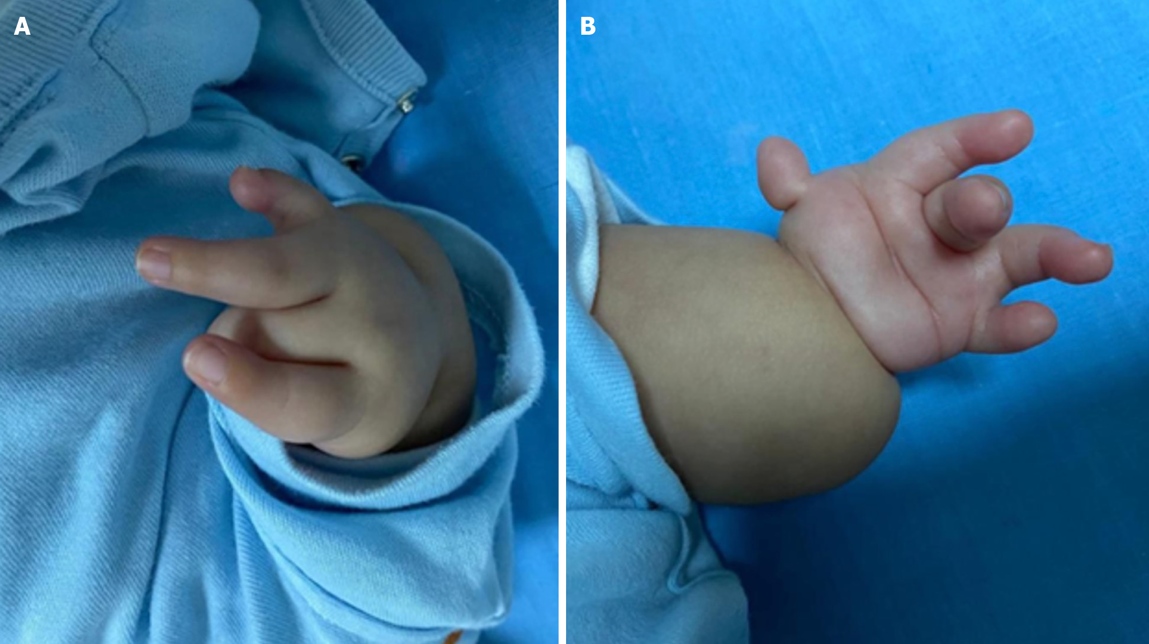
Figure 2 Depiction of bilateral symmetrical phocomelia, oligodactyly, clinodactyly, and thumb anomalies.
A: Right-handed phocomelia, oligodactyly, clinodactyly, and thumb aplasia; B: Left-handed phocomelia and thumb hypoplasia.
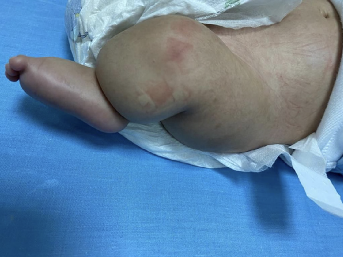
Figure 3 Depiction of lower limb anomalies.
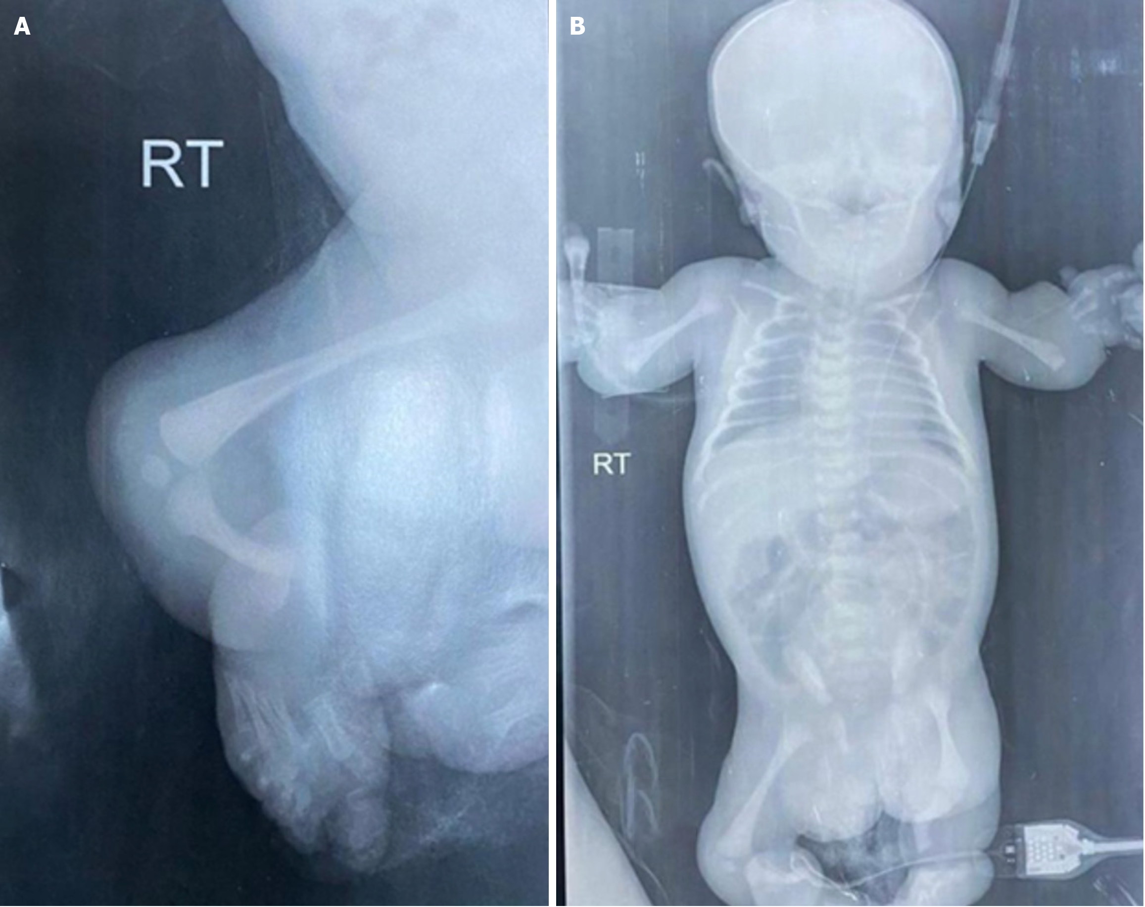
Figure 4 Radiographic findings of bony abnormalities in Roberts syndrome.
A: Right lower limb radiographic findings; B: Whole body bone survey. RT: Right.
- Citation: Sulaiman SA, Kaylani L, Manaseer Q, Mohammed DK. Exploring Roberts syndrome, unique manifestations in a four-month-old infant and genetic findings: A case report. World J Clin Pediatr 2025; 14(4): 110750
- URL: https://www.wjgnet.com/2219-2808/full/v14/i4/110750.htm
- DOI: https://dx.doi.org/10.5409/wjcp.v14.i4.110750


