Copyright
©The Author(s) 2024.
World J Clin Pediatr. Jun 9, 2024; 13(2): 92263
Published online Jun 9, 2024. doi: 10.5409/wjcp.v13.i2.92263
Published online Jun 9, 2024. doi: 10.5409/wjcp.v13.i2.92263
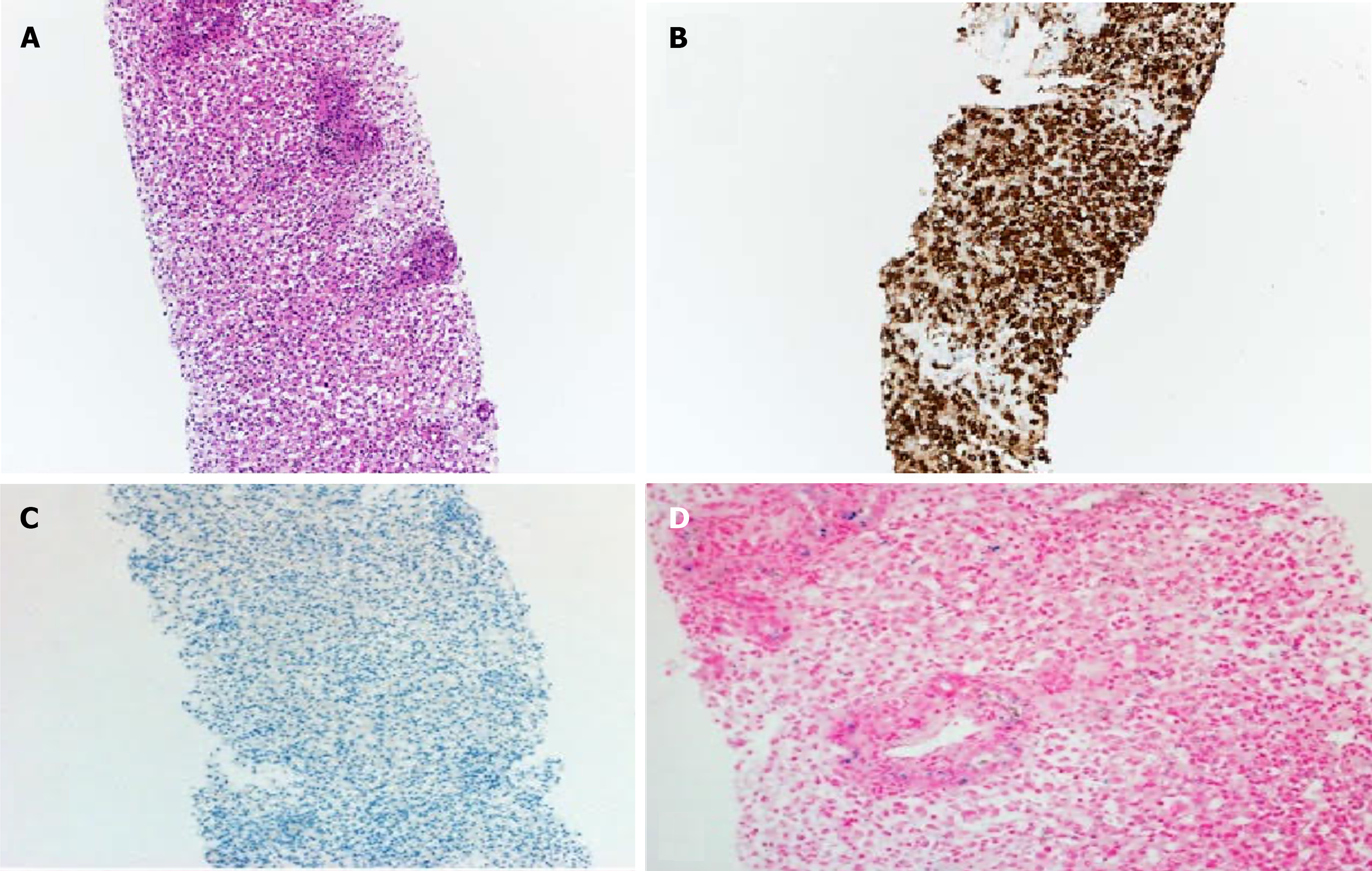
Figure 1 The liver biopsy.
A: The liver biopsy shows prominent histiocytic proliferation between the portal areas (hematoxylin and eosin, × 100); B: The proliferating cells are diffusely positive for CD68 (a histiocytic marker) (CD68 stain, × 100); C: The cells are negative for hepPar confirming the absence of hepatocytes ( × 100); D: Iron stain shows mild iron deposition in the proliferating ductules and histiocytes (iron stain, × 200).
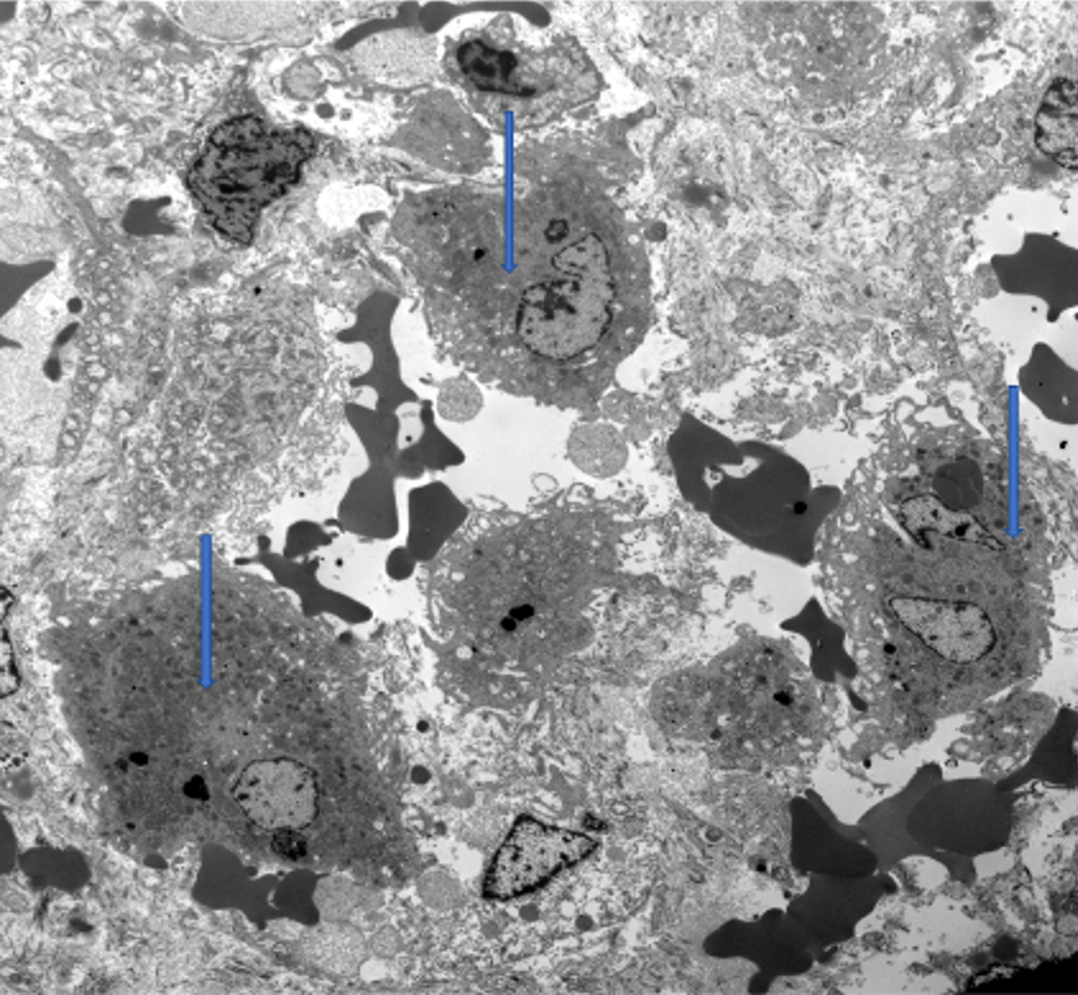
Figure 2
Electron microscopy highlights the presence of abundant histiocytes (highlighted by arrows) with no hepatocytes.
- Citation: Al Atrash E, Azaz A, Said S, Miqdady M. Unique presentation of neonatal liver failure: A case report. World J Clin Pediatr 2024; 13(2): 92263
- URL: https://www.wjgnet.com/2219-2808/full/v13/i2/92263.htm
- DOI: https://dx.doi.org/10.5409/wjcp.v13.i2.92263


