Copyright
©The Author(s) 2024.
World J Clin Pediatr. Mar 9, 2024; 13(1): 89091
Published online Mar 9, 2024. doi: 10.5409/wjcp.v13.i1.89091
Published online Mar 9, 2024. doi: 10.5409/wjcp.v13.i1.89091
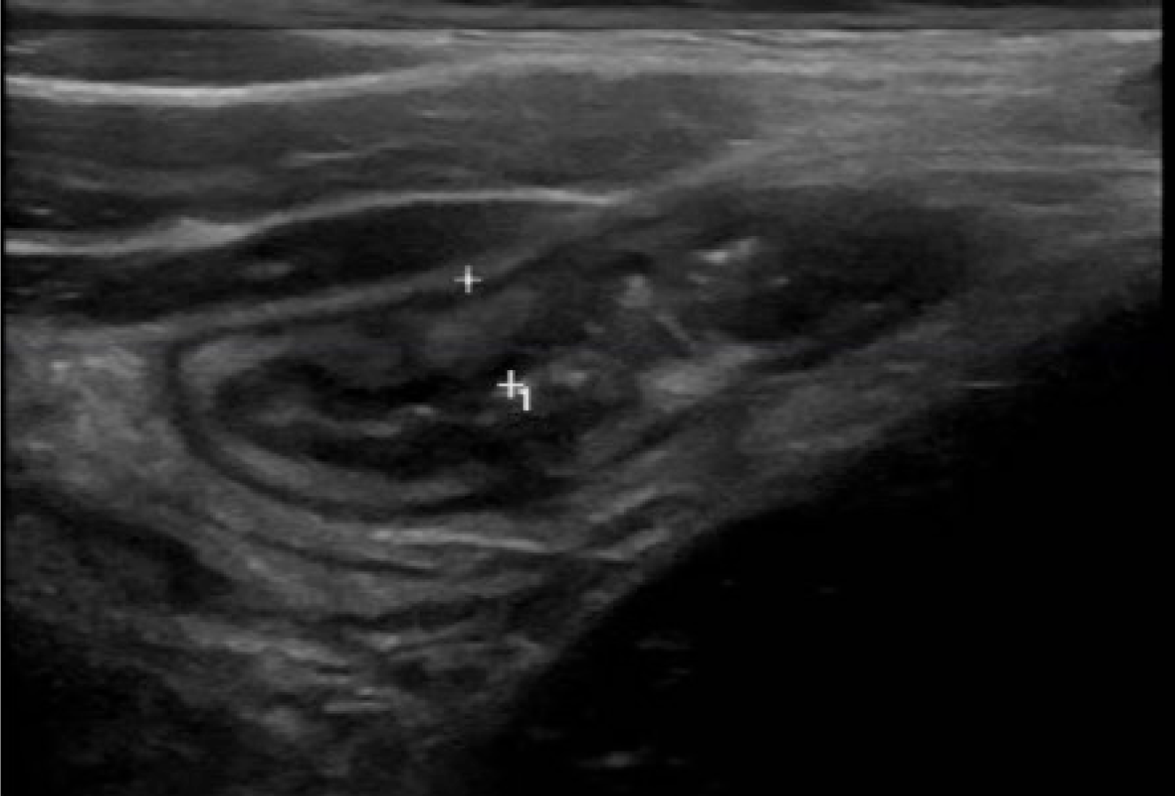
Figure 1
A transabdominal intestinal ultrasound image demonstrating an inflamed, thickened bowel wall of the terminal ileum in a pediatric patient with Crohn’s disease.
Figure 2 Endoscopic images demonstrating a through-the-scope balloon dilatation of an intestinal stricture in a pediatric patient with Crohn’s disease.
A: Predilatation; B: Balloon dilatation; C: Postdilatation.
- Citation: Hudson AS, Wahbeh GT, Zheng HB. Imaging and endoscopic tools in pediatric inflammatory bowel disease: What’s new? World J Clin Pediatr 2024; 13(1): 89091
- URL: https://www.wjgnet.com/2219-2808/full/v13/i1/89091.htm
- DOI: https://dx.doi.org/10.5409/wjcp.v13.i1.89091


