©The Author(s) 2023.
World J Clin Pediatr. Sep 9, 2023; 12(4): 220-229
Published online Sep 9, 2023. doi: 10.5409/wjcp.v12.i4.220
Published online Sep 9, 2023. doi: 10.5409/wjcp.v12.i4.220
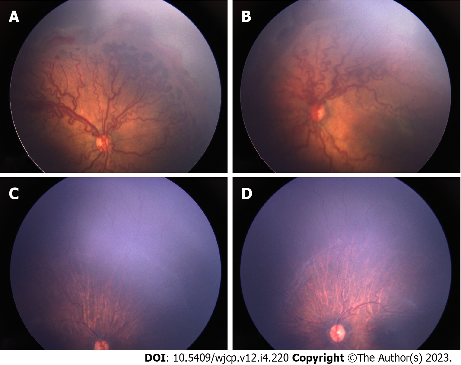
Figure 1 Pre and post intravitreal anti-vascular endothelial growth factor injection fundus pictures showing disease regression.
A: Fundus picture of right eye and; B: Fundus picture of left eye showing severe Zone 1 Aggressive retinopathy of prematurity with extensive fibro vascular proliferation; C: Fundus picture of right eye and; D: Fundus picture of left eye taken 4 months following anti-vascular endothelial growth factor injection showing marked resolution of disease with minimal residual fibrous tissue.
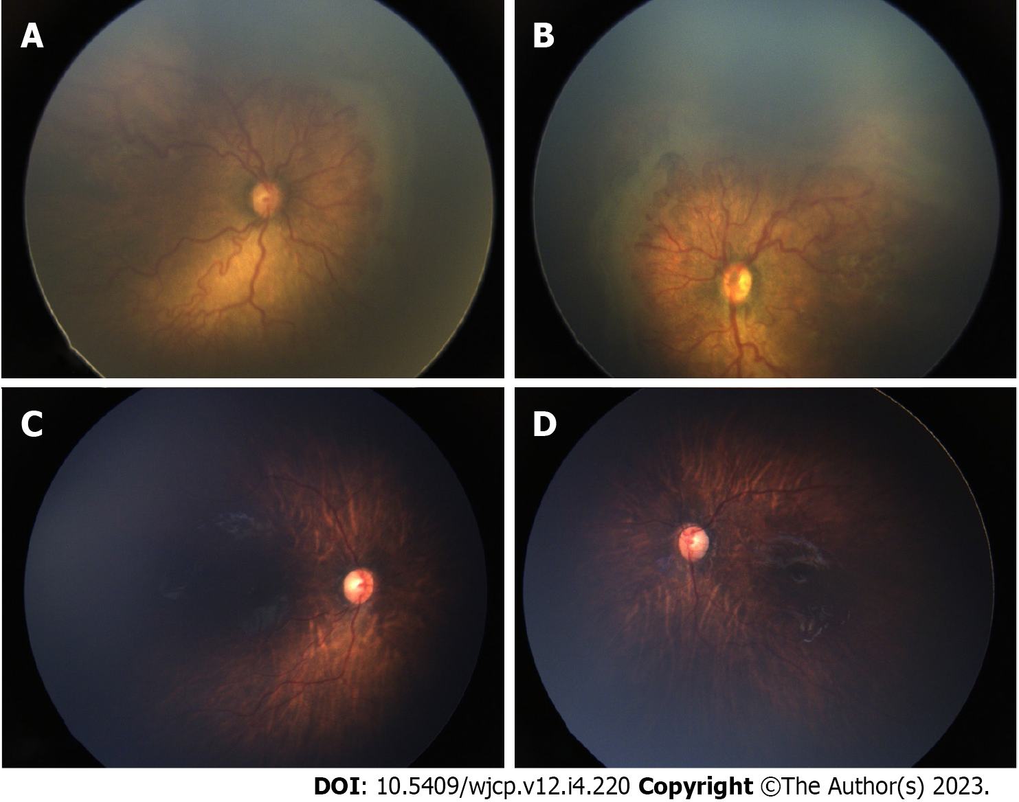
Figure 2 Pre and post intravitreal anti-vascular endothelial growth factor injection fundus pictures showing disease regression.
A: Fundus picture of right eye and; B: Fundus picture of left eye showing severe Zone 1 Aggressive retinopathy of prematurity with extensive fibro vascular proliferation; C: Fundus picture of right eye and; D: Fundus picture of left eye taken 3 months following anti-vascular endothelial growth factor injection showing marked resolution of disease.
- Citation: Maitra P, Prema S, Narendran V, Shah PK. Safety and efficacy of intravitreal anti vascular endothelial growth factor for severe posterior retinopathy of prematurity with flat fibrovascular proliferation. World J Clin Pediatr 2023; 12(4): 220-229
- URL: https://www.wjgnet.com/2219-2808/full/v12/i4/220.htm
- DOI: https://dx.doi.org/10.5409/wjcp.v12.i4.220


