Copyright
©The Author(s) 2022.
World J Clin Pediatr. May 9, 2022; 11(3): 295-306
Published online May 9, 2022. doi: 10.5409/wjcp.v11.i3.295
Published online May 9, 2022. doi: 10.5409/wjcp.v11.i3.295
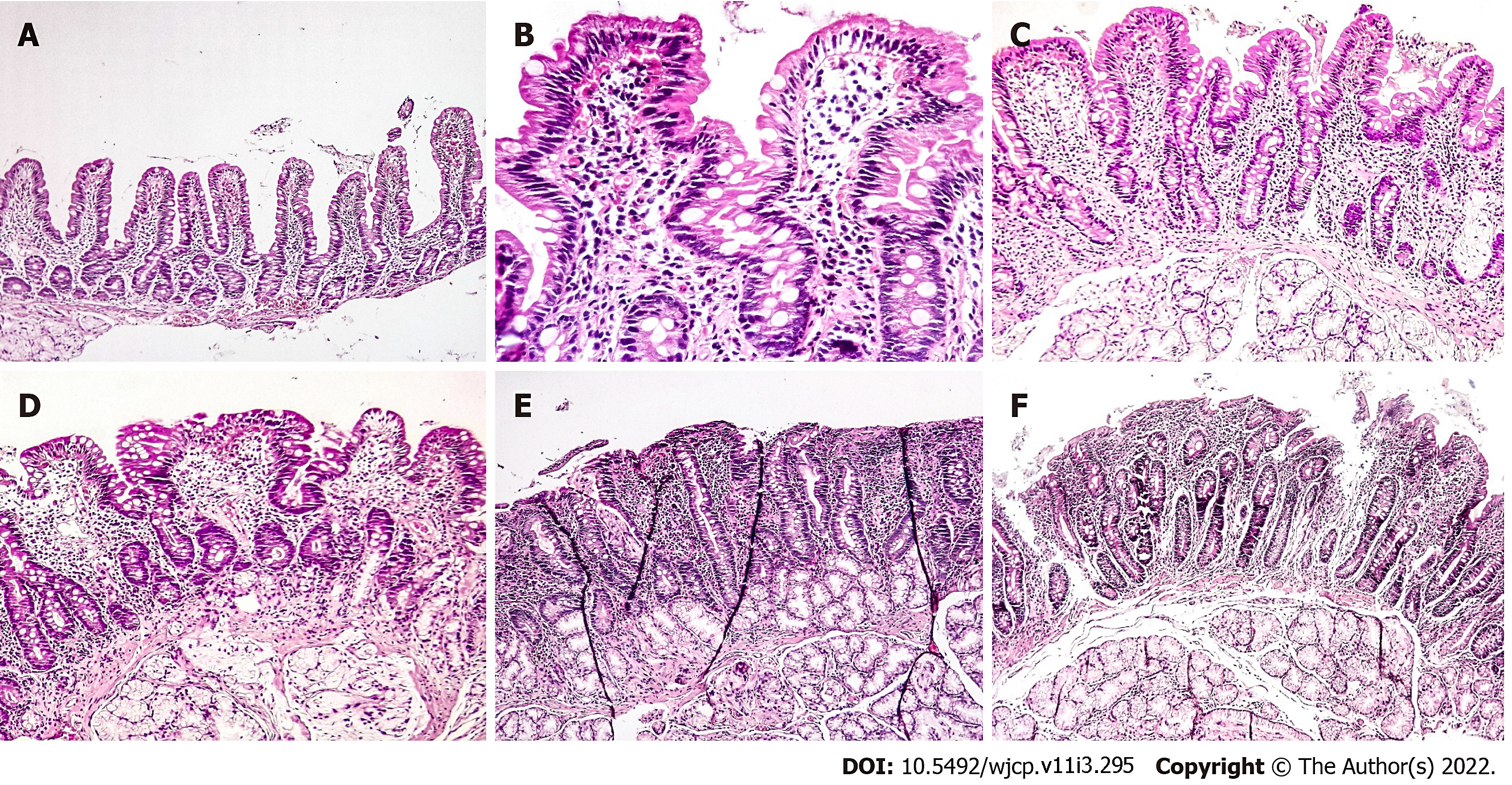
Figure 1 Photomicrographs of duodenal mucosal biopsies.
A: Preserved villi with increased intraepithelial lymphocytes (Marsh-Oberhuber type 1); B and C: Mild villous shortening and crypt hyperplasia with increased intraepithelial lymphocytes (Marsh-Oberhuber type 3a); D: Moderate villous atrophy and crypt hyperplasia with increased intraepithelial lymphocytes (Marsh-Oberhuber type 3b); E and F: Complete villous atrophy (Marsh-Oberhuber type 3c). Hematoxylin and eosin stained sections, original magnification ×40, ×200, ×100, ×100, ×100, ×100 respectively.
- Citation: Mansour HH, Mohsen NA, El-Shabrawi MH, Awad SM, Abd El-Kareem D. Serologic, endoscopic and pathologic findings in pediatric celiac disease: A single center experience in a low/middle income country. World J Clin Pediatr 2022; 11(3): 295-306
- URL: https://www.wjgnet.com/2219-2808/full/v11/i3/295.htm
- DOI: https://dx.doi.org/10.5409/wjcp.v11.i3.295


