Copyright
©2014 Baishideng Publishing Group Co.
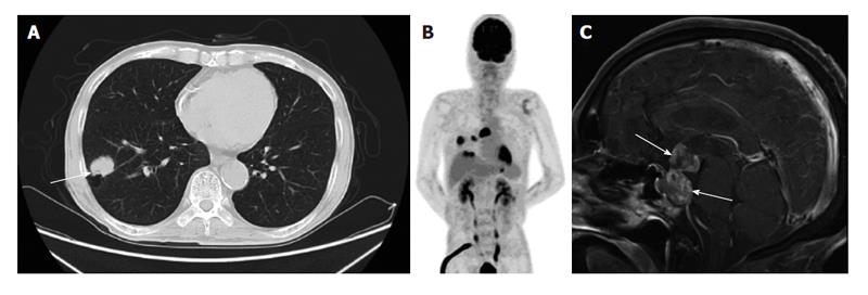
Figure 1 A 74-year-old man with a smoking history presented with anorexia, vomiting, fever and thirst.
A: Chest computed tomography reveals a mass on the right upper lobe (white arrow); B: Fluoro-2-deoxy-D-glucose (FDG) positron emission tomography image shows FDG accumulation at the primary site, hilar and mediastinal lymph nodes, and liver; C: Sagittal view of gadolinium-enhanced brain magnetic resonance imaging shows irregularly enhanced dumbbell-shaped tumor in the intrasellar and suprasellar areas (white arrows).
- Citation: Watanabe T, Kaira K, Mizuide M, Sunaga N, Shibusawa N, Hisada T, Satoh T, Mori M, Yamada M. Solitary pituitary metastasis resulting from pulmonary large cell neuroendocrine carcinoma. World J Respirol 2014; 4(1): 8-10
- URL: https://www.wjgnet.com/2218-6255/full/v4/i1/8.htm
- DOI: https://dx.doi.org/10.5320/wjr.v4.i1.8


