Copyright
©The Author(s) 2016.
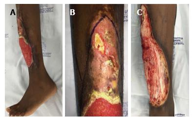
Figure 1 Ulcer on lower leg of the patient with sickle cell disease.
A: Soft tissue defect; B: Detail of bone exposure; C: After extensive bone and soft tissue debridement.
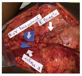
Figure 2 Vascular anatomy of the free flap.
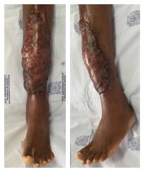
Figure 3 Immediate postoperative result.
The muscle is shown from two angles covered with the split-thickness skin graft.
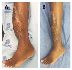
Figure 4 Follow-up postoperative results.
A: After 3 mo of follow-up; B: After 8 mo of follow-up.
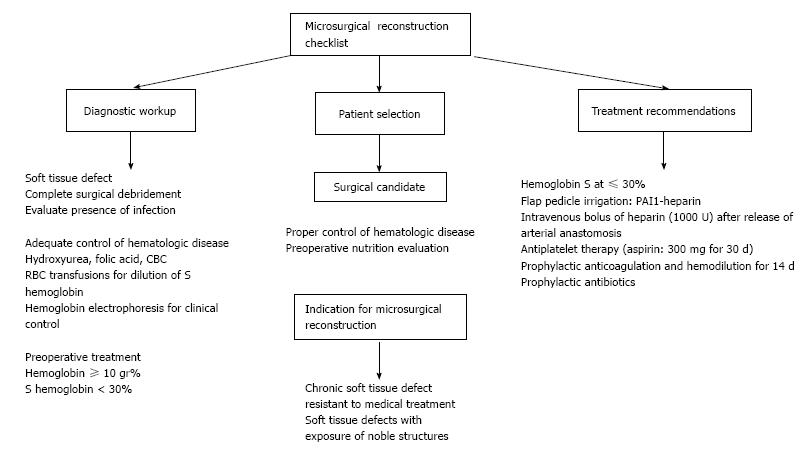
Figure 5 Algorithm treatment strategies for sickle cell disease.
- Citation: Posso C, Cuéllar-Ambrosi F. Successful lower leg microsurgical reconstruction in homozygous sickle cell disease: Case report. World J Hematol 2016; 5(4): 94-98
- URL: https://www.wjgnet.com/2218-6204/full/v5/i4/94.htm
- DOI: https://dx.doi.org/10.5315/wjh.v5.i4.94


