Copyright
©2014 Baishideng Publishing Group Inc.
World J Hematol. Aug 6, 2014; 3(3): 115-117
Published online Aug 6, 2014. doi: 10.5315/wjh.v3.i3.115
Published online Aug 6, 2014. doi: 10.5315/wjh.v3.i3.115
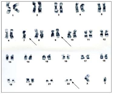
Figure 1 Karyotyping of the patient with mixed phenotype acute leukemia showing (arrows) 45, XY, monosomy 7, t(9;22) (q34;q11.
2)[18]/46,XY[2].
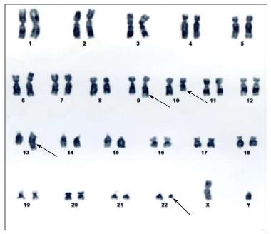
Figure 2 Karyotyping of the patient with chronic myeloid leukemia showing (arrows) 46 XY, t(9;22)(q34;11.
2), t(10;13)(q23;q34)[20].
- Citation: Sharma SK, Handoo A, Choudhary D, Gupta N. Unusual cytogenetic abnormalities associated with Philadelphia chromosome. World J Hematol 2014; 3(3): 115-117
- URL: https://www.wjgnet.com/2218-6204/full/v3/i3/115.htm
- DOI: https://dx.doi.org/10.5315/wjh.v3.i3.115


