Copyright
©The Author(s) 2015.
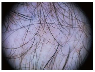
Figure 1 Male androgenetic alopecia.
Hair shaft thickness heterogeneity and predominance of follicular units with only one hair (Dermlite photo®).
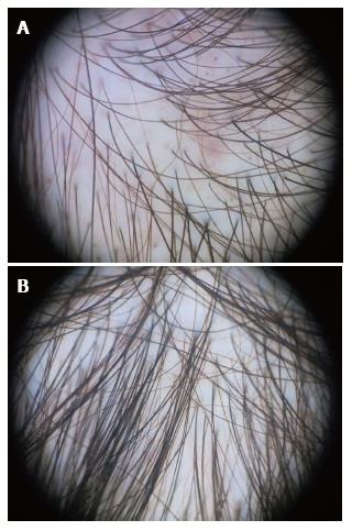
Figure 2 Female androgenetic alopecia.
Significant (> 20%) diversity of hair shaft diameter. Note also yellow dots. Higher hair density and less variability in the occipital area (B) compared to the frontal area (A) (Dermlite photo®).
Figure 3 Alopecia areata.
Exclamation mark signs and black dots (Dermlite photo®).
Figure 4 Telogen effluvium.
Short regrowing hair (videodermoscopy, 70 x magnification). Photo courtesy of Prof. Lidia Rudnicka.
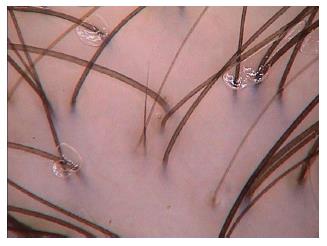
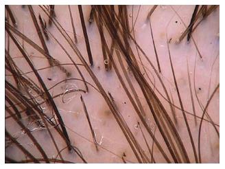
Figure 5 Trichotillomania.
Hairs broken at differents lengths, with trichoptilosis (split end) and black dots (videodermoscopy, 70 × magnification). Photo courtesy of Prof. Lidia Rudnicka.
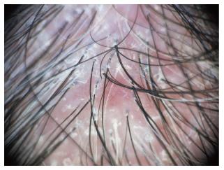
Figure 6 Lichen planopilaris.
Intense perifollicular scaling and tubular structures (Dermlite Photo®).
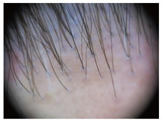
Figure 7 Frontal fibrosing alopecia.
Absence of follicular openings, predominance of follicular units with only 1 hair, mild perifollicular scaling and perifollicular erythema (Dermlite photo®).
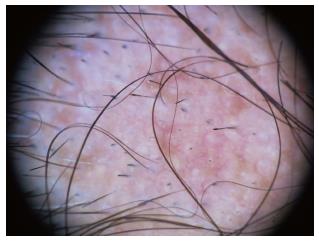
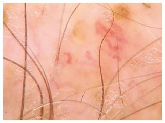
Figure 8 Discoid lupus erythematosus.
Characteristic large yellow dots and arborizing vessels (videodermoscopy, 70 x magnification). Photo courtesy of Prof. Lidia Rudnicka.
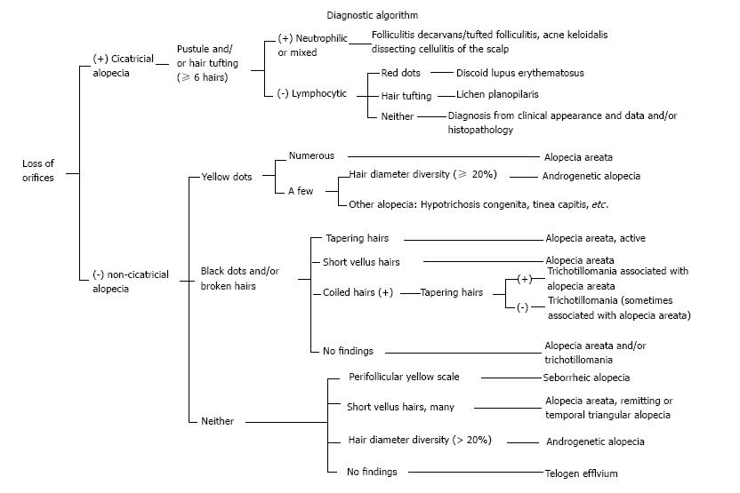
Figure 9 Diagnostic algorithm for trichoscopic findings of hair loss diseases.
From Inui S. Expert Rev Dermatol 2012: 7.
- Citation: Romero JAM, Grimalt R. Trichoscopy: Essentials for the dermatologist. World J Dermatol 2015; 4(2): 63-68
- URL: https://www.wjgnet.com/2218-6190/full/v4/i2/63.htm
- DOI: https://dx.doi.org/10.5314/wjd.v4.i2.63


