Copyright
©2014 Baishideng Publishing Group Co.
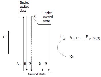
Figure 1 Jablonski diagram showing the various modes of excitation and relaxation in a chromophore.
A: Excitation; B: Fluorescence; C: Intersystem crossing; D: Phosphorescence; E: Non-radiative transfer of energy to singlet oxygen; F: Substrate oxidation by singlet oxygen; G: Internal conversion (adapted from Macdonald et al[1]).
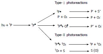
Figure 2 Cellular damage mechanism.
Type-I and Type-II photoreactions, where 1P is a photosensitizer in a singlet ground state, 3P1 is a photosensitizer in a triplet excited state, S is a substrate molecule, Pÿ is reduced photosensitizer molecule, S is an oxidized substrate molecule, O2 is molecular oxygen (triplet ground state), Oÿ2 is the superoxide anion, O2 is the superoxide radical, P is the oxidized photosensitizer, 3O2 is triplet ground-state oxygen, 1O2 is oxygen in a singlet excited state, and S (O) is an oxygen adduct of a substrate (adapted from Macdonald et al[1]).
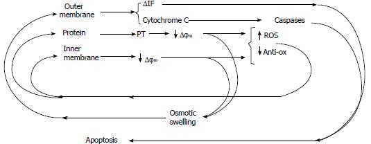
Figure 3 Summary of the pathways and feedback loops involved in mitochondrially controlled apoptosis.
Star bursts represent possible points at which photodynamic therapy initiates mitochondrially controlled apoptosis (adapted from Macdonald et al[1]).
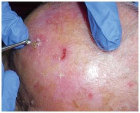
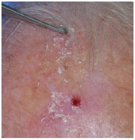
Figure 4 Lesion is treated with a curette to selectively remove the corneal layer.
Figure 5 After curettage no bleeding might be present.
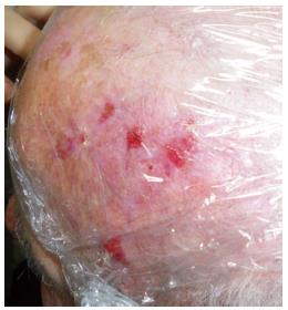
Figure 6 Cream is applied on the lesion and an occlusive dressing is then performed.
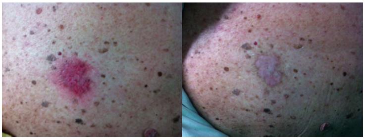
Figure 7 Patient affected by multiple actinic keratosis of the scalp treated with photodynamic therapy with aminolevulinic acid; result after 3 wk from the 1st application.
- Citation: Negosanti L, Pinto V, Sgarzani R, Negosanti F, Zannetti G, Cipriani R. Photodynamic therapy with topical aminolevulinic acid. World J Dermatol 2014; 3(2): 6-14
- URL: https://www.wjgnet.com/2218-6190/full/v3/i2/6.htm
- DOI: https://dx.doi.org/10.5314/wjd.v3.i2.6


