Published online Oct 18, 2016. doi: 10.5312/wjo.v7.i10.695
Peer-review started: February 28, 2016
First decision: June 12, 2016
Revised: June 25, 2016
Accepted: August 15, 2016
Article in press: August 16, 2016
Published online: October 18, 2016
Processing time: 228 Days and 11.6 Hours
Although laminar screw fixation is often used at the C2 and C7 levels, only few previous case reports have presented the use of laminar screws at the C3-C6 levels. Here, we report a novel fixation method involving the use of practical laminar screws in the subaxial spine. We used laminar screws in the subaxial cervical spine in two cases to prevent vertebral artery injury and in one case to minimize exposure of the lamina. This laminar screw technique was successful in all three cases with adequate spinal rigidity, which was achieved without complications. The use of laminar screws in the subaxial cervical spine is a useful option for posterior fusion of the cervical spine.
Core tip: Laminar screw fixation is often used at the C2 and C7 levels, however, only few previous case reports have presented the use of laminar screws at the C3-C6 levels. In this article, the authors describe a novel fixation method involving the use of laminar screws in the subaxial spine with adequate spinal rigidity, which was achieved without complications.
- Citation: Tanabe H, Aota Y, Saito T. Laminar screw fixation in the subaxial cervical spine: A report on three cases. World J Orthop 2016; 7(10): 695-699
- URL: https://www.wjgnet.com/2218-5836/full/v7/i10/695.htm
- DOI: https://dx.doi.org/10.5312/wjo.v7.i10.695
Posterior surgical stabilization for instability of the cervical spine can be achieved using various techniques. Traditionally, posterior wiring methods have been used[1,2]. Recently, methods involving the use of screws and rods, including pedicle[3], lateral mass[4], and transarticular screws[5], have been widely applied because of the resulting biomechanically rigid stabilization. However, screw insertion is associated with a high risk of injury to the vertebral artery because of differences in the position of the foramen transversarium of the cervical spine, the size of the pedicle of the vertebral arch, and the course of the vertebral arteries among patients. Furthermore, the malposition of these screws can cause serious complications, including massive hemorrhage, neurological deficits, and possible death[6-8]. Here, we describe a novel fixation method involving the use of laminar screws in the subaxial spine for posterior fusion of the cervical spine.
A 32-year-old man with neck pain had an asymptomatic dumbbell-shaped schwannoma with highly destructive changes observed on radiography (Figure 1). Preoperative magnetic resonance imaging (MRI) showed narrowing of the right vertebral artery due to pressure from the tumor. Subsequently, tumor resection was performed using a posterior approach with total facetectomies of the right C2-3 through C6-7 levels. Reconstruction was achieved using C2-C6 laminar screw fixation (3.5 mm in diameter); the screws were placed in the left lamina to avoid injuring the vertebral artery on the dominant side. Minimal canal invasion by the screw at the C6 level without any resulting neurological deficit was noted. The remaining screws were appropriately placed (Figure 1).
The patient’s post-surgical course was uneventful. At 5 years postoperatively, his neck pain disappeared and solid bone fusion was achieved with no instrumentation failure (Figure 1).
A 15-year-old girl had a Ewing’s sarcoma that involved the right C7 and C8 nerve roots. She began to feel right arm numbness at 12 years of age, and she was diagnosed with a cervical tumor on MRI. In the same year, hemilaminectomy of the right C6 and C7 levels, medial facetectomies of the right C6-7 and Th1 levels, and tumor resection were performed. Pathology of the resected tissues confirmed Ewing’s sarcoma. Postoperatively, chemotherapy and radiotherapy were repeated.
Two years postoperatively, recurrence of the tumor was detected on MRI. However, no neurological deficits were observed. The tumor was dumbbell-shaped and was located in the intra- and extra-foraminal areas of the C6-7 and C7-Th1 levels (Figure 2). Revision surgery was performed after embolization of the right vertebral artery. Using the posterior approach, laminar screws were inserted at the left C6-Th1 levels to avert the risk of injuring the dominant vertebral artery (Figure 2). Total facetectomies of the right C6-7 and Th1 levels and tumor resection of the right C7 and C8 nerve roots were performed. Additional tumor resection using an anterolateral approach was performed according to the method described by Hodgson[9].
From the day after the operation, she could walk with a soft neck brace, and she exhibited no neurological deficits other than a dropped finger. Two years after the second operation, recurrence of the tumor was detected, and it was treated with chemotherapy. The laminar screws continued to remain rigidly fixed.
A 61-year-old woman experienced neck pain and loss of fine motor control of her hand, and 1 year later, she was referred to our hospital for treatment. She had a history of tuberculosis at 8 years of age, which was conservatively treated for 4 years. She had cervical myelopathy due to an unstable C2-3 joint with marked kyphosis at the C3-6 levels (Figure 3). Because the laminae were very thick, a laminar screw system was selected for cervical fixation. After decompression with partial laminectomy at the C2-3 level, posterior cervical spinal arthrodesis was performed using occipital plates, C2 pedicles, and laminar screws at the C3-C6 levels (3.5 mm screws) with less exposure outside of the lateral mass.
Postoperatively, the patient was mildly immobilized with a soft neck brace, and her post-surgical course was uneventful. At 4 years postoperatively, her neck pain greatly improved and excellent postoperative stability was noted (Figure 3).
Here, we reported a novel fixation method involving the use of practical laminar screws in the subaxial spine. Posterior cervical fixation has often been used for stabilizing the cervical spine, correcting deformities, and easing the symptoms of degenerative diseases. Recently, fixation methods involving screws and rods have become standard, because greater advantages with regard to stabilization and fusion rates of the posterior cervical spine were noted with screw fixation methods than with posterior wiring methods[3,10-12].
Although cervical pedicle screws are the most biomechanically stable screws[13], their use requires an advanced surgical technique and they are associated with the risk of neurovascular complications[14]. To prevent injuring the vertebral artery, pedicle screws should not be used in cases involving the dominant side (e.g., cases 1 and 2). Although the risk of vertebral arterial injury is lower with lateral mass and transarticular screws than with pedicle screws, lateral mass and transarticular screws are difficult to use after facetectomy. Moreover, unilateral use of these screws does not provide sufficient rigidity.
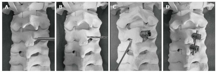
A laminar screw method, which was first reported by Wright[15] in 2004, uses two screws that are inserted crosswise into the lamina of the axis to prevent vertebral artery injuries and safely perform fixation. Although some authors have reported the use of this method in the subaxial lamina of C7[16-18], only two previous studies are present on the use of laminar screws elsewhere in the cervical spine[19,20]. These previous studies did not report sufficient rigidity because thin 1.6 or 2.0 mm mini-screws[19] were used with a mini-plate for fixing the open lamina at the C3-6 levels or an auxiliary laminar screw[20] was inserted at the tip of the C3 lamina accompanied with rigid bilateral C1 lateral mass screws. To our knowledge, our case report is the first to describe the use of practical laminar screws in the subaxial cervical spine. Surgical procedures of subaxial laminar screwing are illustrated in Figure 4.
This new laminar screw technique has four advantages. First, it precludes the risk to the vertebral artery, because the path of the screw is present only in the posterior elements. Second, it is less invasive because of the limited lateral cervical exposure. Third, biomechanical stability with laminar screws is similar to that with pedicle screws, as determined previously by the measurement of pullout forces[16]. Additionally, surgeons can obtain good rigidity by penetrating the facet joints as shown in cases 1 and 2, and using laminar screws with lateral mass screws as shown in case 3. Fourth, intraoperative navigation systems are not needed, because the screws can be inserted into the lamina under direct vision.
Nakanishi et al[21] have pointed out that the laminar screw technique poses a risk to the ventrally located spinal canal that is not easily observed. However, we believe that this risk may be reduced by using a Penfield dissector to detect canal violation following the removal of the flavum ligaments between the laminae. Another disadvantage is that laminar screws with a diameter of 3.5 mm cannot be used at the C3-7 levels in all patients. Cardoso et al[16] measured the diameter of the vertebral arch using a computed tomography (CT) navigation system and reported that the insertion of screws with a diameter of 3 mm was only possible in 2%-39% of male and 0%-26% of female patients at the C3-7 cervical spine[21]. However, other authors reported significantly greater C7 laminar thickness with caliper measurements than CT measurements. Therefore, the underestimation of laminar thickness using CT may provide a margin of safety when placing screws into laminae that measure close to 3.5 mm on CT.
Preoperative measurements of the laminar diameter and evaluation of the vertebral arteries by using CT and magnetic resonance angiography are important. We believe that the use of laminar screws in the subaxial cervical spine is a viable salvage option for cases that have failed pedicle screw fixation. The accumulation of further data from the treatment of additional cases is required to clarify the indications for and limitations of using the laminar screw technique at the C3-6 levels.
In conclusion, the findings in our cases suggest that the use of the laminar screw technique in the subaxial cervical spine is feasible, as it provides sufficient spinal rigidity. Laminar screws are considered useful for avoiding arterial injuries, and the laminar screw technique is a viable salvage technique.
Three cases are discussed with reports of posterior surgical stabilization for instability of the cervical spine.
Tumor was diagnosed for the first two cases. Cervical myelopathy was the clinical diagnosis for the third case.
Magnetic resonance imaging was done to for all cases to establish the cause of the problem.
Pathology analysis was conducted for the 15-year-old female case for confirming the diagnosis of Ewing’s sarcoma.
For the first case, a 32-year-old male, tumor resection was performed and reconstruction was done using screw fixation. For the 15-year-old female case, tumor resection was performed and postoperatively chemotherapy and radiotherapy was performed. For the 61-year-old female case, cervical fixation was done.
A novel fixation method has been reported with use of practical laminar screws in the subaxial cervical spine. This method reduces the risk to the vertebral artery, is less invasive, provides biomechanical stability and the screws can be inserted with direct vision. Additional cases are needed to clarify the indications and limitations for using laminar screw technique.
This is a good case report with medium term result in one patient and will add to the body of literature for posterior cervical fusion.
Manuscript source: Invited manuscript
Specialty type: Orthopedics
Country of origin: Japan
Peer-review report classification
Grade A (Excellent): 0
Grade B (Very good): B, B
Grade C (Good): 0
Grade D (Fair): 0
Grade E (Poor): 0
P- Reviewer: Guerado E, Sarda P S- Editor: Qiu S L- Editor: A E- Editor: Lu YJ
| 1. | Brooks AL, Jenkins EB. Atlanto-axial arthrodesis by the wedge compression method. J Bone Joint Surg Am. 1978;60:279-284. [PubMed] |
| 2. | Dickman CA, Sonntag VK, Papadopoulos SM, Hadley MN. The interspinous method of posterior atlantoaxial arthrodesis. J Neurosurg. 1991;74:190-198. [RCA] [PubMed] [DOI] [Full Text] [Cited by in Crossref: 272] [Cited by in RCA: 236] [Article Influence: 6.7] [Reference Citation Analysis (0)] |
| 3. | Abumi K, Itoh H, Taneichi H, Kaneda K. Transpedicular screw fixation for traumatic lesions of the middle and lower cervical spine: description of the techniques and preliminary report. J Spinal Disord. 1994;7:19-28. [RCA] [PubMed] [DOI] [Full Text] [Cited by in Crossref: 307] [Cited by in RCA: 309] [Article Influence: 9.7] [Reference Citation Analysis (0)] |
| 4. | Taniguchi M, Maruo S. Posterior Lower Cervical Spine Arthrodesis with Lateral Mass Fixation. Spine & Spinal Cord. 1998;11:217-224. |
| 5. | Jeanneret B, Magerl F. Primary posterior fusion C1/2 in odontoid fractures: indications, technique, and results of transarticular screw fixation. J Spinal Disord. 1992;5:464-475. [RCA] [PubMed] [DOI] [Full Text] [Cited by in Crossref: 359] [Cited by in RCA: 314] [Article Influence: 9.2] [Reference Citation Analysis (0)] |
| 6. | Coric D, Branch CL, Wilson JA, Robinson JC. Arteriovenous fistula as a complication of C1-2 transarticular screw fixation. Case report and review of the literature. J Neurosurg. 1996;85:340-343. [RCA] [PubMed] [DOI] [Full Text] [Cited by in Crossref: 104] [Cited by in RCA: 91] [Article Influence: 3.0] [Reference Citation Analysis (0)] |
| 7. | Madawi AA, Casey AT, Solanki GA, Tuite G, Veres R, Crockard HA. Radiological and anatomical evaluation of the atlantoaxial transarticular screw fixation technique. J Neurosurg. 1997;86:961-968. [RCA] [PubMed] [DOI] [Full Text] [Cited by in Crossref: 396] [Cited by in RCA: 350] [Article Influence: 12.1] [Reference Citation Analysis (0)] |
| 8. | Wright NM, Lauryssen C. Vertebral artery injury in C1-2 transarticular screw fixation: results of a survey of the AANS/CNS section on disorders of the spine and peripheral nerves. American Association of Neurological Surgeons/Congress of Neurological Surgeons. J Neurosurg. 1998;88:634-640. [RCA] [PubMed] [DOI] [Full Text] [Cited by in Crossref: 6] [Cited by in RCA: 8] [Article Influence: 0.3] [Reference Citation Analysis (0)] |
| 9. | Hodgson AR. An approach to the cervical spine (C-3 TO C-7). Clin Orthop Relat Res. 1965;39:129-134. [RCA] [PubMed] [DOI] [Full Text] [Cited by in Crossref: 2] [Cited by in RCA: 3] [Article Influence: 0.1] [Reference Citation Analysis (0)] |
| 10. | An HS, Gordin R, Renner K. Anatomic considerations for plate-screw fixation of the cervical spine. Spine (Phila Pa 1976). 1991;16:S548-S551. [RCA] [PubMed] [DOI] [Full Text] [Cited by in Crossref: 256] [Cited by in RCA: 214] [Article Influence: 6.1] [Reference Citation Analysis (0)] |
| 11. | Harris BM, Hilibrand AS, Nien YH, Nachwalter R, Vaccaro A, Albert TJ, Siegler S. A comparison of three screw types for unicortical fixation in the lateral mass of the cervical spine. Spine (Phila Pa 1976). 2001;26:2427-2431. [RCA] [PubMed] [DOI] [Full Text] [Cited by in Crossref: 19] [Cited by in RCA: 19] [Article Influence: 0.8] [Reference Citation Analysis (0)] |
| 12. | Ludwig SC, Kramer DL, Vaccaro AR, Albert TJ. Transpedicle screw fixation of the cervical spine. Clin Orthop Relat Res. 1999;77-88. [RCA] [PubMed] [DOI] [Full Text] [Cited by in Crossref: 99] [Cited by in RCA: 103] [Article Influence: 3.8] [Reference Citation Analysis (0)] |
| 13. | Jones EL, Heller JG, Silcox DH, Hutton WC. Cervical pedicle screws versus lateral mass screws. Anatomic feasibility and biomechanical comparison. Spine (Phila Pa 1976). 1997;22:977-982. [RCA] [PubMed] [DOI] [Full Text] [Cited by in Crossref: 341] [Cited by in RCA: 352] [Article Influence: 12.1] [Reference Citation Analysis (0)] |
| 14. | Taneichi H. Placement technique of cervical screws and prevention of its complications. Spine & Spinal Cord. 2005;18:1043-1052. |
| 15. | Wright NM. Posterior C2 fixation using bilateral, crossing C2 laminar screws: case series and technical note. J Spinal Disord Tech. 2004;17:158-162. [RCA] [PubMed] [DOI] [Full Text] [Cited by in Crossref: 344] [Cited by in RCA: 315] [Article Influence: 14.3] [Reference Citation Analysis (0)] |
| 16. | Cardoso MJ, Dmitriev AE, Helgeson MD, Stephens F, Campbell V, Lehman RA, Cooper P, Rosner MK. Using lamina screws as a salvage technique at C-7: computed tomography and biomechanical analysis using cadaveric vertebrae. Laboratory investigation. J Neurosurg Spine. 2009;11:28-33. [PubMed] [DOI] [Full Text] |
| 17. | Hong JT, Yi JS, Kim JT, Ji C, Ryu KS, Park CK. Clinical and radiologic outcome of laminar screw at C2 and C7 for posterior instrumentation--review of 25 cases and comparison of C2 and C7 intralaminar screw fixation. World Neurosurg. 2010;73:112-118; discussion e15. [RCA] [PubMed] [DOI] [Full Text] [Cited by in Crossref: 26] [Cited by in RCA: 34] [Article Influence: 2.0] [Reference Citation Analysis (0)] |
| 18. | Şenoğlu M, Özkan F, Çelik M. Placement of C-7 intralaminar screws: a quantitative anatomical and morphometric evaluation. J Neurosurg Spine. 2012;16:509-512. [RCA] [PubMed] [DOI] [Full Text] [Cited by in Crossref: 2] [Cited by in RCA: 4] [Article Influence: 0.3] [Reference Citation Analysis (0)] |
| 19. | Hong JT, Sung JH, Son BC, Lee SW, Park CK. Significance of laminar screw fixation in the subaxial cervical spine. Spine (Phila Pa 1976). 2008;33:1739-1743. [RCA] [PubMed] [DOI] [Full Text] [Cited by in Crossref: 52] [Cited by in RCA: 58] [Article Influence: 3.2] [Reference Citation Analysis (0)] |
| 20. | Jea A, Johnson KK, Whitehead WE, Luerssen TG. Translaminar screw fixation in the subaxial pediatric cervical spine. J Neurosurg Pediatr. 2008;2:386-390. [RCA] [PubMed] [DOI] [Full Text] [Cited by in Crossref: 20] [Cited by in RCA: 23] [Article Influence: 1.3] [Reference Citation Analysis (0)] |
| 21. | Nakanishi K, Tanaka M, Sugimoto Y, Misawa H, Takigawa T, Fujiwara K, Nishida K, Ozaki T. Application of laminar screws to posterior fusion of cervical spine: measurement of the cervical vertebral arch diameter with a navigation system. Spine (Phila Pa 1976). 2008;33:620-623. [RCA] [PubMed] [DOI] [Full Text] [Cited by in Crossref: 19] [Cited by in RCA: 17] [Article Influence: 0.9] [Reference Citation Analysis (0)] |


