Copyright
©The Author(s) 2017.
World J Orthop. Sep 18, 2017; 8(9): 741-746
Published online Sep 18, 2017. doi: 10.5312/wjo.v8.i9.741
Published online Sep 18, 2017. doi: 10.5312/wjo.v8.i9.741
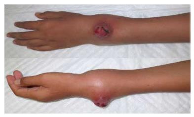
Figure 1 Clinical photograph of the patient.
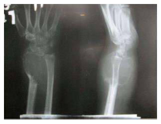
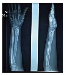
Figure 2 Preoperative X-ray of the patient.
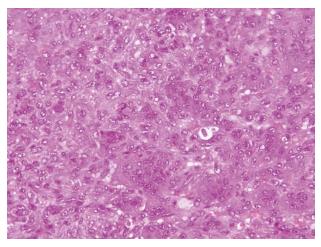
Figure 3 Histopathology examination of the tumor.
Multinucleated giant cells were observed from the examination with background of mononuclear cells.
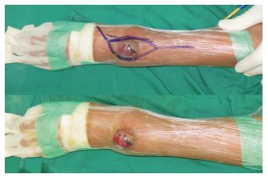
Figure 4 Intra-operative photograph of wide excision with posterior approach.
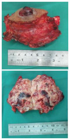
Figure 5 Excision of tumor.
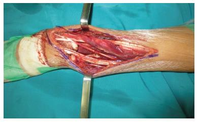
Figure 6 Intra-operative photograph showing large defect due to wide excision.
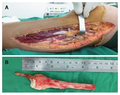
Figure 7 Proximal fibula harvested via lateral approach.
The asterisk showing the intact of peroneal nerve (A) including head of fibula and bicep tendon (B).
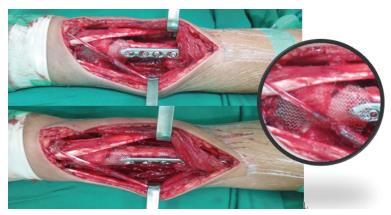
Figure 8 Fibular graft implantation fixed with 3.
5 locking plate and covered with hernia mesh on radioulnar joint and volar aspect of radius. The inset showing the hernia mesh.
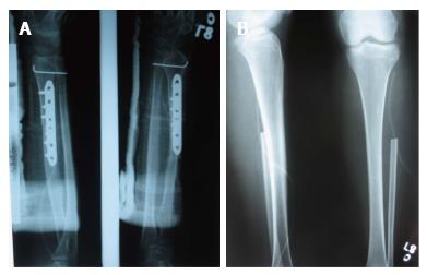
Figure 9 The post operative X-ray after wide excision of giant cell tumor of distal radius (A) and defect in fibula (B).
Figure 10 The follow-up X-ray 5 years after the wide excision of giant cell tumor distal radius and fibular autograft and after removal of plate and screw.
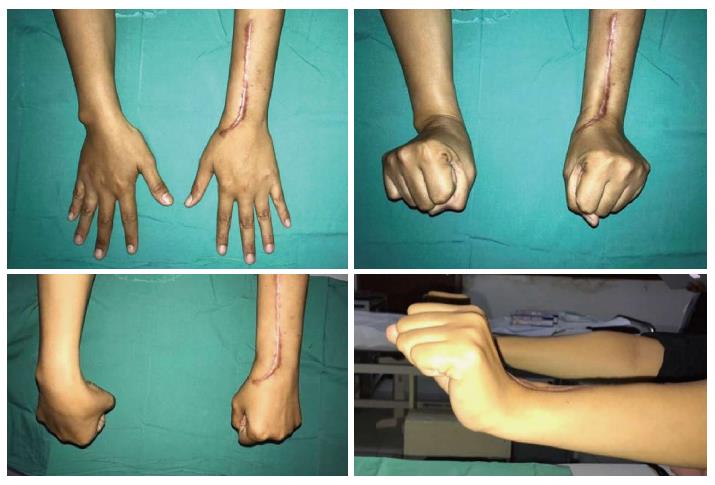
Figure 11 The clinical picture of functional outcome after five years of follow-up.
The pictures showing a range of motion of 75%-99% of normal side, and grip strength of 100% compared with normal hand.
- Citation: Wiratnaya IGE, Budiartha IGBAM, Setiawan IGNY, Sindhughosa DA, Kawiyana IKS, Astawa P. Hernia mesh prevent dislocation after wide excision and reconstruction of giant cell tumor distal radius. World J Orthop 2017; 8(9): 741-746
- URL: https://www.wjgnet.com/2218-5836/full/v8/i9/741.htm
- DOI: https://dx.doi.org/10.5312/wjo.v8.i9.741


