Copyright
©The Author(s) 2017.
World J Orthop. Mar 18, 2017; 8(3): 242-255
Published online Mar 18, 2017. doi: 10.5312/wjo.v8.i3.242
Published online Mar 18, 2017. doi: 10.5312/wjo.v8.i3.242
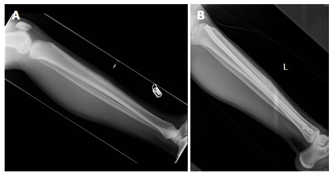
Figure 1 The management of an anterior tibial diaphyseal stress fracture.
A: Pre-operative lateral radiograph; B: Post-operative lateral radiograph.
Figure 2 The management of a postero-medial tibial diaphyseal stress fracture.
Diagnostic lateral radiograph.
Figure 3 The management of a minimally-displaced tension sided femoral neck stress fracture.
A: Pre-operative antero-posterior radiograph; B: Post-operative antero-posterior radiograph.
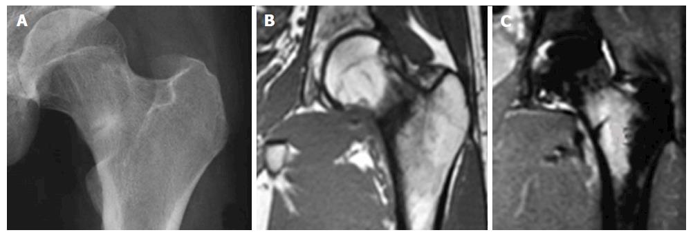
Figure 4 The management of an undisplaced compression sided femoral neck stress fracture.
A: Diagnostic antero-posterior radiograph; B: Diagnostic T1 sequence coronal-plane magnetic resonance imaging (MRI) view; C: Diagnostic short tau inversion recovery sequence coronal-plane MRI view.
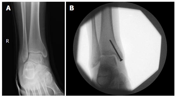
Figure 5 The management of an undisplaced completed medial malleolar stress fracture.
A: Pre-operative antero-posterior radiograph; B: Intra-operative antero-posterior radiograph.
- Citation: Robertson GAJ, Wood AM. Lower limb stress fractures in sport: Optimising their management and outcome. World J Orthop 2017; 8(3): 242-255
- URL: https://www.wjgnet.com/2218-5836/full/v8/i3/242.htm
- DOI: https://dx.doi.org/10.5312/wjo.v8.i3.242


