©The Author(s) 2017.
World J Orthop. Dec 18, 2017; 8(12): 861-873
Published online Dec 18, 2017. doi: 10.5312/wjo.v8.i12.861
Published online Dec 18, 2017. doi: 10.5312/wjo.v8.i12.861
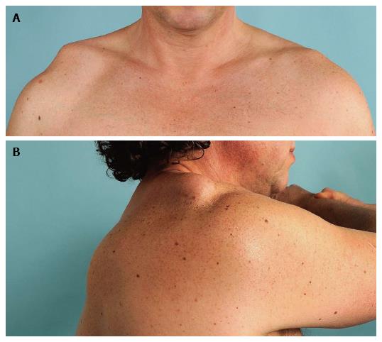
Figure 1 Digital pictures of a patient with a type-V acromioclavicular dislocation.
A: Anterior view; B: Lateral view: The shoulder is passively adducted in the horizontal plain to test horizontal stability. Note the horizontal instability in this case.
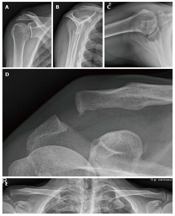
Figure 2 Standard radiographic series of the shoulder.
A: A true anterior-posterior view; B: Scapular Y lateral view; C: Axillary view; D: Zanca view; E: In case of acromioclavicular separation, a bilateral Zanca view can be useful.
Figure 3 Rockwood classification (Case courtesy of Dr Roberto Schubert, Radiopaedia.
org, rID: 19124).
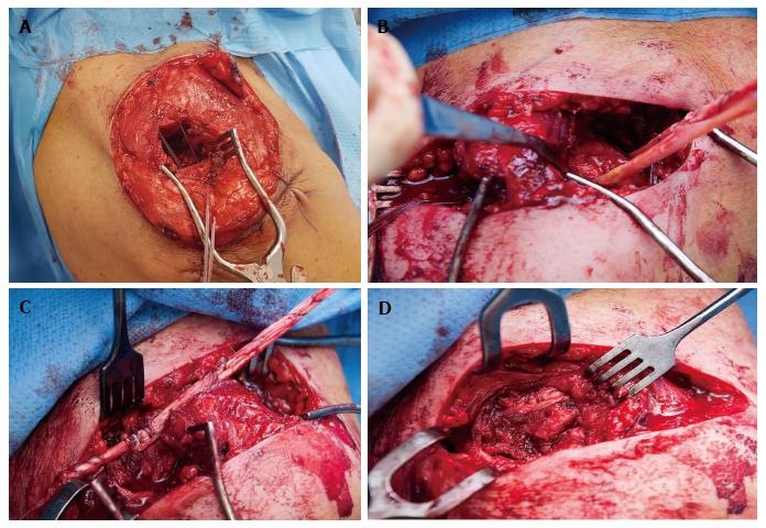
Figure 4 Intra-operative pictures of an autograft tendon reconstruction technique of the coracoclavicular joint without bone tunnels in combination with direct suture fixation of the acromioclavicular joint.
A: The lateral clavicle is resected, and a double nonabsorbale suture is used for AC joint repair; B-D: A semitendinosus tendon is passed under the coracoid and over the clavicle for CC joint repair. AC: Acromioclavicular; CC: Coracoclavicular.
- Citation: van Bergen CJA, van Bemmel AF, Alta TDW, van Noort A. New insights in the treatment of acromioclavicular separation. World J Orthop 2017; 8(12): 861-873
- URL: https://www.wjgnet.com/2218-5836/full/v8/i12/861.htm
- DOI: https://dx.doi.org/10.5312/wjo.v8.i12.861


