Copyright
©The Author(s) 2016.
World J Orthop. Sep 18, 2016; 7(9): 539-545
Published online Sep 18, 2016. doi: 10.5312/wjo.v7.i9.539
Published online Sep 18, 2016. doi: 10.5312/wjo.v7.i9.539
Figure 1 Well-molded cast.
A: Pre-reduction X-rays of pediatric forearm demonstrating a completely displaced distal both bone forearm fracture. Immediate post-reduction X-rays; B: Immediate post-reduction X-rays demonstrating adequate reduction with a flexion mold to prevent fracture from dorsal displacement; C: Three-months follow-up X-rays demonstrating completely healed fracture with near-anatomic alignment.
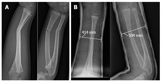
Figure 2 Cast index.
A: Initial X-rays of a pediatric forearm fracture with angulation; B: Immediate post-reduction X-rays demonstrating an appropriate cast application based on the cast index (< 0.8). Cast index = Saggital width/coronal width = 41.4 mm/54.4 mm = 0.76.
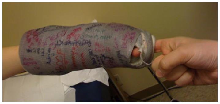
Figure 3 Loose cast.
Pediatric cast that was applied too loosely. The physician’s hand (right) was able to pull the cast off the child easily.
Figure 4 Cast spacers.
Cast spacers with four different size settings (3, 6, 9 and 12 mm).
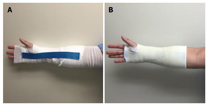
Figure 5 Saw Stop© protective strip (A and B).
This protective strip is placed on top of the webril layer before applying the fiberglass cast roll. The strip provides a durable protective layer for the cast saw to cut over.
- Citation: Nguyen S, McDowell M, Schlechter J. Casting: Pearls and pitfalls learned while caring for children’s fractures. World J Orthop 2016; 7(9): 539-545
- URL: https://www.wjgnet.com/2218-5836/full/v7/i9/539.htm
- DOI: https://dx.doi.org/10.5312/wjo.v7.i9.539


