Copyright
©The Author(s) 2015.
World J Orthop. Oct 18, 2015; 6(9): 688-704
Published online Oct 18, 2015. doi: 10.5312/wjo.v6.i9.688
Published online Oct 18, 2015. doi: 10.5312/wjo.v6.i9.688
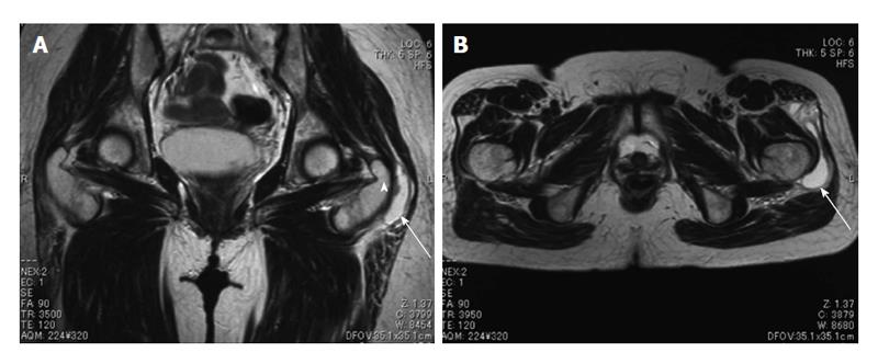
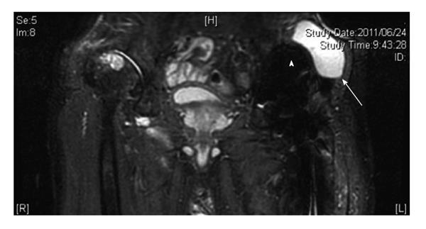
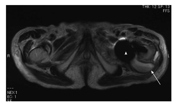
Figure 1 A 58-year-old female with greater trochanteric bursitis.
Coronal (A) and axial (B) T2-weighted magnetic resonance images of a cyst located at the characteristic position of the left greater trochanteric bursa (arrow). Arrowhead: Greater trochanter.
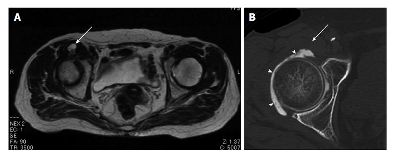
Figure 2 A 81-year-old female with femoral neuropathy caused by distended iliopsoas bursa.
Axial T2-weighted magnetic resonance image shows a cystic lesion, which extends anteriorly into the iliopsoas muscle (A). Computed tomography arthrography demonstrates no communication between the cyst (arrow) and right hip joint (B). Arrowheads: Contrast material.
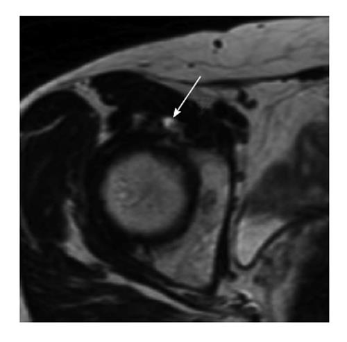
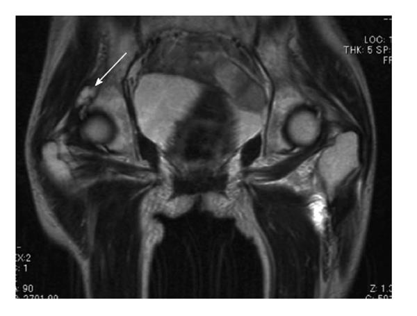
Figure 3 A 50-year-old female with a right hip pain.
Axial T2-weighted magnetic resonance image shows a labral tear and paralabral cyst (arrow) at the anterior aspect of the right hip joint.
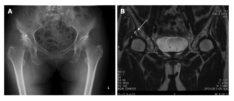
Figure 4 A 70-year-old female with a cystic lesion associated with osteoarthritis.
Plain radiograph of the lower pelvis reveals the secondary osteoarthritis of right hip due to acetabular dysplasia (arrowhead) (A). Coronal T2-weighted magnetic resonance image shows a cystic mass (arrow) located superiorly, connected to the right hip joint (B).
Figure 5 A 76-year-old male with a pseudotumor following cemented total hip arthroplasty, which was implanted 18 years earlier.
Coronal short tau inversion recovery magnetic resonance image shows a cystic lesion (arrow) adjacent to the acetabular component (arrowhead).
Figure 6 A 93-year-old male with an infection after hemiarthroplasty for the left femoral neck fracture.
Axial T2-weighted image reveals a cystic lesion with a fluid-fluid level (arrow) adjacent to the posterior aspect of the endoprosthesis (arrowhead).
Figure 7 A 71-year-old female with the left femoral neck fracture.
Coronal T2-weighted magnetic resonance image shows an asymptomatic multilocular cystic mass (arrow), which is communicated with the right hip joint.
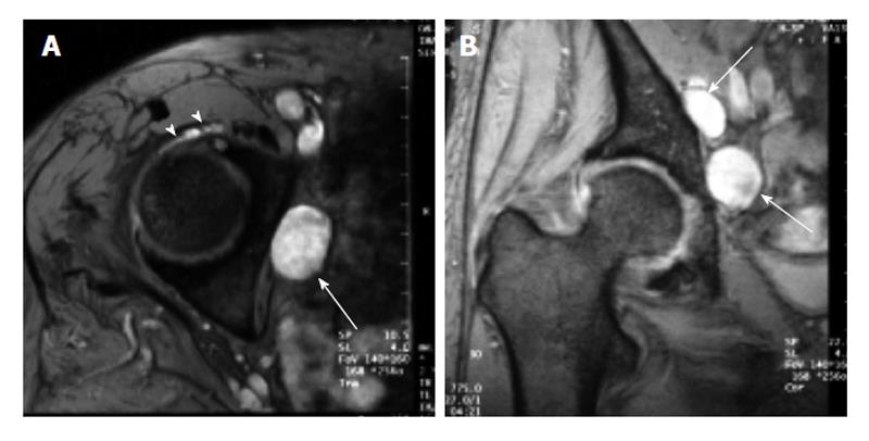
Figure 8 A 75-year-old female with obturator neuropathy.
Axial (A) and coronal (B) short tau inversion recovery magnetic resonance images show that the location of the cystic masses (arrows) is consistent with the site of obturator nerve (Ref: [26]). The stalk of the cyst was connected to the anterior joint capsule (arrowheads).
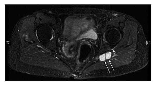
Figure 9 A 67-year-old female with a sciatica.
Axial short tau inversion recovery magnetic resonance image demonstrates cysts at the posterior aspect of the left acetubulum (arrows). The stalk is connected to the posterior joint capsule (arrowhead).
- Citation: Yukata K, Nakai S, Goto T, Ikeda Y, Shimaoka Y, Yamanaka I, Sairyo K, Hamawaki JI. Cystic lesion around the hip joint. World J Orthop 2015; 6(9): 688-704
- URL: https://www.wjgnet.com/2218-5836/full/v6/i9/688.htm
- DOI: https://dx.doi.org/10.5312/wjo.v6.i9.688


