Copyright
©The Author(s) 2015.
World J Orthop. Jul 18, 2015; 6(6): 469-482
Published online Jul 18, 2015. doi: 10.5312/wjo.v6.i6.469
Published online Jul 18, 2015. doi: 10.5312/wjo.v6.i6.469
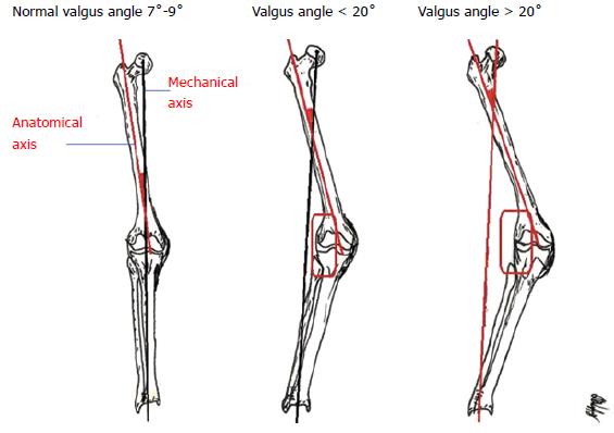
Figure 1 Mechanical and anatomical axis of the normal and valgus knee with deformity less or more of 20˚.
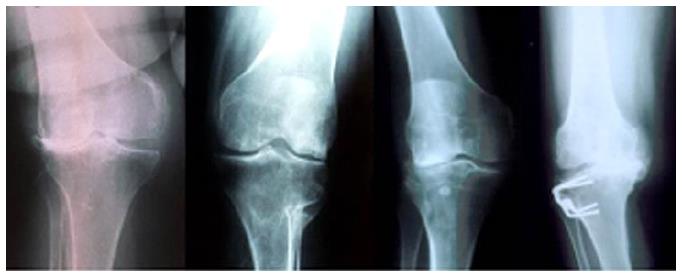
Figure 2 Pre-operative images of different valgus knees.
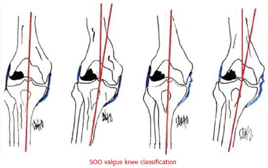
Figure 3 Societe d’Orthopedie de l’Ouest valgus knee classification.
SOO: Societe d’Orthopedie de l’Ouest.
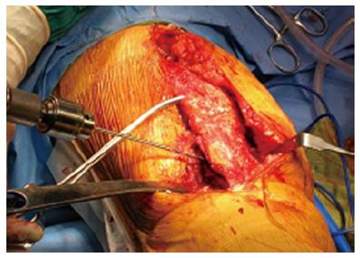
Figure 4 Anterolateral approach with tibial tubercle osteotomy.
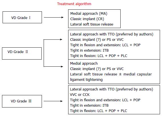
Figure 5 Treatment algorithm in valgus knee arthroplasty.
MA: Medial approach; CR: Cruciate retaining; TTO: Tibial tubercle osteotomy; PS: Posterior stabilize; VVC: Varus-valgus constrained; CCK: Constrained condylar knee; ITB: Iliotibial band; LCL: Lateral collateral ligament; POP: Popliteus tendon; PLC: Posterolateral corner.
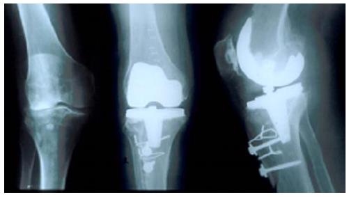
Figure 6 Pre- and Post-operative X-rays in valgus knee (18˚) with lateral approach and tibial tubercle osteotomy.
- Citation: Nikolopoulos D, Michos I, Safos G, Safos P. Current surgical strategies for total arthroplasty in valgus knee. World J Orthop 2015; 6(6): 469-482
- URL: https://www.wjgnet.com/2218-5836/full/v6/i6/469.htm
- DOI: https://dx.doi.org/10.5312/wjo.v6.i6.469


