©The Author(s) 2015.
World J Orthop. Mar 18, 2015; 6(2): 290-297
Published online Mar 18, 2015. doi: 10.5312/wjo.v6.i2.290
Published online Mar 18, 2015. doi: 10.5312/wjo.v6.i2.290
Figure 1 Forest plot for outlier incidence using magnetic resonance imaging.
Figure 2 Forest plot for outlier incidence using computerised tomography.
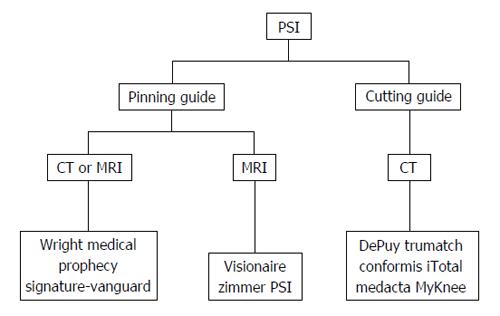
Figure 3 Summary of currently available patient-specific instrumentation systems.
PSI: Patient-specific instrumentation; CT: Computerised tomography; MRI: Magnetic resonance imaging.
-
Citation: Stirling P, Valsalan Mannambeth R, Soler A, Batta V, Malhotra RK, Kalairajah Y. Computerised tomography
vs magnetic resonance imaging for modeling of patient-specific instrumentation in total knee arthroplasty. World J Orthop 2015; 6(2): 290-297 - URL: https://www.wjgnet.com/2218-5836/full/v6/i2/290.htm
- DOI: https://dx.doi.org/10.5312/wjo.v6.i2.290


